+ データを開く
- 基本情報
基本情報
| 登録情報 | データベース: PDB / ID: 2brd | |||||||||
|---|---|---|---|---|---|---|---|---|---|---|
| タイトル | CRYSTAL STRUCTURE OF BACTERIORHODOPSIN IN PURPLE MEMBRANE | |||||||||
 要素 要素 | BACTERIORHODOPSIN | |||||||||
 キーワード キーワード | PHOTORECEPTOR / PROTON PUMP / MEMBRANE PROTEIN / RETINAL PROTEIN / TWO-DIMENSIONAL CRYSTAL | |||||||||
| 機能・相同性 |  機能・相同性情報 機能・相同性情報light-driven active monoatomic ion transmembrane transporter activity / photoreceptor activity / phototransduction / monoatomic ion channel activity / proton transmembrane transport / plasma membrane 類似検索 - 分子機能 | |||||||||
| 生物種 |  Halobacterium salinarum (好塩性) Halobacterium salinarum (好塩性) | |||||||||
| 手法 | 電子線結晶学 / クライオ電子顕微鏡法 / 解像度: 3.5 Å | |||||||||
 データ登録者 データ登録者 | Henderson, R. / Grigorieff, N. | |||||||||
 引用 引用 |  ジャーナル: J Mol Biol / 年: 1996 ジャーナル: J Mol Biol / 年: 1996タイトル: Electron-crystallographic refinement of the structure of bacteriorhodopsin. 著者: N Grigorieff / T A Ceska / K H Downing / J M Baldwin / R Henderson /  要旨: Using electron diffraction data corrected for diffuse scattering together with additional phase information from 30 new images of tilted specimens, an improved experimental density map has been ...Using electron diffraction data corrected for diffuse scattering together with additional phase information from 30 new images of tilted specimens, an improved experimental density map has been calculated for bacteriorhodopsin. The atomic model has then been rebuilt into this new map with particular attention to the surface loops. All the residues from 7 to 227 as well as ten lipid molecules are now included, although a few amino acid residues in three of the six surface loops, about half of the lipid hydrophobic chains and all of the lipid head groups are disordered. The model has then been refined against the experimental diffraction amplitudes to an R-factor of 28% at 3.5 angstrom resolution with strict geometry (0.005 angstrom) bond length deviation) using the improvement of the "free" phase residual between calculated and experimental phases from images as an objective criterion of accuracy. For the refinement some new programs were developed to restrain the number of parameters, to be compatible with the limited resolution of our data. In the final refined model of the protein (2BRD), compared with earlier co-ordinates (1BRD), helix D has been moved towards the cytoplasm by almost 4 angstrom, and the overall accuracy of the co-ordinates of residues in the other six helices has been improved. As a result the positions of nearly all the important residues in bacteriorhodopsin are now well determined. In particular, the buried, protonated Asp115 is 7 angstrom from, and so not in contact with, the retinal and Met118 forms a cap on the pocket occupied by the beta-ionone ring. No clear density exists for the side-chain of Arg82, which forms a central part of the extracellular half-channel. The only arginine side-chain built into good density is that of Arg134 at the extracellular end of helix E, the others being disordered near one of the two surfaces. The interpretation of the end of helix F on the extracellular surface is now clearer; an extra loose helical turn has been built bringing the side-chain of Glu194 close to Arg134 to form a probable salt bridge. The model provides an improved framework for understanding the mechanism of the light-driven proton pumping. A number of cavities that could contain water molecules were found by searching the refined model, most of them above or below the Schiff base in the half-channels leading to the two surfaces. The ordered and disordered regions of the structure are described by the temperature factor distribution. #1:  ジャーナル: J.Mol.Biol. / 年: 1990 ジャーナル: J.Mol.Biol. / 年: 1990タイトル: Analysis of High-Resolution Electron Diffraction Patterns from Purple Membrane Labelled with Heavy-Atoms 著者: Ceska, T.A. / Henderson, R. #2:  ジャーナル: J.Mol.Biol. / 年: 1990 ジャーナル: J.Mol.Biol. / 年: 1990タイトル: Model for the Structure of Bacteriorhodopsin Based on High-Resolution Electron Cryo-Microscopy 著者: Henderson, R. / Baldwin, J.M. / Ceska, T.A. / Zemlin, F. / Beckmann, E. / Downing, K.H. #3:  ジャーナル: Nature / 年: 1975 ジャーナル: Nature / 年: 1975タイトル: Three-Dimensional Model of Purple Membrane Obtained by Electron Microscopy 著者: Henderson, R. / Unwin, P.N. | |||||||||
| 履歴 |
|
- 構造の表示
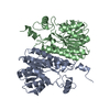
構造の表示
| ムービー |
 ムービービューア ムービービューア |
|---|---|
| 構造ビューア | 分子:  Molmil Molmil Jmol/JSmol Jmol/JSmol |
- ダウンロードとリンク
- ダウンロード
| PDBx/mmCIF形式 |  2brd.cif.gz 2brd.cif.gz | 70 KB | 表示 |  PDBx/mmCIF形式 PDBx/mmCIF形式 |
|---|---|---|---|---|
| PDB形式 |  pdb2brd.ent.gz pdb2brd.ent.gz | 54.1 KB | 表示 |  PDB形式 PDB形式 |
| PDBx/mmJSON形式 |  2brd.json.gz 2brd.json.gz | ツリー表示 |  PDBx/mmJSON形式 PDBx/mmJSON形式 | |
| その他 |  その他のダウンロード その他のダウンロード |
-検証レポート
| 文書・要旨 |  2brd_validation.pdf.gz 2brd_validation.pdf.gz | 904.3 KB | 表示 |  wwPDB検証レポート wwPDB検証レポート |
|---|---|---|---|---|
| 文書・詳細版 |  2brd_full_validation.pdf.gz 2brd_full_validation.pdf.gz | 942.2 KB | 表示 | |
| XML形式データ |  2brd_validation.xml.gz 2brd_validation.xml.gz | 14.5 KB | 表示 | |
| CIF形式データ |  2brd_validation.cif.gz 2brd_validation.cif.gz | 18.1 KB | 表示 | |
| アーカイブディレクトリ |  https://data.pdbj.org/pub/pdb/validation_reports/br/2brd https://data.pdbj.org/pub/pdb/validation_reports/br/2brd ftp://data.pdbj.org/pub/pdb/validation_reports/br/2brd ftp://data.pdbj.org/pub/pdb/validation_reports/br/2brd | HTTPS FTP |
-関連構造データ
- リンク
リンク
- 集合体
集合体
| 登録構造単位 | 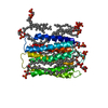
| ||||||||
|---|---|---|---|---|---|---|---|---|---|
| 1 | 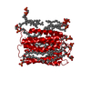
| ||||||||
| 単位格子 |
|
- 要素
要素
| #1: タンパク質 | 分子量: 26797.381 Da / 分子数: 1 / 由来タイプ: 組換発現 / 由来: (組換発現)  Halobacterium salinarum (好塩性) / 株: R1 / 参照: UniProt: P02945 Halobacterium salinarum (好塩性) / 株: R1 / 参照: UniProt: P02945 | ||||
|---|---|---|---|---|---|
| #2: 化合物 | ChemComp-DPG / #3: 化合物 | ChemComp-RET / | Has protein modification | Y | |
-実験情報
-実験
| 実験 | 手法: 電子線結晶学 |
|---|---|
| EM実験 | 試料の集合状態: 2D ARRAY / 3次元再構成法: 電子線結晶学 |
- 試料調製
試料調製
| 構成要素 | 名称: BACTERIORHODOPSIN CRYSTAL / タイプ: COMPLEX |
|---|---|
| 試料 | 包埋: YES / シャドウイング: NO / 染色: NO / 凍結: YES |
| 結晶 | マシュー密度: 4.24 Å3/Da / 溶媒含有率: 70.97 % |
| 結晶化 | *PLUS 手法: other / 詳細: electron-crystallographic method |
-データ収集
| EM imaging | Specimen-ID: 1
| ||||||||||||||||||||||||||||||||||||||||
|---|---|---|---|---|---|---|---|---|---|---|---|---|---|---|---|---|---|---|---|---|---|---|---|---|---|---|---|---|---|---|---|---|---|---|---|---|---|---|---|---|---|
| 撮影 |
| ||||||||||||||||||||||||||||||||||||||||
| 放射光源 | 波長: 0.033 | ||||||||||||||||||||||||||||||||||||||||
| 検出器 | 検出器: FILM / 日付: 1986年1月1日 | ||||||||||||||||||||||||||||||||||||||||
| 放射 | 単色(M)・ラウエ(L): M / 散乱光タイプ: electron | ||||||||||||||||||||||||||||||||||||||||
| 放射波長 | 波長: 0.033 Å / 相対比: 1 | ||||||||||||||||||||||||||||||||||||||||
| 反射 | 解像度: 2.8→54 Å / Num. obs: 6750 / % possible obs: 62.3 % / Observed criterion σ(I): 0 / 冗長度: 18 % / Rmerge(I) obs: 0.15 |
- 解析
解析
| ソフトウェア |
| ||||||||||||||||||||||||||||||||||||||||||||||||||||||||||||||||||||||||||||||||||||
|---|---|---|---|---|---|---|---|---|---|---|---|---|---|---|---|---|---|---|---|---|---|---|---|---|---|---|---|---|---|---|---|---|---|---|---|---|---|---|---|---|---|---|---|---|---|---|---|---|---|---|---|---|---|---|---|---|---|---|---|---|---|---|---|---|---|---|---|---|---|---|---|---|---|---|---|---|---|---|---|---|---|---|---|---|---|
| 3次元再構成 | 対称性のタイプ: 2D CRYSTAL | ||||||||||||||||||||||||||||||||||||||||||||||||||||||||||||||||||||||||||||||||||||
| 精密化 | 解像度: 3.5→30 Å / σ(F): 0 /
| ||||||||||||||||||||||||||||||||||||||||||||||||||||||||||||||||||||||||||||||||||||
| 原子変位パラメータ | Biso mean: 114 Å2 | ||||||||||||||||||||||||||||||||||||||||||||||||||||||||||||||||||||||||||||||||||||
| 精密化ステップ | サイクル: LAST / 解像度: 3.5→30 Å
| ||||||||||||||||||||||||||||||||||||||||||||||||||||||||||||||||||||||||||||||||||||
| 拘束条件 |
| ||||||||||||||||||||||||||||||||||||||||||||||||||||||||||||||||||||||||||||||||||||
| ソフトウェア | *PLUS 名称: PROLSQ / 分類: refinement | ||||||||||||||||||||||||||||||||||||||||||||||||||||||||||||||||||||||||||||||||||||
| 精密化 | *PLUS Rfactor obs: 0.18 | ||||||||||||||||||||||||||||||||||||||||||||||||||||||||||||||||||||||||||||||||||||
| 溶媒の処理 | *PLUS | ||||||||||||||||||||||||||||||||||||||||||||||||||||||||||||||||||||||||||||||||||||
| 原子変位パラメータ | *PLUS |
 ムービー
ムービー コントローラー




 PDBj
PDBj


 FIELD EMISSION GUN