 Movie
Movie Controller

[English] 日本語
 Yorodumi
- PDB-2a89: Monomeric Sarcosine Oxidase: Structure of a covalently flavinylat... -
+ Open data

- Basic information
| Entry | Database: PDB / ID: 2a89 | ||||||
|---|---|---|---|---|---|---|---|
| Title | Monomeric Sarcosine Oxidase: Structure of a covalently flavinylated amine oxidizing enzyme | ||||||
 Components Components | Monomeric sarcosine oxidase | ||||||
 Keywords Keywords | OXIDOREDUCTASE / FLAVOPROTEIN / OXIDASE | ||||||
| Function / homology |  Function and homology information Function and homology informationsarcosine oxidase (formaldehyde-forming) / sarcosine oxidase activity / flavin adenine dinucleotide binding / cytoplasm Similarity search - Function | ||||||
| Biological species |  | ||||||
| Method |  X-RAY DIFFRACTION / MOLECULAR REPLACEMENT / Resolution: 1.85 Å X-RAY DIFFRACTION / MOLECULAR REPLACEMENT / Resolution: 1.85 Å | ||||||
 Authors Authors | Chen, Z.-W. / Zhao, G. / Martinovic, S. / Jorns, M.S. / Mathews, F.S. | ||||||
 Citation Citation | Journal: Biochemistry / Year: 2005 Title: Structure of the sodium borohydride-reduced N-(cyclopropyl)glycine adduct of the flavoenzyme monomeric sarcosine oxidase. Authors: Chen, Z.-W. / Zhao, G. / Martinovic, S. / Jorns, M.S. / Mathews, F.S. #1: Journal: Structure / Year: 1999Title: Monomeric Sarcosine Oxidase: Structure of a covalently flavinylated amine oxidizing enzyme Authors: Trickey, P. / Wagner, M.A. / Jorn, M.S. / Mathews, F.S. | ||||||
| History |
|
- Structure visualization
| Structure viewer | Molecule: MolmilJmol/JSmol |
|---|
- Downloads & links
-Download
| PDBx/mmCIF format | 2a89.cif.gz | 190.7 KB | Display | PDBx/mmCIF format |
|---|---|---|---|---|
| PDB format | pdb2a89.ent.gz | 146.9 KB | Display | PDB format |
| PDBx/mmJSON format | 2a89.json.gz | Tree view | PDBx/mmJSON format | |
| Others |  Other downloads Other downloads |
-Validation report
| Summary document | 2a89_validation.pdf.gz | 927.7 KB | Display | wwPDB validaton report |
|---|---|---|---|---|
| Full document | 2a89_full_validation.pdf.gz | 949.3 KB | Display | |
| Data in XML | 2a89_validation.xml.gz | 42 KB | Display | |
| Data in CIF | 2a89_validation.cif.gz | 64.1 KB | Display | |
| Arichive directory | https://data.pdbj.org/pub/pdb/validation_reports/a8/2a89ftp://data.pdbj.org/pub/pdb/validation_reports/a8/2a89 | HTTPS FTP |
-Related structure data
| Related structure data |  1l9f S: Starting model for refinement |
|---|---|
| Similar structure data |


-Links
 PDBj
PDBj- Assembly
| Deposited unit | 
| ||||||||
|---|---|---|---|---|---|---|---|---|---|
| 1 | 
| ||||||||
| 2 | 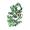
| ||||||||
| Unit cell |
| ||||||||
| Details | The biological assembly is monomer. |
-Components
| #1: Protein | Mass: 43101.355 Da / Num. of mol.: 2 Source method: isolated from a genetically manipulated source Source: (gene. exp.) References: UniProt: P40859, sarcosine oxidase (formaldehyde-forming) #2: Chemical |   Mass: 35.453 Da / Num. of mol.: 3 / Source method: obtained synthetically / Formula: Cl Mass: 35.453 Da / Num. of mol.: 3 / Source method: obtained synthetically / Formula: Cl#3: Chemical |   Mass: 847.661 Da / Num. of mol.: 2 / Source method: obtained synthetically / Formula: C30H43N9O16P2 Mass: 847.661 Da / Num. of mol.: 2 / Source method: obtained synthetically / Formula: C30H43N9O16P2#4: Chemical | ChemComp-PO4 / |   Mass: 94.971 Da / Num. of mol.: 1 / Source method: obtained synthetically / Formula: PO4 Mass: 94.971 Da / Num. of mol.: 1 / Source method: obtained synthetically / Formula: PO4#5: Water | ChemComp-HOH / |  Mass: 18.015 Da / Num. of mol.: 980 / Source method: isolated from a natural source / Formula: H2O Mass: 18.015 Da / Num. of mol.: 980 / Source method: isolated from a natural source / Formula: H2OHas protein modification | Y | |
|---|
-Experimental details
-Experiment
| Experiment | Method: X-RAY DIFFRACTION / Number of used crystals: 1 |
|---|
- Sample preparation
| Crystal | Density Matthews: 2.12 Å3/Da / Density % sol: 42 % |
|---|---|
| Crystal grow | Temperature: 296 K / Method: vapor diffusion, sitting drop / pH: 6.7 Details: Na/K phosphate, pH 6.7, VAPOR DIFFUSION, SITTING DROP, temperature 296K |
-Data collection
| Diffraction | Mean temperature: 100 K |
|---|---|
| Diffraction source | Source: ROTATING ANODE / Type: RIGAKU RU200 / Wavelength: 1.5418 Å |
| Detector | Type: RIGAKU RAXIS IV / Detector: IMAGE PLATE / Date: Jul 7, 2000 |
| Radiation | Monochromator: GRAPHITE / Protocol: SINGLE WAVELENGTH / Monochromatic (M) / Laue (L): M / Scattering type: x-ray |
| Radiation wavelength | Wavelength: 1.5418 Å / Relative weight: 1 |
| Reflection | Resolution: 1.85→40 Å / Num. all: 62473 / Num. obs: 55726 / % possible obs: 89.2 % / Observed criterion σ(F): 0 / Observed criterion σ(I): 0 / Redundancy: 3.85 % / Biso Wilson estimate: 5.5 Å2 / Rmerge(I) obs: 0.068 / Net I/σ(I): 12.4 |
| Reflection shell | Resolution: 1.85→1.92 Å / Redundancy: 2.7 % / Rmerge(I) obs: 0.254 / Mean I/σ(I) obs: 2.4 / Num. unique all: 3058 / % possible all: 49.4 |
- Processing
| Software |
| |||||||||||||||||||||||||||||||||||||||||||||||||
|---|---|---|---|---|---|---|---|---|---|---|---|---|---|---|---|---|---|---|---|---|---|---|---|---|---|---|---|---|---|---|---|---|---|---|---|---|---|---|---|---|---|---|---|---|---|---|---|---|---|---|
| Refinement | Method to determine structure: MOLECULAR REPLACEMENT Starting model: PDB ENTRY 1L9F 1l9f Resolution: 1.85→40 Å / Isotropic thermal model: Isotropic / Cross valid method: THROUGHOUT / σ(F): 0 / σ(I): 0 / Stereochemistry target values: Engh & Huber
| |||||||||||||||||||||||||||||||||||||||||||||||||
| Displacement parameters | Biso mean: 16.6 Å2 | |||||||||||||||||||||||||||||||||||||||||||||||||
| Refine analyze |
| |||||||||||||||||||||||||||||||||||||||||||||||||
| Refinement step | Cycle: LAST / Resolution: 1.85→40 Å
| |||||||||||||||||||||||||||||||||||||||||||||||||
| Refine LS restraints |
| |||||||||||||||||||||||||||||||||||||||||||||||||
| LS refinement shell |
|
