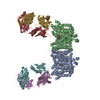
+ データを開く
- 基本情報
基本情報
| 登録情報 |  | |||||||||
|---|---|---|---|---|---|---|---|---|---|---|
| タイトル | Band 3-Glycophorin A complex, outward facing | |||||||||
 マップデータ マップデータ | Main map used for building/refinement. Density modified using phenix.resolve_cryo_em and cropped to minimal box, then resampled to 2x finer spacing to aid visualization (original pixel size was 0.83) | |||||||||
 試料 試料 |
| |||||||||
 キーワード キーワード | Membrane Protein / Anion Exchange / Erythrocyte / Glycoprotein / TRANSPORT PROTEIN | |||||||||
| 機能・相同性 |  機能・相同性情報 機能・相同性情報pH elevation / Defective SLC4A1 causes hereditary spherocytosis type 4 (HSP4), distal renal tubular acidosis (dRTA) and dRTA with hemolytic anemia (dRTA-HA) / negative regulation of urine volume / Bicarbonate transporters / intracellular monoatomic ion homeostasis / ankyrin-1 complex / plasma membrane phospholipid scrambling / monoatomic anion transmembrane transporter activity / chloride:bicarbonate antiporter activity / solute:inorganic anion antiporter activity ...pH elevation / Defective SLC4A1 causes hereditary spherocytosis type 4 (HSP4), distal renal tubular acidosis (dRTA) and dRTA with hemolytic anemia (dRTA-HA) / negative regulation of urine volume / Bicarbonate transporters / intracellular monoatomic ion homeostasis / ankyrin-1 complex / plasma membrane phospholipid scrambling / monoatomic anion transmembrane transporter activity / chloride:bicarbonate antiporter activity / solute:inorganic anion antiporter activity / bicarbonate transport / bicarbonate transmembrane transporter activity / monoatomic anion transport / chloride transport / chloride transmembrane transporter activity / ankyrin binding / negative regulation of glycolytic process through fructose-6-phosphate / hemoglobin binding / cortical cytoskeleton / erythrocyte development / protein-membrane adaptor activity / chloride transmembrane transport / protein localization to plasma membrane / regulation of intracellular pH / Cell surface interactions at the vascular wall / Erythrocytes take up oxygen and release carbon dioxide / Erythrocytes take up carbon dioxide and release oxygen / transmembrane transport / Z disc / cytoplasmic side of plasma membrane / blood coagulation / virus receptor activity / basolateral plasma membrane / blood microparticle / protein homodimerization activity / extracellular exosome / nucleoplasm / identical protein binding / membrane / plasma membrane / cytosol 類似検索 - 分子機能 | |||||||||
| 生物種 |  Homo sapiens (ヒト) Homo sapiens (ヒト) | |||||||||
| 手法 | 単粒子再構成法 / クライオ電子顕微鏡法 / 解像度: 2.35 Å | |||||||||
 データ登録者 データ登録者 | Vallese F / Kim K / Yen LY / Johnston JD / Noble AJ / Cali T / Clarke OB | |||||||||
| 資金援助 | 1件
| |||||||||
 引用 引用 |  ジャーナル: Nat Struct Mol Biol / 年: 2022 ジャーナル: Nat Struct Mol Biol / 年: 2022タイトル: Architecture of the human erythrocyte ankyrin-1 complex. 著者: Francesca Vallese / Kookjoo Kim / Laura Y Yen / Jake D Johnston / Alex J Noble / Tito Calì / Oliver Biggs Clarke /   要旨: The stability and shape of the erythrocyte membrane is provided by the ankyrin-1 complex, but how it tethers the spectrin-actin cytoskeleton to the lipid bilayer and the nature of its association ...The stability and shape of the erythrocyte membrane is provided by the ankyrin-1 complex, but how it tethers the spectrin-actin cytoskeleton to the lipid bilayer and the nature of its association with the band 3 anion exchanger and the Rhesus glycoproteins remains unknown. Here we present structures of ankyrin-1 complexes purified from human erythrocytes. We reveal the architecture of a core complex of ankyrin-1, the Rhesus proteins RhAG and RhCE, the band 3 anion exchanger, protein 4.2, glycophorin A and glycophorin B. The distinct T-shaped conformation of membrane-bound ankyrin-1 facilitates recognition of RhCE and, unexpectedly, the water channel aquaporin-1. Together, our results uncover the molecular details of ankyrin-1 association with the erythrocyte membrane, and illustrate the mechanism of ankyrin-mediated membrane protein clustering. | |||||||||
| 履歴 |
|
- 構造の表示
構造の表示
| 添付画像 |
|---|
- ダウンロードとリンク
-EMDBアーカイブ
| マップデータ |  emd_26874.map.gz emd_26874.map.gz | 224.3 MB |  EMDBマップデータ形式 EMDBマップデータ形式 | |
|---|---|---|---|---|
| ヘッダ (付随情報) |  emd-26874-v30.xml emd-26874-v30.xml emd-26874.xml emd-26874.xml | 37.5 KB 37.5 KB | 表示 表示 |  EMDBヘッダ EMDBヘッダ |
| FSC (解像度算出) |  emd_26874_fsc.xml emd_26874_fsc.xml | 11.3 KB | 表示 |  FSCデータファイル FSCデータファイル |
| 画像 |  emd_26874.png emd_26874.png | 108.3 KB | ||
| Filedesc metadata |  emd-26874.cif.gz emd-26874.cif.gz | 7.7 KB | ||
| その他 |  emd_26874_additional_1.map.gz emd_26874_additional_1.map.gz emd_26874_additional_2.map.gz emd_26874_additional_2.map.gz emd_26874_additional_3.map.gz emd_26874_additional_3.map.gz emd_26874_additional_4.map.gz emd_26874_additional_4.map.gz emd_26874_additional_5.map.gz emd_26874_additional_5.map.gz emd_26874_half_map_1.map.gz emd_26874_half_map_1.map.gz emd_26874_half_map_2.map.gz emd_26874_half_map_2.map.gz | 115.7 MB 118 MB 115.7 MB 395.3 KB 62.5 MB 223 MB 223 MB | ||
| アーカイブディレクトリ |  http://ftp.pdbj.org/pub/emdb/structures/EMD-26874 http://ftp.pdbj.org/pub/emdb/structures/EMD-26874 ftp://ftp.pdbj.org/pub/emdb/structures/EMD-26874 ftp://ftp.pdbj.org/pub/emdb/structures/EMD-26874 | HTTPS FTP |
-検証レポート
| 文書・要旨 |  emd_26874_validation.pdf.gz emd_26874_validation.pdf.gz | 872.7 KB | 表示 |  EMDB検証レポート EMDB検証レポート |
|---|---|---|---|---|
| 文書・詳細版 |  emd_26874_full_validation.pdf.gz emd_26874_full_validation.pdf.gz | 872.2 KB | 表示 | |
| XML形式データ |  emd_26874_validation.xml.gz emd_26874_validation.xml.gz | 21.4 KB | 表示 | |
| CIF形式データ |  emd_26874_validation.cif.gz emd_26874_validation.cif.gz | 27.3 KB | 表示 | |
| アーカイブディレクトリ |  https://ftp.pdbj.org/pub/emdb/validation_reports/EMD-26874 https://ftp.pdbj.org/pub/emdb/validation_reports/EMD-26874 ftp://ftp.pdbj.org/pub/emdb/validation_reports/EMD-26874 ftp://ftp.pdbj.org/pub/emdb/validation_reports/EMD-26874 | HTTPS FTP |
-関連構造データ
| 関連構造データ |  7uz3MC  7uzeC  7uzqC  7uzsC 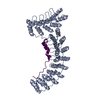 7uzuC  7uzvC  7v07C  7v0kC 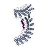 7v0mC  7v0qC  7v0sC  7v0tC  7v0uC 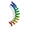 7v0xC  7v0yC  7v19C  8crqC  8crrC  8crtC  8cs9C  8cslC 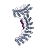 8csvC  8cswC  8csxC  8csyC  8ct2C  8ct3C  8cteC C: 同じ文献を引用 ( M: このマップから作成された原子モデル |
|---|---|
| 類似構造データ | 類似検索 - 機能・相同性  F&H 検索 F&H 検索 |
- リンク
リンク
| EMDBのページ |  EMDB (EBI/PDBe) / EMDB (EBI/PDBe) /  EMDataResource EMDataResource |
|---|
- マップ
マップ
| ファイル |  ダウンロード / ファイル: emd_26874.map.gz / 形式: CCP4 / 大きさ: 240.5 MB / タイプ: IMAGE STORED AS FLOATING POINT NUMBER (4 BYTES) ダウンロード / ファイル: emd_26874.map.gz / 形式: CCP4 / 大きさ: 240.5 MB / タイプ: IMAGE STORED AS FLOATING POINT NUMBER (4 BYTES) | ||||||||||||||||||||||||||||||||||||
|---|---|---|---|---|---|---|---|---|---|---|---|---|---|---|---|---|---|---|---|---|---|---|---|---|---|---|---|---|---|---|---|---|---|---|---|---|---|
| 注釈 | Main map used for building/refinement. Density modified using phenix.resolve_cryo_em and cropped to minimal box, then resampled to 2x finer spacing to aid visualization (original pixel size was 0.83) | ||||||||||||||||||||||||||||||||||||
| 投影像・断面図 | 画像のコントロール
画像は Spider により作成 | ||||||||||||||||||||||||||||||||||||
| ボクセルのサイズ | X=Y=Z: 0.415 Å | ||||||||||||||||||||||||||||||||||||
| 密度 |
| ||||||||||||||||||||||||||||||||||||
| 対称性 | 空間群: 1 | ||||||||||||||||||||||||||||||||||||
| 詳細 | EMDB XML:
|
-添付データ
-追加マップ: Unfiltered half map 1. Used for FSC calculation.
| ファイル | emd_26874_additional_1.map | ||||||||||||
|---|---|---|---|---|---|---|---|---|---|---|---|---|---|
| 注釈 | Unfiltered half map 1. Used for FSC calculation. | ||||||||||||
| 投影像・断面図 |
| ||||||||||||
| 密度ヒストグラム |
-追加マップ: Map auto-sharpened with B=48 (not cropped).
| ファイル | emd_26874_additional_2.map | ||||||||||||
|---|---|---|---|---|---|---|---|---|---|---|---|---|---|
| 注釈 | Map auto-sharpened with B=48 (not cropped). | ||||||||||||
| 投影像・断面図 |
| ||||||||||||
| 密度ヒストグラム |
-追加マップ: Unfiltered half map 2. Used for FSC calculation.
| ファイル | emd_26874_additional_3.map | ||||||||||||
|---|---|---|---|---|---|---|---|---|---|---|---|---|---|
| 注釈 | Unfiltered half map 2. Used for FSC calculation. | ||||||||||||
| 投影像・断面図 |
| ||||||||||||
| 密度ヒストグラム |
-追加マップ: Mask used for FSC calculation.
| ファイル | emd_26874_additional_4.map | ||||||||||||
|---|---|---|---|---|---|---|---|---|---|---|---|---|---|
| 注釈 | Mask used for FSC calculation. | ||||||||||||
| 投影像・断面図 |
| ||||||||||||
| 密度ヒストグラム |
-追加マップ: Unsharpened map (not cropped).
| ファイル | emd_26874_additional_5.map | ||||||||||||
|---|---|---|---|---|---|---|---|---|---|---|---|---|---|
| 注釈 | Unsharpened map (not cropped). | ||||||||||||
| 投影像・断面図 |
| ||||||||||||
| 密度ヒストグラム |
-ハーフマップ: Unfiltered half map 2, cropped and resampled to...
| ファイル | emd_26874_half_map_1.map | ||||||||||||
|---|---|---|---|---|---|---|---|---|---|---|---|---|---|
| 注釈 | Unfiltered half map 2, cropped and resampled to match the main map and model, but otherwise unaltered. Original half maps and FSC mask are provided as additional maps. | ||||||||||||
| 投影像・断面図 |
| ||||||||||||
| 密度ヒストグラム |
-ハーフマップ: Unfiltered half map 1, cropped and resampled to...
| ファイル | emd_26874_half_map_2.map | ||||||||||||
|---|---|---|---|---|---|---|---|---|---|---|---|---|---|
| 注釈 | Unfiltered half map 1, cropped and resampled to match the main map and model, but otherwise unaltered. Original half maps are provided as additional maps. | ||||||||||||
| 投影像・断面図 |
| ||||||||||||
| 密度ヒストグラム |
- 試料の構成要素
-全体 : Band 3 anion exchanger complexed with glycophorin A, in outward f...
| 全体 | 名称: Band 3 anion exchanger complexed with glycophorin A, in outward facing state. |
|---|---|
| 要素 |
|
-超分子 #1: Band 3 anion exchanger complexed with glycophorin A, in outward f...
| 超分子 | 名称: Band 3 anion exchanger complexed with glycophorin A, in outward facing state. タイプ: complex / ID: 1 / 親要素: 0 / 含まれる分子: #1-#2 詳細: Particle set isolated by 3D classification from mixture mostly containing ankyrin complexes. |
|---|---|
| 由来(天然) | 生物種:  Homo sapiens (ヒト) / 器官: Blood / 組織: Erythrocytes / 細胞中の位置: Plasma membrane Homo sapiens (ヒト) / 器官: Blood / 組織: Erythrocytes / 細胞中の位置: Plasma membrane |
-分子 #1: Glycophorin-A
| 分子 | 名称: Glycophorin-A / タイプ: protein_or_peptide / ID: 1 / コピー数: 2 / 光学異性体: LEVO |
|---|---|
| 由来(天然) | 生物種:  Homo sapiens (ヒト) / 器官: Blood / 組織: Erythrocytes Homo sapiens (ヒト) / 器官: Blood / 組織: Erythrocytes |
| 分子量 | 理論値: 16.348433 KDa |
| 配列 | 文字列: MYGKIIFVLL LSEIVSISAS STTGVAMHTS TSSSVTKSYI SSQTNDTHKR DTYAATPRAH EVSEISVRTV YPPEEETGER VQLAHHFSE PEITLIIFGV MAGVIGTILL ISYGIRRLIK KSPSDVKPLP SPDTDVPLSS VEIENPETSD Q UniProtKB: Glycophorin-A |
-分子 #2: Band 3 anion transport protein
| 分子 | 名称: Band 3 anion transport protein / タイプ: protein_or_peptide / ID: 2 / コピー数: 2 / 光学異性体: LEVO |
|---|---|
| 由来(天然) | 生物種:  Homo sapiens (ヒト) / 器官: Blood / 組織: Erythrocytes Homo sapiens (ヒト) / 器官: Blood / 組織: Erythrocytes |
| 分子量 | 理論値: 101.883859 KDa |
| 配列 | 文字列: MEELQDDYED MMEENLEQEE YEDPDIPESQ MEEPAAHDTE ATATDYHTTS HPGTHKVYVE LQELVMDEKN QELRWMEAAR WVQLEENLG ENGAWGRPHL SHLTFWSLLE LRRVFTKGTV LLDLQETSLA GVANQLLDRF IFEDQIRPQD REELLRALLL K HSHAGELE ...文字列: MEELQDDYED MMEENLEQEE YEDPDIPESQ MEEPAAHDTE ATATDYHTTS HPGTHKVYVE LQELVMDEKN QELRWMEAAR WVQLEENLG ENGAWGRPHL SHLTFWSLLE LRRVFTKGTV LLDLQETSLA GVANQLLDRF IFEDQIRPQD REELLRALLL K HSHAGELE ALGGVKPAVL TRSGDPSQPL LPQHSSLETQ LFCEQGDGGT EGHSPSGILE KIPPDSEATL VLVGRADFLE QP VLGFVRL QEAAELEAVE LPVPIRFLFV LLGPEAPHID YTQLGRAAAT LMSERVFRID AYMAQSRGEL LHSLEGFLDC SLV LPPTDA PSEQALLSLV PVQRELLRRR YQSSPAKPDS SFYKGLDLNG GPDDPLQQTG QLFGGLVRDI RRRYPYYLSD ITDA FSPQV LAAVIFIYFA ALSPAITFGG LLGEKTRNQM GVSELLISTA VQGILFALLG AQPLLVVGFS GPLLVFEEAF FSFCE TNGL EYIVGRVWIG FWLILLVVLV VAFEGSFLVR FISRYTQEIF SFLISLIFIY ETFSKLIKIF QDHPLQKTYN YNVLMV PKP QGPLPNTALL SLVLMAGTFF FAMMLRKFKN SSYFPGKLRR VIGDFGVPIS ILIMVLVDFF IQDTYTQKLS VPDGFKV SN SSARGWVIHP LGLRSEFPIW MMFASALPAL LVFILIFLES QITTLIVSKP ERKMVKGSGF HLDLLLVVGM GGVAALFG M PWLSATTVRS VTHANALTVM GKASTPGAAA QIQEVKEQRI SGLLVAVLVG LSILMEPILS RIPLAVLFGI FLYMGVTSL SGIQLFDRIL LLFKPPKYHP DVPYVKRVKT WRMHLFTGIQ IICLAVLWVV KSTPASLALP FVLILTVPLR RVLLPLIFRN VELQCLDAD DAKATFDEEE GRDEYDEVAM PV UniProtKB: Band 3 anion transport protein |
-分子 #4: CHOLESTEROL
| 分子 | 名称: CHOLESTEROL / タイプ: ligand / ID: 4 / コピー数: 4 / 式: CLR |
|---|---|
| 分子量 | 理論値: 386.654 Da |
| Chemical component information |  ChemComp-CLR: |
-分子 #5: DIUNDECYL PHOSPHATIDYL CHOLINE
| 分子 | 名称: DIUNDECYL PHOSPHATIDYL CHOLINE / タイプ: ligand / ID: 5 / コピー数: 2 / 式: PLC |
|---|---|
| 分子量 | 理論値: 622.834 Da |
| Chemical component information |  ChemComp-PLC: |
-分子 #6: [(2R)-2-octanoyloxy-3-[oxidanyl-[(1R,2R,3S,4R,5R,6S)-2,3,6-tris(o...
| 分子 | 名称: [(2R)-2-octanoyloxy-3-[oxidanyl-[(1R,2R,3S,4R,5R,6S)-2,3,6-tris(oxidanyl)-4,5-diphosphonooxy-cyclohexyl]oxy-phosphoryl]oxy-propyl] octanoate タイプ: ligand / ID: 6 / コピー数: 2 / 式: PIO |
|---|---|
| 分子量 | 理論値: 746.566 Da |
| Chemical component information | 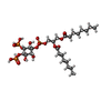 ChemComp-PIO: |
-分子 #7: water
| 分子 | 名称: water / タイプ: ligand / ID: 7 / コピー数: 153 / 式: HOH |
|---|---|
| 分子量 | 理論値: 18.015 Da |
| Chemical component information |  ChemComp-HOH: |
-実験情報
-構造解析
| 手法 | クライオ電子顕微鏡法 |
|---|---|
 解析 解析 | 単粒子再構成法 |
| 試料の集合状態 | particle |
- 試料調製
試料調製
| 濃度 | 8 mg/mL |
|---|---|
| 緩衝液 | pH: 7.4 詳細: Final gel filtration buffer contained 0.05 % (w/v) digitonin, 130mM KCl, 20mM HEPES pH 7.4, 1mM ATP, 1mM MgCl2, 1mM PMSF. Peak fractions were concentrated to 8mg/mL, and 0.01% (w/v) of ...詳細: Final gel filtration buffer contained 0.05 % (w/v) digitonin, 130mM KCl, 20mM HEPES pH 7.4, 1mM ATP, 1mM MgCl2, 1mM PMSF. Peak fractions were concentrated to 8mg/mL, and 0.01% (w/v) of glycyrrhizic acid was added immediately prior to vitrification. |
| グリッド | モデル: UltrAuFoil R0.6/1 / 材質: GOLD / メッシュ: 300 / 前処理 - タイプ: GLOW DISCHARGE |
| 凍結 | 凍結剤: ETHANE / チャンバー内湿度: 100 % / チャンバー内温度: 277 K / 装置: FEI VITROBOT MARK IV / 詳細: 4-6 seconds, wait time 30 seconds.. |
| 詳細 | Ankyrin complex mixture, purified from digitonin-solubilized erythrocyte ghost membranes. |
- 電子顕微鏡法
| 顕微鏡 | FEI TITAN KRIOS |
|---|---|
| 特殊光学系 | エネルギーフィルター - 名称: GIF Bioquantum / エネルギーフィルター - スリット幅: 20 eV |
| 撮影 | フィルム・検出器のモデル: GATAN K3 (6k x 4k) / 撮影したグリッド数: 2 / 実像数: 14464 / 平均露光時間: 2.5 sec. / 平均電子線量: 58.0 e/Å2 / 詳細: Two grids were imaged in a single session. |
| 電子線 | 加速電圧: 300 kV / 電子線源:  FIELD EMISSION GUN FIELD EMISSION GUN |
| 電子光学系 | 照射モード: FLOOD BEAM / 撮影モード: BRIGHT FIELD / Cs: 2.7 mm / 最大 デフォーカス(公称値): 1.5 µm / 最小 デフォーカス(公称値): 0.5 µm |
| 試料ステージ | 試料ホルダーモデル: FEI TITAN KRIOS AUTOGRID HOLDER ホルダー冷却材: NITROGEN |
| 実験機器 |  モデル: Titan Krios / 画像提供: FEI Company |
 ムービー
ムービー コントローラー


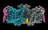


 Z (Sec.)
 Y (Row.)
 X (Col.)


