 Movie
Movie Controller


+ Open data

- Basic information
| Entry | Database: EMDB / ID: EMD-24512 | |||||||||
|---|---|---|---|---|---|---|---|---|---|---|
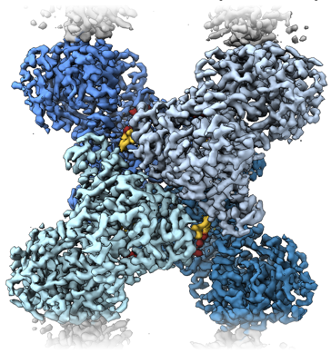
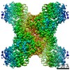
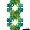
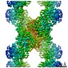
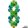
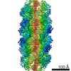
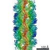
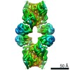
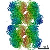
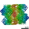
| Title | Yeast CTP Synthase (URA8) Filament bound to ATP/UTP at low pH | |||||||||
 Map data Map data | Yeast CTP Synthase (URA8) filament bound to ATP/UTP at low pH | |||||||||
 Sample Sample |
| |||||||||
 Keywords Keywords | glutaminase / amino-ligase / nucleotide metabolism / PROTEIN FIBRIL | |||||||||
| Function / homology |  Function and homology information Function and homology informationCTP synthase (glutamine hydrolysing) / CTP synthase activity / cytoophidium / 'de novo' CTP biosynthetic process / pyrimidine nucleobase biosynthetic process / L-glutamine metabolic process / ATP binding / identical protein binding / cytosol Similarity search - Function | |||||||||
| Biological species |  | |||||||||
| Method | single particle reconstruction / cryo EM / Resolution: 2.8 Å | |||||||||
 Authors Authors | Hansen JM / Lynch EM | |||||||||
| Funding support |  United States, 2 items United States, 2 items
| |||||||||
 Citation Citation | Journal: Elife / Year: 2021 Title: Cryo-EM structures of CTP synthase filaments reveal mechanism of pH-sensitive assembly during budding yeast starvation. Authors: Jesse M Hansen / Avital Horowitz / Eric M Lynch / Daniel P Farrell / Joel Quispe / Frank DiMaio / Justin M Kollman / Abstract: Many metabolic enzymes self-assemble into micron-scale filaments to organize and regulate metabolism. The appearance of these assemblies often coincides with large metabolic changes as in ...Many metabolic enzymes self-assemble into micron-scale filaments to organize and regulate metabolism. The appearance of these assemblies often coincides with large metabolic changes as in development, cancer, and stress. Yeast undergo cytoplasmic acidification upon starvation, triggering the assembly of many metabolic enzymes into filaments. However, it is unclear how these filaments assemble at the molecular level and what their role is in the yeast starvation response. CTP Synthase (CTPS) assembles into metabolic filaments across many species. Here, we characterize in vitro polymerization and investigate in vivo consequences of CTPS assembly in yeast. Cryo-EM structures reveal a pH-sensitive assembly mechanism and highly ordered filament bundles that stabilize an inactive state of the enzyme, features unique to yeast CTPS. Disruption of filaments in cells with non-assembly or pH-insensitive mutations decreases growth rate, reflecting the importance of regulated CTPS filament assembly in homeotstasis. | |||||||||
| History |
|
- Structure visualization
| Movie |
Movie viewer |
|---|---|
| Structure viewer | EM map: SurfViewMolmilJmol/JSmol |
| Supplemental images |
- Downloads & links
-EMDB archive
| Map data | emd_24512.map.gz | 13.9 MB | EMDB map data format | |
|---|---|---|---|---|
| Header (meta data) | emd-24512-v30.xmlemd-24512.xml | 13.1 KB 13.1 KB | Display Display | EMDB header |
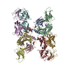
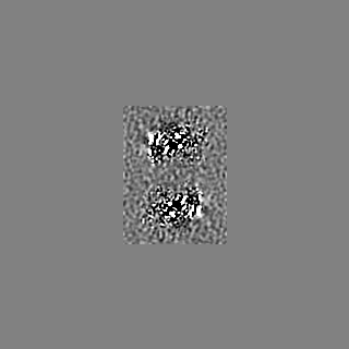
| Images |  emd_24512.png emd_24512.png | 222.1 KB | ||
| Filedesc metadata | emd-24512.cif.gz | 6 KB | ||
| Archive directory |  http://ftp.pdbj.org/pub/emdb/structures/EMD-24512ftp://ftp.pdbj.org/pub/emdb/structures/EMD-24512 http://ftp.pdbj.org/pub/emdb/structures/EMD-24512ftp://ftp.pdbj.org/pub/emdb/structures/EMD-24512 | HTTPS FTP |
-Related structure data
| Related structure data |  7rl0MC 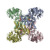 7rkhC  7rl5C  7rmcC  7rmfC 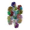 7rmkC 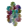 7rmoC  7rmvC 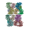 7rnlC 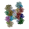 7rnrC M: atomic model generated by this map C: citing same article ( |
|---|---|
| Similar structure data |
-Links
| EMDB pages | EMDB (EBI/PDBe) / EMDataResource |
|---|---|
| Related items in Molecule of the Month |


-Map
| File | Download / File: emd_24512.map.gz / Format: CCP4 / Size: 125 MB / Type: IMAGE STORED AS FLOATING POINT NUMBER (4 BYTES) | ||||||||||||||||||||||||||||||||||||||||||||||||||||||||||||||||||||
|---|---|---|---|---|---|---|---|---|---|---|---|---|---|---|---|---|---|---|---|---|---|---|---|---|---|---|---|---|---|---|---|---|---|---|---|---|---|---|---|---|---|---|---|---|---|---|---|---|---|---|---|---|---|---|---|---|---|---|---|---|---|---|---|---|---|---|---|---|---|
| Annotation | Yeast CTP Synthase (URA8) filament bound to ATP/UTP at low pH | ||||||||||||||||||||||||||||||||||||||||||||||||||||||||||||||||||||
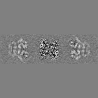
| Projections & slices | Image control
Images are generated by Spider. | ||||||||||||||||||||||||||||||||||||||||||||||||||||||||||||||||||||
| Voxel size | X=Y=Z: 1.05 Å | ||||||||||||||||||||||||||||||||||||||||||||||||||||||||||||||||||||
| Density |
| ||||||||||||||||||||||||||||||||||||||||||||||||||||||||||||||||||||
| Symmetry | Space group: 1 | ||||||||||||||||||||||||||||||||||||||||||||||||||||||||||||||||||||
| Details | EMDB XML:
CCP4 map header:
| ||||||||||||||||||||||||||||||||||||||||||||||||||||||||||||||||||||
 X (Sec.)
 Y (Row.)
 Z (Col.)


-Supplemental data
- Sample components
-Entire : Yeast CTP Synthase (URA8) filament bound to ATP/UTP at low pH
| Entire | Name: Yeast CTP Synthase (URA8) filament bound to ATP/UTP at low pH |
|---|---|
| Components |
|
-Supramolecule #1: Yeast CTP Synthase (URA8) filament bound to ATP/UTP at low pH
| Supramolecule | Name: Yeast CTP Synthase (URA8) filament bound to ATP/UTP at low pH type: complex / ID: 1 / Parent: 0 / Macromolecule list: #1 |
|---|---|
| Source (natural) | Organism: |
| Molecular weight | Theoretical: 256 kDa/nm |
-Macromolecule #1: CTP synthase
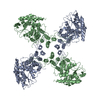
| Macromolecule | Name: CTP synthase / type: protein_or_peptide / ID: 1 / Number of copies: 12 / Enantiomer: LEVO / EC number: CTP synthase (glutamine hydrolysing) |
|---|---|
| Source (natural) | Organism: |
| Molecular weight | Theoretical: 62.439168 KDa |
| Recombinant expression | Organism:  |
| Sequence | String: MKYVVVSGGV ISGIGKGVLA SSTGMLLKTL GLKVTSIKID PYMNIDAGTM SPLEHGECFV LDDGGETDLD LGNYERYLGI TLSRDHNIT TGKIYSHVIS RERRGDYLGK TVQIVPHLTN AIQDWIQRVS KIPVDDTGLE PDVCIIELGG TVGDIESAPF V EALRQFQF ...String: MKYVVVSGGV ISGIGKGVLA SSTGMLLKTL GLKVTSIKID PYMNIDAGTM SPLEHGECFV LDDGGETDLD LGNYERYLGI TLSRDHNIT TGKIYSHVIS RERRGDYLGK TVQIVPHLTN AIQDWIQRVS KIPVDDTGLE PDVCIIELGG TVGDIESAPF V EALRQFQF EVGRENFALI HVSLVPVIHG EQKTKPTQAA IKDLRSLGLI PDMIACRCSE ELNRSTIDKI AMFCHVGPEQ VV NVHDVNS TYHVPLLLLK QHMIDYLHSR LKLGEVPLTL EDKERGSQLL TNWENMTKNL DDSDDVVKIA LVGKYTNLKD SYL SVTKSL EHASMKCRRQ LEILWVEASN LEPETQEVDK NKFHDSWNKL SSADGILVPG GFGTRGIEGM ILAAKWARES GVPF LGVCL GLQVAAIEFA RNVIGRPNSS STEFLDETLL APEDQVVITM RLGLRPTIFQ PNSEWSNIRK LYGEVNEVHE RHRHR YEIN PKIVNDMESR GFIFVGKDET GQRCEIFELK GHPYYVGTQY HPEYTSKVLE PSRPFWGLVA AASGTLGEVI KDINL UniProtKB: CTP synthase |
-Macromolecule #2: MAGNESIUM ION
| Macromolecule | Name: MAGNESIUM ION / type: ligand / ID: 2 / Number of copies: 24 / Formula: MG |
|---|---|
| Molecular weight | Theoretical: 24.305 Da |
-Macromolecule #3: ADENOSINE-5'-TRIPHOSPHATE
| Macromolecule | Name: ADENOSINE-5'-TRIPHOSPHATE / type: ligand / ID: 3 / Number of copies: 12 / Formula: ATP |
|---|---|
| Molecular weight | Theoretical: 507.181 Da |
| Chemical component information |  ChemComp-ATP: |
-Macromolecule #4: URIDINE 5'-TRIPHOSPHATE
| Macromolecule | Name: URIDINE 5'-TRIPHOSPHATE / type: ligand / ID: 4 / Number of copies: 12 / Formula: UTP |
|---|---|
| Molecular weight | Theoretical: 484.141 Da |
| Chemical component information |  ChemComp-UTP: |
-Experimental details
-Structure determination
| Method | cryo EM |
|---|---|
 Processing Processing | single particle reconstruction |
| Aggregation state | particle |
-Sample preparation
| Buffer | pH: 6 |
|---|---|
| Grid | Model: C-flat / Support film - Material: CARBON / Support film - topology: HOLEY |
| Vitrification | Cryogen name: ETHANE / Chamber humidity: 100 % / Chamber temperature: 277 K / Instrument: FEI VITROBOT MARK IV |
- Electron microscopy
| Microscope | FEI TITAN KRIOS |
|---|---|
| Specialist optics | Energy filter - Name: GIF Bioquantum / Energy filter - Slit width: 20 eV |
| Image recording | Film or detector model: GATAN K2 SUMMIT (4k x 4k) / Detector mode: COUNTING / Average electron dose: 90.0 e/Å2 |
| Electron beam | Acceleration voltage: 300 kV / Electron source:  FIELD EMISSION GUN FIELD EMISSION GUN |
| Electron optics | Illumination mode: FLOOD BEAM / Imaging mode: BRIGHT FIELD / Cs: 2.7 mm / Nominal defocus max: 1.9000000000000001 µm / Nominal defocus min: 0.4 µm / Nominal magnification: 130000 |
| Sample stage | Specimen holder model: FEI TITAN KRIOS AUTOGRID HOLDER / Cooling holder cryogen: NITROGEN |
| Experimental equipment |  Model: Titan Krios / Image courtesy: FEI Company |
-Image processing
| Startup model | Type of model: PDB ENTRY |
|---|---|
| Final reconstruction | Applied symmetry - Point group: D2 (2x2 fold dihedral) / Resolution.type: BY AUTHOR / Resolution: 2.8 Å / Resolution method: OTHER / Software - Name: RELION (ver. 3.1) / Details: FSCref0.5 (Phenix Density Modification) / Number images used: 40474 |
| Initial angle assignment | Type: MAXIMUM LIKELIHOOD |
| Final angle assignment | Type: MAXIMUM LIKELIHOOD / Software - Name: RELION (ver. 3.1) |
-Atomic model buiding 1
| Refinement | Overall B value: 32 |
|---|---|
| Output model | PDB-7rl0: |
