 ムービー
ムービー コントローラー


+ データを開く

- 基本情報
基本情報
| 登録情報 | データベース: EMDB / ID: EMD-2411 | |||||||||
|---|---|---|---|---|---|---|---|---|---|---|
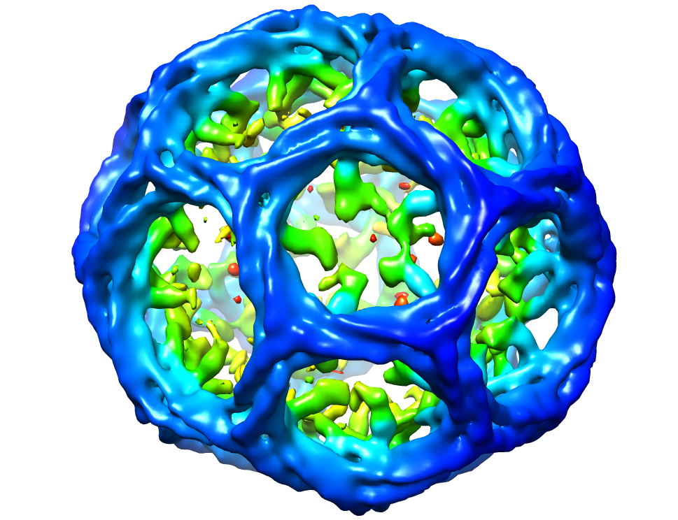
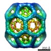
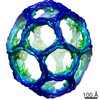
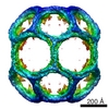
| タイトル | Hsc70-induced Changes in Clathrin-Auxilin Cage Structure Suggest a Role for Clathrin Light Chains in Cage Disassembly | |||||||||
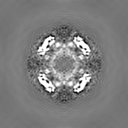
 マップデータ マップデータ | Map of Clathrin-Auxilin complex. Sharpened to match a Fourier amplitude profile derived from a model of the hexagonal barrel (the biological unit) created from the crystal structure atomic model pdb id1XI5. Fourier filtered to 30 Angstroms. | |||||||||
 試料 試料 |
| |||||||||
 キーワード キーワード | Endocytosis / coated vesicles / clathrin / auxilin | |||||||||
| 生物種 |  | |||||||||
| 手法 | 単粒子再構成法 / クライオ電子顕微鏡法 / 解像度: 31.0 Å | |||||||||
 データ登録者 データ登録者 | Young A / Stoilova-McPhie S / Rothnie A / Vallis Y / Harvey-Smith P / Ranson N / Kent H / Brodsky FM / Pearse BM / Roseman A / Smith CJ | |||||||||
 引用 引用 | ジャーナル: J Mol Biol / 年: 2004 タイトル: Location of auxilin within a clathrin cage. 著者: Corinne J Smith / Timothy R Dafforn / Helen Kent / Catherine A Sims / Kavita Khubchandani-Aswani / Lin Zhang / Helen R Saibil / Barbara M F Pearse /  要旨: The Dna J homologue, auxilin, acts as a co-chaperone for Hsc70 in the uncoating of clathrin-coated vesicles during endocytosis. Biochemical studies have aided understanding of the uncoating mechanism ...The Dna J homologue, auxilin, acts as a co-chaperone for Hsc70 in the uncoating of clathrin-coated vesicles during endocytosis. Biochemical studies have aided understanding of the uncoating mechanism but until now there was no structural information on how auxilin interacts with the clathrin cage. Here we have determined the three-dimensional structure of a complex of auxilin with clathrin cages by cryo-electron microscopy and single particle analysis. We show that auxilin forms a discrete shell of density on the inside of the clathrin cage. Peptide competition assays confirm that a candidate clathrin box motif in auxilin, LLGLE, can bind to a clathrin construct containing the beta-propeller domain and also displace the well-characterised LLNLD clathrin box motif derived from the beta-adaptin hinge region. The means by which auxilin could both aid clathrin coat assembly and displace clathrin from AP2 during uncoating is discussed. | |||||||||
| 履歴 |
|
- 構造の表示
構造の表示
| ムービー |
 ムービービューア ムービービューア |
|---|---|
| 構造ビューア | EMマップ: SurfViewMolmilJmol/JSmol |
| 添付画像 |
- ダウンロードとリンク
-EMDBアーカイブ
| マップデータ | emd_2411.map.gz | 58.8 MB | EMDBマップデータ形式 | |
|---|---|---|---|---|
| ヘッダ (付随情報) | emd-2411-v30.xmlemd-2411.xml | 10.2 KB 10.2 KB | 表示 表示 | EMDBヘッダ |
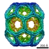
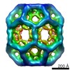
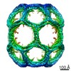
| 画像 |  emd_2411.png emd_2411.png | 530.7 KB | ||
| アーカイブディレクトリ |  http://ftp.pdbj.org/pub/emdb/structures/EMD-2411ftp://ftp.pdbj.org/pub/emdb/structures/EMD-2411 http://ftp.pdbj.org/pub/emdb/structures/EMD-2411ftp://ftp.pdbj.org/pub/emdb/structures/EMD-2411 | HTTPS FTP |
-検証レポート
| 文書・要旨 | emd_2411_validation.pdf.gz | 246.6 KB | 表示 | EMDB検証レポート |
|---|---|---|---|---|
| 文書・詳細版 | emd_2411_full_validation.pdf.gz | 245.7 KB | 表示 | |
| XML形式データ | emd_2411_validation.xml.gz | 6.9 KB | 表示 | |
| アーカイブディレクトリ | https://ftp.pdbj.org/pub/emdb/validation_reports/EMD-2411ftp://ftp.pdbj.org/pub/emdb/validation_reports/EMD-2411 | HTTPS FTP |
-関連構造データ

-リンク
| EMDBのページ | EMDB (EBI/PDBe) / EMDataResource |
|---|
-マップ
| ファイル | ダウンロード / ファイル: emd_2411.map.gz / 形式: CCP4 / 大きさ: 62.5 MB / タイプ: IMAGE STORED AS FLOATING POINT NUMBER (4 BYTES) | ||||||||||||||||||||||||||||||||||||||||||||||||||||||||||||||||||||
|---|---|---|---|---|---|---|---|---|---|---|---|---|---|---|---|---|---|---|---|---|---|---|---|---|---|---|---|---|---|---|---|---|---|---|---|---|---|---|---|---|---|---|---|---|---|---|---|---|---|---|---|---|---|---|---|---|---|---|---|---|---|---|---|---|---|---|---|---|---|
| 注釈 | Map of Clathrin-Auxilin complex. Sharpened to match a Fourier amplitude profile derived from a model of the hexagonal barrel (the biological unit) created from the crystal structure atomic model pdb id1XI5. Fourier filtered to 30 Angstroms. | ||||||||||||||||||||||||||||||||||||||||||||||||||||||||||||||||||||
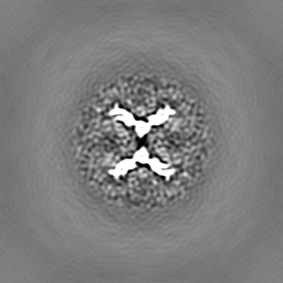
| 投影像・断面図 | 画像のコントロール
画像は Spider により作成 | ||||||||||||||||||||||||||||||||||||||||||||||||||||||||||||||||||||
| ボクセルのサイズ | X=Y=Z: 6.4 Å | ||||||||||||||||||||||||||||||||||||||||||||||||||||||||||||||||||||
| 密度 |
| ||||||||||||||||||||||||||||||||||||||||||||||||||||||||||||||||||||
| 対称性 | 空間群: 1 | ||||||||||||||||||||||||||||||||||||||||||||||||||||||||||||||||||||
| 詳細 | EMDB XML:
CCP4マップ ヘッダ情報:
| ||||||||||||||||||||||||||||||||||||||||||||||||||||||||||||||||||||
 Z (Sec.)
 Y (Row.)
 X (Col.)


-添付データ
- 試料の構成要素
-全体 : Clathrin bound to auxilin
| 全体 | 名称: Clathrin bound to auxilin |
|---|---|
| 要素 |
|
-超分子 #1000: Clathrin bound to auxilin
| 超分子 | 名称: Clathrin bound to auxilin / タイプ: sample / ID: 1000 / Number unique components: 2 |
|---|
-分子 #1: Clathrin
| 分子 | 名称: Clathrin / タイプ: protein_or_peptide / ID: 1 / 組換発現: No |
|---|---|
| 由来(天然) | 生物種: |
-分子 #2: Auxilin
| 分子 | 名称: Auxilin / タイプ: protein_or_peptide / ID: 2 / 組換発現: No |
|---|---|
| 由来(天然) | 生物種: |
-実験情報
-構造解析
| 手法 | クライオ電子顕微鏡法 |
|---|---|
 解析 解析 | 単粒子再構成法 |
| 試料の集合状態 | particle |
-試料調製
| 緩衝液 | pH: 6 詳細: 20 mM Mes (pH 6.0), 2 mM magnesium acetate, 25 mM KCl, 10 mM (NH4)2SO4, 1 mM DTT |
|---|---|
| 凍結 | 凍結剤: ETHANE / 装置: HOMEMADE PLUNGER |
- 電子顕微鏡法 #1
| Microscopy ID | 1 |
|---|---|
| 顕微鏡 | FEI/PHILIPS CM200FEG |
| 日付 | 2000年2月2日 |
| 撮影 | デジタル化 - スキャナー: ZEISS SCAI |
| 電子線 | 加速電圧: 200 kV / 電子線源:  FIELD EMISSION GUN FIELD EMISSION GUN |
| 電子光学系 | 照射モード: FLOOD BEAM / 撮影モード: BRIGHT FIELD |
| 試料ステージ | 試料ホルダーモデル: GATAN LIQUID NITROGEN |
-電子顕微鏡法 #2
| Microscopy ID | 2 |
|---|---|
| 顕微鏡 | FEI/PHILIPS CM200FEG |
| 日付 | 2000年2月23日 |
| 撮影 | デジタル化 - スキャナー: ZEISS SCAI |
| 電子線 | 加速電圧: 200 kV / 電子線源: FIELD EMISSION GUN |
| 電子光学系 | 照射モード: FLOOD BEAM / 撮影モード: BRIGHT FIELD |
| 試料ステージ | 試料ホルダーモデル: GATAN LIQUID NITROGEN |
-画像解析
| CTF補正 | 詳細: Whole micrographs |
|---|---|
| 最終 再構成 | 想定した対称性 - 点群: D6 (2回x6回 2面回転対称) アルゴリズム: OTHER / 解像度のタイプ: BY AUTHOR / 解像度: 31.0 Å / 解像度の算出法: FSC 0.143 CUT-OFF / ソフトウェア - 名称: MRC, SPIDER, FREALIGN 詳細: Sharpened to match a Fourier amplitude profile derived from a model of the hexagonal barrel (the biological unit) created from the crystal structure atomic model pdb id1XI5. Fourier filtered to 30 Angstroms. 使用した粒子像数: 1745 |
