[English] 日本語
 Yorodumi
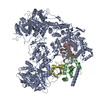
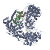
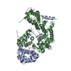
- EMDB-23807: Structure of yeast Ubr1 in complex with Ubc2 and monoubiquitinate... -
+ Open data
- Basic information
| Entry | Database: EMDB / ID: EMD-23807 | |||||||||
|---|---|---|---|---|---|---|---|---|---|---|
| Title | Structure of yeast Ubr1 in complex with Ubc2 and monoubiquitinated N-degron | |||||||||
 Map data Map data | ||||||||||
 Sample Sample |
| |||||||||
 Keywords Keywords | Ubiquitin E3 ligase / ubiquitination / Ubr1 / Ubc2 / Degron / N-end rule / TRANSFERASE | |||||||||
| Function / homology |  Function and homology information Function and homology informationMUB1-RAD6-UBR2 ubiquitin ligase complex / RAD6-UBR2 ubiquitin ligase complex / Rad6-Rad18 complex / regulation of dipeptide transport / UBR1-RAD6 ubiquitin ligase complex / sno(s)RNA transcription / proteasome regulatory particle binding / HULC complex / error-free postreplication DNA repair / stress-induced homeostatically regulated protein degradation pathway ...MUB1-RAD6-UBR2 ubiquitin ligase complex / RAD6-UBR2 ubiquitin ligase complex / Rad6-Rad18 complex / regulation of dipeptide transport / UBR1-RAD6 ubiquitin ligase complex / sno(s)RNA transcription / proteasome regulatory particle binding / HULC complex / error-free postreplication DNA repair / stress-induced homeostatically regulated protein degradation pathway / : / meiotic DNA double-strand break formation / ubiquitin-dependent protein catabolic process via the N-end rule pathway / mitochondria-associated ubiquitin-dependent protein catabolic process / cytoplasm protein quality control by the ubiquitin-proteasome system / : / telomere maintenance via recombination / proteasome regulatory particle, base subcomplex / hypothalamus gonadotrophin-releasing hormone neuron development / female meiosis I / ribosome-associated ubiquitin-dependent protein catabolic process / positive regulation of protein monoubiquitination / fat pad development / mitochondrion transport along microtubule / E2 ubiquitin-conjugating enzyme / error-free translesion synthesis / sporulation resulting in formation of a cellular spore / female gonad development / seminiferous tubule development / proteasome binding / ubiquitin conjugating enzyme activity / male meiosis I / Antigen processing: Ubiquitination & Proteasome degradation / positive regulation of intrinsic apoptotic signaling pathway by p53 class mediator / cellular response to unfolded protein / protein monoubiquitination / error-prone translesion synthesis / ubiquitin ligase complex / subtelomeric heterochromatin formation / energy homeostasis / regulation of neuron apoptotic process / neuron projection morphogenesis / mitotic G1 DNA damage checkpoint signaling / ERAD pathway / regulation of proteasomal protein catabolic process / Maturation of protein E / Maturation of protein E / ER Quality Control Compartment (ERQC) / Myoclonic epilepsy of Lafora / FLT3 signaling by CBL mutants / Constitutive Signaling by NOTCH1 HD Domain Mutants / IRAK2 mediated activation of TAK1 complex / Prevention of phagosomal-lysosomal fusion / Alpha-protein kinase 1 signaling pathway / Glycogen synthesis / IRAK1 recruits IKK complex / IRAK1 recruits IKK complex upon TLR7/8 or 9 stimulation / Endosomal Sorting Complex Required For Transport (ESCRT) / Membrane binding and targetting of GAG proteins / Negative regulation of FLT3 / Regulation of TBK1, IKKε (IKBKE)-mediated activation of IRF3, IRF7 / PTK6 Regulates RTKs and Their Effectors AKT1 and DOK1 / Regulation of TBK1, IKKε-mediated activation of IRF3, IRF7 upon TLR3 ligation / IRAK2 mediated activation of TAK1 complex upon TLR7/8 or 9 stimulation / NOTCH2 Activation and Transmission of Signal to the Nucleus / TICAM1,TRAF6-dependent induction of TAK1 complex / TICAM1-dependent activation of IRF3/IRF7 / APC/C:Cdc20 mediated degradation of Cyclin B / Regulation of FZD by ubiquitination / Downregulation of ERBB4 signaling / APC-Cdc20 mediated degradation of Nek2A / p75NTR recruits signalling complexes / InlA-mediated entry of Listeria monocytogenes into host cells / TRAF6 mediated IRF7 activation in TLR7/8 or 9 signaling / TRAF6-mediated induction of TAK1 complex within TLR4 complex / Regulation of pyruvate metabolism / NF-kB is activated and signals survival / Regulation of innate immune responses to cytosolic DNA / Pexophagy / Downregulation of ERBB2:ERBB3 signaling / NRIF signals cell death from the nucleus / Activated NOTCH1 Transmits Signal to the Nucleus / Regulation of PTEN localization / VLDLR internalisation and degradation / Regulation of BACH1 activity / Synthesis of active ubiquitin: roles of E1 and E2 enzymes / MAP3K8 (TPL2)-dependent MAPK1/3 activation / Translesion synthesis by REV1 / TICAM1, RIP1-mediated IKK complex recruitment / Translesion synthesis by POLK / InlB-mediated entry of Listeria monocytogenes into host cell / positive regulation of protein ubiquitination / Activation of IRF3, IRF7 mediated by TBK1, IKKε (IKBKE) / JNK (c-Jun kinases) phosphorylation and activation mediated by activated human TAK1 / Josephin domain DUBs / Downregulation of TGF-beta receptor signaling / Translesion synthesis by POLI / Gap-filling DNA repair synthesis and ligation in GG-NER / IKK complex recruitment mediated by RIP1 / Regulation of activated PAK-2p34 by proteasome mediated degradation Similarity search - Function | |||||||||
| Biological species |   Homo sapiens (human) / Homo sapiens (human) /  | |||||||||
| Method | single particle reconstruction / cryo EM / Resolution: 3.67 Å | |||||||||
 Authors Authors | Pan M / Zheng Q | |||||||||
| Funding support | 1 items
| |||||||||
 Citation Citation |  Journal: Nature / Year: 2021 Journal: Nature / Year: 2021Title: Structural insights into Ubr1-mediated N-degron polyubiquitination. Authors: Man Pan / Qingyun Zheng / Tian Wang / Lujun Liang / Junxiong Mao / Chong Zuo / Ruichao Ding / Huasong Ai / Yuan Xie / Dong Si / Yuanyuan Yu / Lei Liu / Minglei Zhao /   Abstract: The N-degron pathway targets proteins that bear a destabilizing residue at the N terminus for proteasome-dependent degradation. In yeast, Ubr1-a single-subunit E3 ligase-is responsible for the Arg/N- ...The N-degron pathway targets proteins that bear a destabilizing residue at the N terminus for proteasome-dependent degradation. In yeast, Ubr1-a single-subunit E3 ligase-is responsible for the Arg/N-degron pathway. How Ubr1 mediates the initiation of ubiquitination and the elongation of the ubiquitin chain in a linkage-specific manner through a single E2 ubiquitin-conjugating enzyme (Ubc2) remains unknown. Here we developed chemical strategies to mimic the reaction intermediates of the first and second ubiquitin transfer steps, and determined the cryo-electron microscopy structures of Ubr1 in complex with Ubc2, ubiquitin and two N-degron peptides, representing the initiation and elongation steps of ubiquitination. Key structural elements, including a Ubc2-binding region and an acceptor ubiquitin-binding loop on Ubr1, were identified and characterized. These structures provide mechanistic insights into the initiation and elongation of ubiquitination catalysed by Ubr1. | |||||||||
| History |
|
- Structure visualization
| Movie |
 Movie viewer Movie viewer |
|---|---|
| Structure viewer | EM map:  SurfView SurfView Molmil Molmil Jmol/JSmol Jmol/JSmol |
| Supplemental images |
- Downloads & links
-EMDB archive
| Map data |  emd_23807.map.gz emd_23807.map.gz | 49.5 MB |  EMDB map data format EMDB map data format | |
|---|---|---|---|---|
| Header (meta data) |  emd-23807-v30.xml emd-23807-v30.xml emd-23807.xml emd-23807.xml | 24.1 KB 24.1 KB | Display Display |  EMDB header EMDB header |
| Images |  emd_23807.png emd_23807.png | 167.9 KB | ||
| Masks |  emd_23807_msk_1.map emd_23807_msk_1.map | 64 MB |  Mask map Mask map | |
| Filedesc metadata |  emd-23807.cif.gz emd-23807.cif.gz | 7.3 KB | ||
| Others |  emd_23807_additional_1.map.gz emd_23807_additional_1.map.gz emd_23807_half_map_1.map.gz emd_23807_half_map_1.map.gz emd_23807_half_map_2.map.gz emd_23807_half_map_2.map.gz | 59.8 MB 49.6 MB 49.8 MB | ||
| Archive directory |  http://ftp.pdbj.org/pub/emdb/structures/EMD-23807 http://ftp.pdbj.org/pub/emdb/structures/EMD-23807 ftp://ftp.pdbj.org/pub/emdb/structures/EMD-23807 ftp://ftp.pdbj.org/pub/emdb/structures/EMD-23807 | HTTPS FTP |
-Validation report
| Summary document |  emd_23807_validation.pdf.gz emd_23807_validation.pdf.gz | 966.3 KB | Display |  EMDB validaton report EMDB validaton report |
|---|---|---|---|---|
| Full document |  emd_23807_full_validation.pdf.gz emd_23807_full_validation.pdf.gz | 965.9 KB | Display | |
| Data in XML |  emd_23807_validation.xml.gz emd_23807_validation.xml.gz | 12.2 KB | Display | |
| Data in CIF |  emd_23807_validation.cif.gz emd_23807_validation.cif.gz | 14.4 KB | Display | |
| Arichive directory |  https://ftp.pdbj.org/pub/emdb/validation_reports/EMD-23807 https://ftp.pdbj.org/pub/emdb/validation_reports/EMD-23807 ftp://ftp.pdbj.org/pub/emdb/validation_reports/EMD-23807 ftp://ftp.pdbj.org/pub/emdb/validation_reports/EMD-23807 | HTTPS FTP |
-Related structure data
| Related structure data |  7meyMC  7mexC C: citing same article ( M: atomic model generated by this map |
|---|---|
| Similar structure data | |
| EM raw data |  EMPIAR-10902 (Title: Single-particle cryoEM data of yeast Ubr1-Ubc2-Ub-Ub-N-degron complex (elongation) EMPIAR-10902 (Title: Single-particle cryoEM data of yeast Ubr1-Ubc2-Ub-Ub-N-degron complex (elongation)Data size: 2.9 TB Data #1: Unaligned movie data of yeast Ubr1-Ubc2-Ub-Ub-N-degron complex (elongation complex) [micrographs - multiframe]) |
- Links
Links
| EMDB pages |  EMDB (EBI/PDBe) / EMDB (EBI/PDBe) /  EMDataResource EMDataResource |
|---|---|
| Related items in Molecule of the Month |
- Map
Map
| File |  Download / File: emd_23807.map.gz / Format: CCP4 / Size: 64 MB / Type: IMAGE STORED AS FLOATING POINT NUMBER (4 BYTES) Download / File: emd_23807.map.gz / Format: CCP4 / Size: 64 MB / Type: IMAGE STORED AS FLOATING POINT NUMBER (4 BYTES) | ||||||||||||||||||||||||||||||||||||||||||||||||||||||||||||||||||||
|---|---|---|---|---|---|---|---|---|---|---|---|---|---|---|---|---|---|---|---|---|---|---|---|---|---|---|---|---|---|---|---|---|---|---|---|---|---|---|---|---|---|---|---|---|---|---|---|---|---|---|---|---|---|---|---|---|---|---|---|---|---|---|---|---|---|---|---|---|---|
| Projections & slices | Image control
Images are generated by Spider. | ||||||||||||||||||||||||||||||||||||||||||||||||||||||||||||||||||||
| Voxel size | X=Y=Z: 1.063 Å | ||||||||||||||||||||||||||||||||||||||||||||||||||||||||||||||||||||
| Density |
| ||||||||||||||||||||||||||||||||||||||||||||||||||||||||||||||||||||
| Symmetry | Space group: 1 | ||||||||||||||||||||||||||||||||||||||||||||||||||||||||||||||||||||
| Details | EMDB XML:
CCP4 map header:
| ||||||||||||||||||||||||||||||||||||||||||||||||||||||||||||||||||||
-Supplemental data
-Mask #1
| File |  emd_23807_msk_1.map emd_23807_msk_1.map | ||||||||||||
|---|---|---|---|---|---|---|---|---|---|---|---|---|---|
| Projections & Slices |
| ||||||||||||
| Density Histograms |
-Additional map: #1
| File | emd_23807_additional_1.map | ||||||||||||
|---|---|---|---|---|---|---|---|---|---|---|---|---|---|
| Projections & Slices |
| ||||||||||||
| Density Histograms |
-Half map: #2
| File | emd_23807_half_map_1.map | ||||||||||||
|---|---|---|---|---|---|---|---|---|---|---|---|---|---|
| Projections & Slices |
| ||||||||||||
| Density Histograms |
-Half map: #1
| File | emd_23807_half_map_2.map | ||||||||||||
|---|---|---|---|---|---|---|---|---|---|---|---|---|---|
| Projections & Slices |
| ||||||||||||
| Density Histograms |
- Sample components
-Entire : yeast Ubr1 in complex with Ubc2 and monoubiquitinated N-degron
| Entire | Name: yeast Ubr1 in complex with Ubc2 and monoubiquitinated N-degron |
|---|---|
| Components |
|
-Supramolecule #1: yeast Ubr1 in complex with Ubc2 and monoubiquitinated N-degron
| Supramolecule | Name: yeast Ubr1 in complex with Ubc2 and monoubiquitinated N-degron type: complex / ID: 1 / Parent: 0 / Macromolecule list: #1-#5 |
|---|---|
| Source (natural) | Organism:  |
-Macromolecule #1: E3 ubiquitin-protein ligase UBR1
| Macromolecule | Name: E3 ubiquitin-protein ligase UBR1 / type: protein_or_peptide / ID: 1 / Number of copies: 1 / Enantiomer: LEVO / EC number: RING-type E3 ubiquitin transferase |
|---|---|
| Source (natural) | Organism:  Strain: ATCC 204508 / S288c |
| Molecular weight | Theoretical: 225.10275 KDa |
| Recombinant expression | Organism:  |
| Sequence | String: MSVADDDLGS LQGHIRRTLR SIHNLPYFRY TRGPTERADM SRALKEFIYR YLYFVISNSG ENLPTLFNAH PKQKLSNPEL TVFPDSLED AVDIDKITSQ QTIPFYKIDE SRIGDVHKHT GRNCGRKFKI GEPLYRCHEC GCDDTCVLCI HCFNPKDHVN H HVCTDICT ...String: MSVADDDLGS LQGHIRRTLR SIHNLPYFRY TRGPTERADM SRALKEFIYR YLYFVISNSG ENLPTLFNAH PKQKLSNPEL TVFPDSLED AVDIDKITSQ QTIPFYKIDE SRIGDVHKHT GRNCGRKFKI GEPLYRCHEC GCDDTCVLCI HCFNPKDHVN H HVCTDICT EFTSGICDCG DEEAWNSPLH CKAEEQENDI SEDPATNADI KEEDVWNDSV NIALVELVLA EVFDYFIDVF NQ NIEPLPT IQKDITIKLR EMTQQGKMYE RAQFLNDLKY ENDYMFDGTT TAKTSPSNSP EASPSLAKID PENYTVIIYN DEY HNYSQA TTALRQGVPD NVHIDLLTSR IDGEGRAMLK CSQDLSSVLG GFFAVQTNGL SATLTSWSEY LHQETCKYII LWIT HCLNI PNSSFQTTFR NMMGKTLCSE YLNATECRDM TPVVEKYFSN KFDKNDPYRY IDLSILADGN QIPLGHHKIL PESST HSLS PLINDVETPT SRTYSNTRLQ HILYFDNRYW KRLRKDIQNV IIPTLASSNL YKPIFCQQVV EIFNHITRSV AYMDRE PQL TAIRECVVQL FTCPTNAKNI FENQSFLDIV WSIIDIFKEF CKVEGGVLIW QRVQKSNLTK SYSISFKQGL YTVETLL SK VHDPNIPLRP KEIISLLTLC KLFNGAWKIK RKEGEHVLHE DQNFISYLEY TTSIYSIIQT AEKVSEKSKD SIDSKLFL N AIRIISSFLG NRSLTYKLIY DSHEVIKFSV SHERVAFMNP LQTMLSFLIE KVSLKDAYEA LEDCSDFLKI SDFSLRSVV LCSQIDVGFW VRNGMSVLHQ ASYYKNNPEL GSYSRDIHLN QLAILWERDD IPRIIYNILD RWELLDWFTG EVDYQHTVYE DKISFIIQQ FIAFIYQILT ERQYFKTFSS LKDRRMDQIK NSIIYNLYMK PLSYSKLLRS VPDYLTEDTT EFDEALEEVS V FVEPKGLA DNGVFKLKAS LYAKVDPLKL LNLENEFESS ATIIKSHLAK DKDEIAKVVL IPQVSIKQLD KDALNLGAFT RN TVFAKVV YKLLQVCLDM EDSTFLNELL HLVHGIFRDD ELINGKDSIP EAYLSKPICN LLLSIANAKS DVFSESIVRK ADY LLEKMI MKKPNELFES LIASFGNQYV NDYKDKKLRQ GVNLQETEKE RKRRLAKKHQ ARLLAKFNNQ QTKFMKEHES EFDE QDNDV DMVGEKVYES EDFTCALCQD SSSTDFFVIP AYHDHSPIFR PGNIFNPNEF MPMWDGFYND DEKQAYIDDD VLEAL KENG SCGSRKVFVS CNHHIHHNCF KRYVQKKRFS SNAFICPLCQ TFSNCTLPLC QTSKANTGLS LDMFLESELS LDTLSR LFK PFTEENYRTI NSIFSLMISQ CQGFDKAVRK RANFSHKDVS LILSVHWANT ISMLEIASRL EKPYSISFFR SREQKYK TL KNILVCIMLF TFVIGKPSME FEPYPQQPDT VWNQNQLFQY IVRSALFSPV SLRQTVTEAL TTFSRQFLRD FLQGLSDA E QVTKLYAKAS KIGDVLKVSE QMLFALRTIS DVRMEGLDSE SIIYDLAYTF LLKSLLPTIR RCLVFIKVLH ELVKDSENE TLVINGHEVE EELEFEDTAE FVNKALKMIT EKESLVDLLT TQESIVSHPY LENIPYEYCG IIKLIDLSKY LNTYVTQSKE IKLREERSQ HMKNADNRLD FKICLTCGVK VHLRADRHEM TKHLNKNCFK PFGAFLMPNS SEVCLHLTQP PSNIFISAPY L NSHGEVGR NAMRRGDLTT LNLKRYEHLN RLWINNEIPG YISRVMGDEF RVTILSNGFL FAFNREPRPR RIPPTDEDDE DM EEGEDGF FTEGNDEMDV DDETGQAANL FGVGAEGIAG GGVRDFFQFF ENFRNTLQPQ GNGDDDAPQN PPPILQFLGP QFD GATIIR NTNPRNLDED DSDDNDDSDE REIW UniProtKB: E3 ubiquitin-protein ligase UBR1 |
-Macromolecule #2: Ubiquitin
| Macromolecule | Name: Ubiquitin / type: protein_or_peptide / ID: 2 / Number of copies: 1 / Enantiomer: LEVO |
|---|---|
| Source (natural) | Organism:  Homo sapiens (human) Homo sapiens (human) |
| Molecular weight | Theoretical: 8.493741 KDa |
| Recombinant expression | Organism:  Homo sapiens (human) Homo sapiens (human) |
| Sequence | String: MQIFVKTLTG KTITLEVEPS DTIENVKAKI QDKEGIPPDQ QRLIFAGCQL EDGRTLSDYN IQKESTLHLV LRLRG UniProtKB: Polyubiquitin-B |
-Macromolecule #3: Ubiquitin-conjugating enzyme E2 2
| Macromolecule | Name: Ubiquitin-conjugating enzyme E2 2 / type: protein_or_peptide / ID: 3 / Number of copies: 1 / Enantiomer: LEVO / EC number: E2 ubiquitin-conjugating enzyme |
|---|---|
| Source (natural) | Organism:  Strain: ATCC 204508 / S288c |
| Molecular weight | Theoretical: 19.725662 KDa |
| Recombinant expression | Organism:  |
| Sequence | String: MSTPARRRLM RDFKRMKEDA PPGVSASPLP DNVMVWNAMI IGPADTPYED GTFRLLLEFD EEYPNKPPHV KFLSEMFHPN VYANGEICL DILQNRWTPT YDVASILTSI QSLFNDPNPA SPANVEAATL FKDHKSQYVK RVKETVEKSW EDDMDDMDDD D DDDDDDDD DEAD UniProtKB: Ubiquitin-conjugating enzyme E2 2 |
-Macromolecule #4: Ubiquitin
| Macromolecule | Name: Ubiquitin / type: protein_or_peptide / ID: 4 / Number of copies: 1 / Enantiomer: LEVO |
|---|---|
| Source (natural) | Organism:  Homo sapiens (human) Homo sapiens (human) |
| Molecular weight | Theoretical: 8.519778 KDa |
| Recombinant expression | Organism:  |
| Sequence | String: MQIFVKTLTG KTITLEVEPS DTIENVKAKI QDKEGIPPDQ QRLIFAGKQL EDGRTLSDYN IQKESTLHLV LRLRG UniProtKB: Polyubiquitin-B |
-Macromolecule #5: Monoubiquitinated N-degron
| Macromolecule | Name: Monoubiquitinated N-degron / type: protein_or_peptide / ID: 5 / Number of copies: 1 / Enantiomer: LEVO |
|---|---|
| Source (natural) | Organism:  |
| Molecular weight | Theoretical: 4.571223 KDa |
| Sequence | String: RHGSGSGAWL LPVSLVKRKT TLAPNTQTAS PPSYRALADS LMQ |
-Macromolecule #6: ZINC ION
| Macromolecule | Name: ZINC ION / type: ligand / ID: 6 / Number of copies: 7 / Formula: ZN |
|---|---|
| Molecular weight | Theoretical: 65.409 Da |
-Macromolecule #7: 2-(ethylamino)ethane-1-thiol
| Macromolecule | Name: 2-(ethylamino)ethane-1-thiol / type: ligand / ID: 7 / Number of copies: 1 / Formula: Z3V |
|---|---|
| Molecular weight | Theoretical: 105.202 Da |
| Chemical component information |  ChemComp-Z3V: |
-Experimental details
-Structure determination
| Method | cryo EM |
|---|---|
 Processing Processing | single particle reconstruction |
| Aggregation state | particle |
- Sample preparation
| Buffer | pH: 8 |
|---|---|
| Vitrification | Cryogen name: ETHANE |
- Electron microscopy
| Microscope | FEI TITAN KRIOS |
|---|---|
| Image recording | Film or detector model: GATAN K3 (6k x 4k) / Average electron dose: 48.0 e/Å2 |
| Electron beam | Acceleration voltage: 300 kV / Electron source:  FIELD EMISSION GUN FIELD EMISSION GUN |
| Electron optics | C2 aperture diameter: 70.0 µm / Illumination mode: FLOOD BEAM / Imaging mode: BRIGHT FIELD / Cs: 2.7 mm |
| Experimental equipment |  Model: Titan Krios / Image courtesy: FEI Company |
+ Image processing
-Atomic model buiding 1
| Refinement | Space: REAL / Protocol: AB INITIO MODEL |
|---|---|
| Output model |  PDB-7mey: |
 Movie
Movie Controller





 Z (Sec.)
 Y (Row.)
 X (Col.)


