National Institutes of Health/National Institute of General Medical Sciences (NIH/NIGMS)
U24GM129564
United States
National Institutes of Health/National Institute of General Medical Sciences (NIH/NIGMS)
R01GM079429
United States
National Institutes of Health/National Institute of General Medical Sciences (NIH/NIGMS)
P41GM103832
United States
Citation
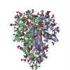
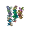
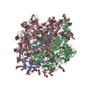
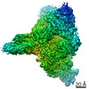
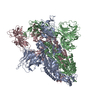
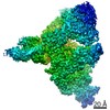
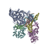
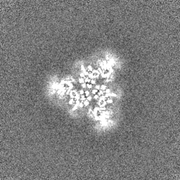
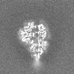
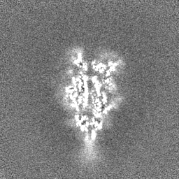
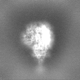
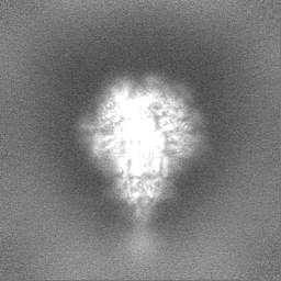
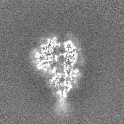
Journal: bioRxiv / Year: 2020 Title: A 3.4-Å cryo-EM structure of the human coronavirus spike trimer computationally derived from vitrified NL63 virus particles. Authors: Kaiming Zhang / Shanshan Li / Grigore Pintilie / David Chmielewski / Michael F Schmid / Graham Simmons / Jing Jin / Wah Chiu Abstract: Human coronavirus NL63 (HCoV-NL63) is an enveloped pathogen of the family that spreads worldwide and causes up to 10% of all annual respiratory diseases. HCoV-NL63 is typically associated with mild ...Human coronavirus NL63 (HCoV-NL63) is an enveloped pathogen of the family that spreads worldwide and causes up to 10% of all annual respiratory diseases. HCoV-NL63 is typically associated with mild upper respiratory symptoms in children, elderly and immunocompromised individuals. It has also been shown to cause severe lower respiratory illness. NL63 shares ACE2 as a receptor for viral entry with SARS-CoV and SARS-CoV-2. Here we present the structure of HCoV-NL63 spike (S) trimer at 3.4-Å resolution by single-particle cryo-EM imaging of vitrified virions without chemical fixative. It is structurally homologous to that obtained previously from the biochemically purified ectodomain of HCoV-NL63 S trimer, which displays a 3-fold symmetric trimer in a single conformation. In addition to previously proposed and observed glycosylation sites, our map shows density at other amino acid positions as well as differences in glycan structures. The domain arrangement within a protomer is strikingly different from that of the SARS-CoV-2 S and may explain their different requirements for activating binding to the receptor. This structure provides the basis for future studies of spike proteins with receptors, antibodies, or drugs, in the native state of the coronavirus particles.
History
Deposition
Oct 24, 2020
-
Header (metadata) release
Nov 11, 2020
-
Map release
Nov 11, 2020
-
Update
Oct 23, 2024
-
Current status
Oct 23, 2024
Processing site: RCSB / Status: Released
-
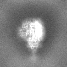
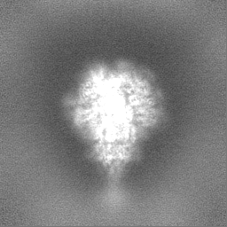
Structure visualization
Movie
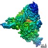
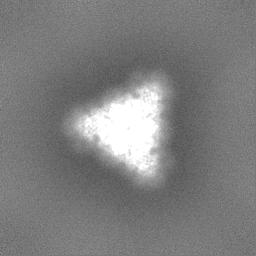
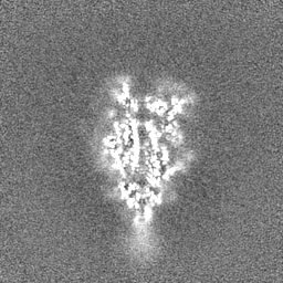
Surface view with section colored by density value
pH: 8 / Details: 20 mM Tris, pH 8.0, 120 mM NaCl, 1 mM EDTA
Grid
Model: Quantifoil / Material: COPPER / Mesh: 300 / Support film - Material: CARBON / Support film - topology: HOLEY
Vitrification
Cryogen name: ETHANE
Details
HCoV-NL63 virions
-
Electron microscopy
Microscope
FEI TITAN KRIOS
Specialist optics
Energy filter - Name: GIF Bioquantum / Energy filter - Slit width: 15 eV
Image recording
Film or detector model: GATAN K3 BIOQUANTUM (6k x 4k) / Number grids imaged: 1 / Number real images: 4138 / Average exposure time: 3.0 sec. / Average electron dose: 48.0 e/Å2
Electron beam
Acceleration voltage: 300 kV / Electron source: FIELD EMISSION GUN
In the structure databanks used in Yorodumi, some data are registered as the other names, "COVID-19 virus" and "2019-nCoV". Here are the details of the virus and the list of structure data.
Jan 31, 2019. EMDB accession codes are about to change! (news from PDBe EMDB page)
EMDB accession codes are about to change! (news from PDBe EMDB page)
The allocation of 4 digits for EMDB accession codes will soon come to an end. Whilst these codes will remain in use, new EMDB accession codes will include an additional digit and will expand incrementally as the available range of codes is exhausted. The current 4-digit format prefixed with “EMD-” (i.e. EMD-XXXX) will advance to a 5-digit format (i.e. EMD-XXXXX), and so on. It is currently estimated that the 4-digit codes will be depleted around Spring 2019, at which point the 5-digit format will come into force.
The EM Navigator/Yorodumi systems omit the EMD- prefix.
Related info.:Q: What is EMD? / ID/Accession-code notation in Yorodumi/EM Navigator
Yorodumi is a browser for structure data from EMDB, PDB, SASBDB, etc.
This page is also the successor to EM Navigator detail page, and also detail information page/front-end page for Omokage search.
The word "yorodu" (or yorozu) is an old Japanese word meaning "ten thousand". "mi" (miru) is to see.
Related info.:EMDB / PDB / SASBDB / Comparison of 3 databanks / Yorodumi Search / Aug 31, 2016. New EM Navigator & Yorodumi / Yorodumi Papers / Jmol/JSmol / Function and homology information / Changes in new EM Navigator and Yorodumi
 Movie
Movie Controller

 Yorodumi
 Open data
 Basic information
 Map data
 Sample
 Keywords
 Function and homology information
 Human coronavirus NL63
 Authors
 United States, 3 items
  Citation
 Structure visualization
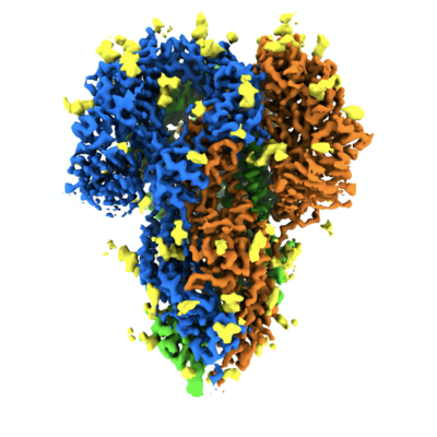
 Downloads & links
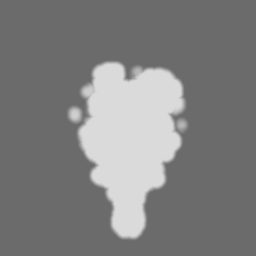
 emd_22889.png
 http://ftp.pdbj.org/pub/emdb/structures/EMD-22889
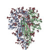


 Z (Sec.)
 Y (Row.)
 X (Col.)


 Sample components
 Processing
 Electron microscopy
 FIELD EMISSION GUN