 Movie
Movie Controller

[English] 日本語
 Yorodumi
- PDB-8fr7: A hinge glycan regulates spike bending and impacts coronavirus in... -
+ Open data

- Basic information
| Entry | Database: PDB / ID: 8fr7 | |||||||||||||||||||||||||||
|---|---|---|---|---|---|---|---|---|---|---|---|---|---|---|---|---|---|---|---|---|---|---|---|---|---|---|---|---|
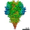
| Title | A hinge glycan regulates spike bending and impacts coronavirus infectivity | |||||||||||||||||||||||||||
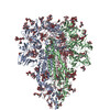
 Components Components | Spike glycoprotein | |||||||||||||||||||||||||||
 Keywords Keywords | VIRAL PROTEIN / NL63 / coronavirus / glycan | |||||||||||||||||||||||||||
| Function / homology |  Function and homology information Function and homology informationhost cell endoplasmic reticulum-Golgi intermediate compartment membrane / receptor-mediated virion attachment to host cell / host cell surface receptor binding / endocytosis involved in viral entry into host cell / fusion of virus membrane with host plasma membrane / fusion of virus membrane with host endosome membrane / viral envelope / virion membrane / membrane Similarity search - Function | |||||||||||||||||||||||||||
| Biological species |  Human coronavirus NL63 Human coronavirus NL63 | |||||||||||||||||||||||||||
| Method | ELECTRON MICROSCOPY / single particle reconstruction / cryo EM / Resolution: 3.39 Å | |||||||||||||||||||||||||||
 Authors Authors | Pintilie, G. / Wilson, E. / Chmielewski, D. / Schmid, M.F. / Jin, J. / Chen, M. / Singharoy, A. / Chiu, W. | |||||||||||||||||||||||||||
| Funding support |  United States, 8items United States, 8items
| |||||||||||||||||||||||||||
 Citation Citation | Journal: Nat Commun / Year: 2023 Title: Structural insights into the modulation of coronavirus spike tilting and infectivity by hinge glycans. Authors: David Chmielewski / Eric A Wilson / Grigore Pintilie / Peng Zhao / Muyuan Chen / Michael F Schmid / Graham Simmons / Lance Wells / Jing Jin / Abhishek Singharoy / Wah Chiu / Abstract: Coronavirus spike glycoproteins presented on the virion surface mediate receptor binding, and membrane fusion during virus entry and constitute the primary target for vaccine and drug development. ...Coronavirus spike glycoproteins presented on the virion surface mediate receptor binding, and membrane fusion during virus entry and constitute the primary target for vaccine and drug development. How the structure dynamics of the full-length spikes incorporated in viral lipid envelope correlates with the virus infectivity remains poorly understood. Here we present structures and distributions of native spike conformations on vitrified human coronavirus NL63 (HCoV-NL63) virions without chemical fixation by cryogenic electron tomography (cryoET) and subtomogram averaging, along with site-specific glycan composition and occupancy determined by mass spectrometry. The higher oligomannose glycan shield on HCoV-NL63 spikes than on SARS-CoV-2 spikes correlates with stronger immune evasion of HCoV-NL63. Incorporation of cryoET-derived native spike conformations into all-atom molecular dynamic simulations elucidate the conformational landscape of the glycosylated, full-length spike that reveals a role of hinge glycans in modulating spike bending. We show that glycosylation at N1242 at the upper portion of the stalk is responsible for the extensive orientational freedom of the spike crown. Subsequent infectivity assays implicated involvement of N1242-glyan in virus entry. Our results suggest a potential therapeutic target site for HCoV-NL63. | |||||||||||||||||||||||||||
| History |
|
- Structure visualization
| Structure viewer | Molecule: MolmilJmol/JSmol |
|---|
- Downloads & links
-Download
| PDBx/mmCIF format | 8fr7.cif.gz | 900.8 KB | Display | PDBx/mmCIF format |
|---|---|---|---|---|
| PDB format | pdb8fr7.ent.gz | Display | PDB format | |
| PDBx/mmJSON format | 8fr7.json.gz | Tree view | PDBx/mmJSON format | |
| Others |  Other downloads Other downloads |
-Validation report
| Arichive directory | https://data.pdbj.org/pub/pdb/validation_reports/fr/8fr7ftp://data.pdbj.org/pub/pdb/validation_reports/fr/8fr7 | HTTPS FTP |
|---|
-Related structure data
| Related structure data | M: map data used to model this data |
|---|---|
| Similar structure data |
-Links
 PDBj
PDBj- Assembly
| Deposited unit | 
|
|---|---|
| 1 |
|
-Components
-Protein , 1 types, 3 molecules ACB
| #1: Protein | Mass: 149954.109 Da / Num. of mol.: 3 Source method: isolated from a genetically manipulated source Source: (gene. exp.) Human coronavirus NL63 / Gene: S, 2 / Cell line (production host): Vero E6 / Organ (production host): kidney / Production host:  Chlorocebus pygerythrus (vervet) / References: UniProt: Q6Q1S2 Chlorocebus pygerythrus (vervet) / References: UniProt: Q6Q1S2 |
|---|
-Sugars , 9 types, 119 molecules
| #2: Polysaccharide | alpha-D-mannopyranose-(1-3)-[alpha-D-mannopyranose-(1-6)]alpha-D-mannopyranose-(1-3)-[alpha-D- ...alpha-D-mannopyranose-(1-3)-[alpha-D-mannopyranose-(1-6)]alpha-D-mannopyranose-(1-3)-[alpha-D-mannopyranose-(1-6)]beta-D-mannopyranose-(1-4)-2-acetamido-2-deoxy-beta-D-glucopyranose-(1-4)-2-acetamido-2-deoxy-beta-D-glucopyranose Type: oligosaccharide / Mass: 1235.105 Da / Num. of mol.: 36 Source method: isolated from a genetically manipulated source #3: Polysaccharide | alpha-D-mannopyranose-(1-3)-[alpha-D-mannopyranose-(1-6)]alpha-D-mannopyranose-(1-6)-[alpha-D- ...alpha-D-mannopyranose-(1-3)-[alpha-D-mannopyranose-(1-6)]alpha-D-mannopyranose-(1-6)-[alpha-D-mannopyranose-(1-3)]beta-D-mannopyranose-(1-4)-2-acetamido-2-deoxy-beta-D-glucopyranose-(1-4)-2-acetamido-2-deoxy-beta-D-glucopyranose Source method: isolated from a genetically manipulated source #4: Polysaccharide | Type: oligosaccharide / Mass: 1559.386 Da / Num. of mol.: 3 Source method: isolated from a genetically manipulated source #5: Polysaccharide | Source method: isolated from a genetically manipulated source #6: Polysaccharide | Source method: isolated from a genetically manipulated source #7: Polysaccharide | Type: oligosaccharide / Mass: 2152.965 Da / Num. of mol.: 3 Source method: isolated from a genetically manipulated source #8: Polysaccharide | Source method: isolated from a genetically manipulated source #9: Polysaccharide | Source method: isolated from a genetically manipulated source #10: Polysaccharide | alpha-D-mannopyranose-(1-4)-[alpha-D-mannopyranose-(1-6)]alpha-D-mannopyranose-(1-6)-[alpha-D- ...alpha-D-mannopyranose-(1-4)-[alpha-D-mannopyranose-(1-6)]alpha-D-mannopyranose-(1-6)-[alpha-D-mannopyranose-(1-4)]beta-D-mannopyranose-(1-4)-2-acetamido-2-deoxy-beta-D-glucopyranose-(1-4)-2-acetamido-2-deoxy-beta-D-glucopyranose Type: oligosaccharide / Mass: 1235.105 Da / Num. of mol.: 8 Source method: isolated from a genetically manipulated source |
|---|
-Details
| Has ligand of interest | Y |
|---|---|
| Has protein modification | Y |
-Experimental details
-Experiment
| Experiment | Method: ELECTRON MICROSCOPY |
|---|---|
| EM experiment | Aggregation state: PARTICLE / 3D reconstruction method: single particle reconstruction |
- Sample preparation
| Component | Name: HCoV-NL63 spike trimer / Type: COMPLEX / Entity ID: #1 / Source: NATURAL |
|---|---|
| Molecular weight | Value: 0.6 MDa / Experimental value: YES |
| Source (natural) | Organism: unidentified human coronavirus |
| Buffer solution | pH: 8 / Details: 20 mM Tris, pH 8.0, 120 mM NaCl, 1 mM EDTA |
| Specimen | Conc.: 4.5 mg/ml / Embedding applied: NO / Shadowing applied: NO / Staining applied: NO / Vitrification applied: YES / Details: HCoV-NL63 virions |
| Specimen support | Grid material: COPPER / Grid mesh size: 300 divisions/in. / Grid type: Quantifoil |
| Vitrification | Cryogen name: ETHANE |
- Electron microscopy imaging
| Experimental equipment |  Model: Titan Krios / Image courtesy: FEI Company |
|---|---|
| Microscopy | Model: FEI TITAN KRIOS |
| Electron gun | Electron source:  FIELD EMISSION GUN / Accelerating voltage: 300 kV / Illumination mode: FLOOD BEAM FIELD EMISSION GUN / Accelerating voltage: 300 kV / Illumination mode: FLOOD BEAM |
| Electron lens | Mode: BRIGHT FIELD / Nominal magnification: 64000 X / Nominal defocus max: 3000 nm / Nominal defocus min: 600 nm / Calibrated defocus min: 400 nm / Calibrated defocus max: 30000 nm / Cs: 2.7 mm / C2 aperture diameter: 70 µm / Alignment procedure: COMA FREE |
| Specimen holder | Cryogen: NITROGEN / Specimen holder model: FEI TITAN KRIOS AUTOGRID HOLDER |
| Image recording | Average exposure time: 3 sec. / Electron dose: 48 e/Å2 / Film or detector model: GATAN K3 BIOQUANTUM (6k x 4k) / Num. of grids imaged: 1 / Num. of real images: 4138 |
| EM imaging optics | Energyfilter name: GIF Bioquantum / Energyfilter slit width: 15 eV |
- Processing
| EM software |
| ||||||||||||||||||||||||||||
|---|---|---|---|---|---|---|---|---|---|---|---|---|---|---|---|---|---|---|---|---|---|---|---|---|---|---|---|---|---|
| CTF correction | Type: PHASE FLIPPING AND AMPLITUDE CORRECTION | ||||||||||||||||||||||||||||
| Particle selection | Num. of particles selected: 944822 | ||||||||||||||||||||||||||||
| 3D reconstruction | Resolution: 3.39 Å / Resolution method: FSC 0.143 CUT-OFF / Num. of particles: 82030 / Symmetry type: POINT | ||||||||||||||||||||||||||||
| Atomic model building | Protocol: FLEXIBLE FIT / Target criteria: Cross-correlation |
