Movie
Movie Controller


+ Open data

- Basic information
| Entry | Database: EMDB / ID: EMD-22454 | |||||||||
|---|---|---|---|---|---|---|---|---|---|---|
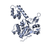
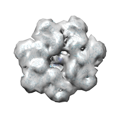
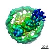
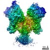
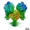
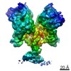
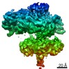
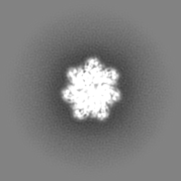
| Title | Adeno-Associated Virus 2 Rep68 HD Heptamer-ssAAVS1 with ATPgS | |||||||||
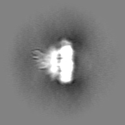
 Map data Map data | Local refined map of heptameric Rep68 HD complex | |||||||||
 Sample Sample |
| |||||||||
 Keywords Keywords | AAV / Protein-DNA / AAA+ / SF3 / HUH / VIRAL PROTEIN / VIRAL PROTEIN-DNA complex | |||||||||
| Function / homology |  Function and homology information Function and homology informationsymbiont-mediated arrest of host cell cycle during G2/M transition / symbiont entry into host cell via permeabilization of host membrane / viral DNA genome replication / symbiont-mediated perturbation of host cell cycle G1/S transition checkpoint / DNA helicase activity / endonuclease activity / DNA helicase / DNA replication / host cell nucleus / ATP hydrolysis activity ...symbiont-mediated arrest of host cell cycle during G2/M transition / symbiont entry into host cell via permeabilization of host membrane / viral DNA genome replication / symbiont-mediated perturbation of host cell cycle G1/S transition checkpoint / DNA helicase activity / endonuclease activity / DNA helicase / DNA replication / host cell nucleus / ATP hydrolysis activity / DNA binding / ATP binding / metal ion binding Similarity search - Function | |||||||||
| Biological species |  Adeno-associated virus - 2 / synthetic construct (others) Adeno-associated virus - 2 / synthetic construct (others) | |||||||||
| Method | single particle reconstruction / cryo EM / Resolution: 4.4 Å | |||||||||
 Authors Authors | Escalante CR | |||||||||
| Funding support |  United States, 1 items United States, 1 items
| |||||||||
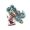
 Citation Citation | Journal: Nucleic Acids Res / Year: 2020 Title: The Cryo-EM structure of AAV2 Rep68 in complex with ssDNA reveals a malleable AAA+ machine that can switch between oligomeric states. Authors: Vishaka Santosh / Faik N Musayev / Rahul Jaiswal / Francisco Zárate-Pérez / Bram Vandewinkel / Caroline Dierckx / Molly Endicott / Kamyar Sharifi / Kelly Dryden / Els Henckaerts / Carlos R Escalante /  Abstract: The adeno-associated virus (AAV) non-structural Rep proteins catalyze all the DNA transactions required for virus viability including, DNA replication, transcription regulation, genome packaging, and ...The adeno-associated virus (AAV) non-structural Rep proteins catalyze all the DNA transactions required for virus viability including, DNA replication, transcription regulation, genome packaging, and during the latent phase, site-specific integration. Rep proteins contain two multifunctional domains: an Origin Binding Domain (OBD) and a SF3 helicase domain (HD). Studies have shown that Rep proteins have a dynamic oligomeric behavior where the nature of the DNA substrate molecule modulates its oligomeric state. In the presence of ssDNA, Rep68 forms a large double-octameric ring complex. To understand the mechanisms underlying AAV Rep function, we investigated the cryo-EM and X-ray structures of Rep68-ssDNA complexes. Surprisingly, Rep68 generates hybrid ring structures where the OBD forms octameric rings while the HD forms heptamers. Moreover, the binding to ATPγS promotes a large conformational change in the entire AAA+ domain that leads the HD to form both heptamer and hexamers. The HD oligomerization is driven by an interdomain linker region that acts as a latch to 'catch' the neighboring HD subunit and is flexible enough to permit the formation of different stoichiometric ring structures. Overall, our studies show the structural basis of AAV Rep's structural flexibility required to fulfill its multifunctional role during the AAV life cycle. | |||||||||
| History |
|
- Structure visualization
| Movie |
Movie viewer |
|---|---|
| Structure viewer | EM map: SurfViewMolmilJmol/JSmol |
| Supplemental images |
- Downloads & links
-EMDB archive
| Map data | emd_22454.map.gz | 59.6 MB | EMDB map data format | |
|---|---|---|---|---|
| Header (meta data) | emd-22454-v30.xmlemd-22454.xml | 15.9 KB 15.9 KB | Display Display | EMDB header |
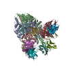
| Images |  emd_22454.png emd_22454.png | 151.3 KB | ||
| Filedesc metadata | emd-22454.cif.gz | 6.5 KB | ||
| Archive directory |  http://ftp.pdbj.org/pub/emdb/structures/EMD-22454ftp://ftp.pdbj.org/pub/emdb/structures/EMD-22454 http://ftp.pdbj.org/pub/emdb/structures/EMD-22454ftp://ftp.pdbj.org/pub/emdb/structures/EMD-22454 | HTTPS FTP |
-Related structure data
| Related structure data |  7jshMC 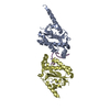 6xb8C  7jseC  7jsfC  7jsgC  7jsiC M: atomic model generated by this map C: citing same article ( |
|---|---|
| Similar structure data |


-Links
| EMDB pages | EMDB (EBI/PDBe) / EMDataResource |
|---|---|
| Related items in Molecule of the Month |


-Map
| File | Download / File: emd_22454.map.gz / Format: CCP4 / Size: 64 MB / Type: IMAGE STORED AS FLOATING POINT NUMBER (4 BYTES) | ||||||||||||||||||||||||||||||||||||||||||||||||||||||||||||||||||||
|---|---|---|---|---|---|---|---|---|---|---|---|---|---|---|---|---|---|---|---|---|---|---|---|---|---|---|---|---|---|---|---|---|---|---|---|---|---|---|---|---|---|---|---|---|---|---|---|---|---|---|---|---|---|---|---|---|---|---|---|---|---|---|---|---|---|---|---|---|---|
| Annotation | Local refined map of heptameric Rep68 HD complex | ||||||||||||||||||||||||||||||||||||||||||||||||||||||||||||||||||||
| Projections & slices | Image control
Images are generated by Spider. | ||||||||||||||||||||||||||||||||||||||||||||||||||||||||||||||||||||
| Voxel size | X=Y=Z: 1.41484 Å | ||||||||||||||||||||||||||||||||||||||||||||||||||||||||||||||||||||
| Density |
| ||||||||||||||||||||||||||||||||||||||||||||||||||||||||||||||||||||
| Symmetry | Space group: 1 | ||||||||||||||||||||||||||||||||||||||||||||||||||||||||||||||||||||
| Details | EMDB XML:
CCP4 map header:
| ||||||||||||||||||||||||||||||||||||||||||||||||||||||||||||||||||||
 Z (Sec.)
 Y (Row.)
 X (Col.)


-Supplemental data
- Sample components
-Entire : Helicase domain heptamer of AAV-2 Rep68 complex bound to AAVS1 ssDNA.
| Entire | Name: Helicase domain heptamer of AAV-2 Rep68 complex bound to AAVS1 ssDNA. |
|---|---|
| Components |
|
-Supramolecule #1: Helicase domain heptamer of AAV-2 Rep68 complex bound to AAVS1 ssDNA.
| Supramolecule | Name: Helicase domain heptamer of AAV-2 Rep68 complex bound to AAVS1 ssDNA. type: complex / ID: 1 / Parent: 0 / Macromolecule list: all Details: Model from local resolution electron density map of helicase domain. |
|---|---|
| Source (natural) | Organism: Adeno-associated virus - 2 |
| Molecular weight | Theoretical: 4.481 kDa/nm |
-Supramolecule #2: AAV-2 Rep68
| Supramolecule | Name: AAV-2 Rep68 / type: complex / ID: 2 / Parent: 1 / Macromolecule list: #1 / Details: Local map includes portion of the helicase domain |
|---|---|
| Source (natural) | Organism: Adeno-associated virus - 2 |
-Supramolecule #3: AAVS1 ssDNA
| Supramolecule | Name: AAVS1 ssDNA / type: complex / ID: 3 / Parent: 1 / Macromolecule list: #2 |
|---|---|
| Source (natural) | Organism: synthetic construct (others) |
-Macromolecule #1: Protein Rep68
| Macromolecule | Name: Protein Rep68 / type: protein_or_peptide / ID: 1 / Number of copies: 7 / Enantiomer: LEVO / EC number: DNA helicase |
|---|---|
| Source (natural) | Organism: Adeno-associated virus - 2 |
| Molecular weight | Theoretical: 61.00582 KDa |
| Recombinant expression | Organism:  |
| Sequence | String: GPPPGFYEIV IKVPSDLDEH LPGISDSFVN WVAEKEWELP PDSDMDLNLI EQAPLTVAEK LQRDFLTEWR RVSKAPEALF FVQFEKGES YFHMHVLVET TGVKSMVLGR FLSQIREKLI QRIYRGIEPT LPNWFAVTKT RNGAGGGNKV VDESYIPNYL L PKTQPELQ ...String: GPPPGFYEIV IKVPSDLDEH LPGISDSFVN WVAEKEWELP PDSDMDLNLI EQAPLTVAEK LQRDFLTEWR RVSKAPEALF FVQFEKGES YFHMHVLVET TGVKSMVLGR FLSQIREKLI QRIYRGIEPT LPNWFAVTKT RNGAGGGNKV VDESYIPNYL L PKTQPELQ WAWTNMEQYL SACLNLTERK RLVAQHLTHV SQTQEQNKEN QNPNSDAPVI RSKTSARYME LVGWLVDKGI TS EKQWIQE DQASYISFNA ASNSRSQIKA ALDNAGKIMS LTKTAPDYLV GQQPVEDISS NRIYKILELN GYDPQYAASV FLG WATKKF GKRNTIWLFG PATTGKTNIA EAIAHTVPFY GCVNWTNENF PFNDCVDKMV IWWEEGKMTA KVVESAKAIL GGSK VRVDQ KCKSSAQIDP TPVIVTSNTN MCAVIDGNST TFEHQQPLQD RMFKFELTRR LDHDFGKVTK QEVKDFFRWA KDHVV EVEH EFYVKKGGAK KRPAPSDADI SEPKRVRESV AQPSTSDAEA SINYADRLAR GHSL UniProtKB: Protein Rep68 |
-Macromolecule #2: DNA (5'-D(P*CP*GP*CP*TP*CP*GP*CP*TP*CP*GP*CP*TP*CP*GP*C)-3')
| Macromolecule | Name: DNA (5'-D(P*CP*GP*CP*TP*CP*GP*CP*TP*CP*GP*CP*TP*CP*GP*C)-3') type: dna / ID: 2 / Number of copies: 1 / Classification: DNA |
|---|---|
| Source (natural) | Organism: synthetic construct (others) |
| Molecular weight | Theoretical: 4.497898 KDa |
| Sequence | String: (DC)(DG)(DC)(DT)(DC)(DG)(DC)(DT)(DC)(DG) (DC)(DT)(DC)(DG)(DC) |
-Experimental details
-Structure determination
| Method | cryo EM |
|---|---|
 Processing Processing | single particle reconstruction |
| Aggregation state | particle |
-Sample preparation
| Concentration | 0.75 mg/mL | ||||||||||||||||||
|---|---|---|---|---|---|---|---|---|---|---|---|---|---|---|---|---|---|---|---|
| Buffer | pH: 7.9 Component:
| ||||||||||||||||||
| Grid | Model: C-flat / Material: COPPER / Mesh: 400 / Support film - Material: CARBON / Support film - topology: HOLEY / Pretreatment - Type: GLOW DISCHARGE / Pretreatment - Time: 40 sec. | ||||||||||||||||||
| Vitrification | Cryogen name: ETHANE / Instrument: FEI VITROBOT MARK IV |
- Electron microscopy
| Microscope | FEI TITAN KRIOS |
|---|---|
| Image recording | Film or detector model: FEI FALCON III (4k x 4k) / Detector mode: COUNTING / Number real images: 2669 / Average electron dose: 50.0 e/Å2 |
| Electron beam | Acceleration voltage: 300 kV / Electron source:  FIELD EMISSION GUN FIELD EMISSION GUN |
| Electron optics | Calibrated magnification: 59000 / Illumination mode: FLOOD BEAM / Imaging mode: BRIGHT FIELD |
| Sample stage | Specimen holder model: FEI TITAN KRIOS AUTOGRID HOLDER |
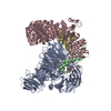
| Experimental equipment |  Model: Titan Krios / Image courtesy: FEI Company |
+Image processing
-Atomic model buiding 1
| Initial model | PDB ID: Chain - Chain ID: A / Chain - Residue range: 225-490 / Chain - Source name: PDB / Chain - Initial model type: experimental model |
|---|---|
| Refinement | Space: REAL / Protocol: RIGID BODY FIT / Target criteria: correlation coefficient |
| Output model | PDB-7jsh: |