[English] 日本語
 Yorodumi
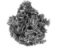
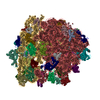
- EMDB-21907: 50S subunit of 70S Ribosome Enterococcus faecalis MultiBody refinement -
+ Open data
- Basic information
| Entry | Database: EMDB / ID: EMD-21907 | |||||||||
|---|---|---|---|---|---|---|---|---|---|---|
| Title | 50S subunit of 70S Ribosome Enterococcus faecalis MultiBody refinement | |||||||||
 Map data Map data | 50S subunit of 70S Ribosome Enterococcus faecalis MultiBody refinement | |||||||||
 Sample Sample |
| |||||||||
 Keywords Keywords | 70S / ribosome / pathogen / antibiotic development / antibiotic resistant | |||||||||
| Function / homology |  Function and homology information Function and homology informationlarge ribosomal subunit / 5S rRNA binding / ribosomal large subunit assembly / large ribosomal subunit rRNA binding / cytosolic large ribosomal subunit / cytoplasmic translation / tRNA binding / negative regulation of translation / rRNA binding / structural constituent of ribosome ...large ribosomal subunit / 5S rRNA binding / ribosomal large subunit assembly / large ribosomal subunit rRNA binding / cytosolic large ribosomal subunit / cytoplasmic translation / tRNA binding / negative regulation of translation / rRNA binding / structural constituent of ribosome / ribosome / translation / ribonucleoprotein complex / mRNA binding / cytoplasm Similarity search - Function | |||||||||
| Biological species |  Enterococcus faecalis OG1RF (bacteria) Enterococcus faecalis OG1RF (bacteria) | |||||||||
| Method | single particle reconstruction / cryo EM / Resolution: 2.9 Å | |||||||||
 Authors Authors | Jogl G / Khayat R | |||||||||
| Funding support |  United States, 2 items United States, 2 items
| |||||||||
 Citation Citation |  Journal: Sci Rep / Year: 2020 Journal: Sci Rep / Year: 2020Title: Cryo-electron microscopy structure of the 70S ribosome from Enterococcus faecalis. Authors: Eileen L Murphy / Kavindra V Singh / Bryant Avila / Torsten Kleffmann / Steven T Gregory / Barbara E Murray / Kurt L Krause / Reza Khayat / Gerwald Jogl /   Abstract: Enterococcus faecalis is a gram-positive organism responsible for serious infections in humans, but as with many bacterial pathogens, resistance has rendered a number of commonly used antibiotics ...Enterococcus faecalis is a gram-positive organism responsible for serious infections in humans, but as with many bacterial pathogens, resistance has rendered a number of commonly used antibiotics ineffective. Here, we report the cryo-EM structure of the E. faecalis 70S ribosome to a global resolution of 2.8 Å. Structural differences are clustered in peripheral and solvent exposed regions when compared with Escherichia coli, whereas functional centres, including antibiotic binding sites, are similar to other bacterial ribosomes. Comparison of intersubunit conformations among five classes obtained after three-dimensional classification identifies several rotated states. Large ribosomal subunit protein bL31, which forms intersubunit bridges to the small ribosomal subunit, assumes different conformations in the five classes, revealing how contacts to the small subunit are maintained throughout intersubunit rotation. A tRNA observed in one of the five classes is positioned in a chimeric pe/E position in a rotated ribosomal state. The 70S ribosome structure of E. faecalis now extends our knowledge of bacterial ribosome structures and may serve as a basis for the development of novel antibiotic compounds effective against this pathogen. | |||||||||
| History |
|
- Structure visualization
| Movie |
 Movie viewer Movie viewer |
|---|---|
| Structure viewer | EM map:  SurfView SurfView Molmil Molmil Jmol/JSmol Jmol/JSmol |
| Supplemental images |
- Downloads & links
-EMDB archive
| Map data |  emd_21907.map.gz emd_21907.map.gz | 302.9 MB |  EMDB map data format EMDB map data format | |
|---|---|---|---|---|
| Header (meta data) |  emd-21907-v30.xml emd-21907-v30.xml emd-21907.xml emd-21907.xml | 40.9 KB 40.9 KB | Display Display |  EMDB header EMDB header |
| Images |  emd_21907.png emd_21907.png | 85 KB | ||
| Filedesc metadata |  emd-21907.cif.gz emd-21907.cif.gz | 9.3 KB | ||
| Archive directory |  http://ftp.pdbj.org/pub/emdb/structures/EMD-21907 http://ftp.pdbj.org/pub/emdb/structures/EMD-21907 ftp://ftp.pdbj.org/pub/emdb/structures/EMD-21907 ftp://ftp.pdbj.org/pub/emdb/structures/EMD-21907 | HTTPS FTP |
-Validation report
| Summary document |  emd_21907_validation.pdf.gz emd_21907_validation.pdf.gz | 691.8 KB | Display |  EMDB validaton report EMDB validaton report |
|---|---|---|---|---|
| Full document |  emd_21907_full_validation.pdf.gz emd_21907_full_validation.pdf.gz | 691.4 KB | Display | |
| Data in XML |  emd_21907_validation.xml.gz emd_21907_validation.xml.gz | 7.4 KB | Display | |
| Data in CIF |  emd_21907_validation.cif.gz emd_21907_validation.cif.gz | 8.4 KB | Display | |
| Arichive directory |  https://ftp.pdbj.org/pub/emdb/validation_reports/EMD-21907 https://ftp.pdbj.org/pub/emdb/validation_reports/EMD-21907 ftp://ftp.pdbj.org/pub/emdb/validation_reports/EMD-21907 ftp://ftp.pdbj.org/pub/emdb/validation_reports/EMD-21907 | HTTPS FTP |
-Related structure data
| Related structure data |  6wu9MC  0656C  0657C  0658C  0659C  0660C  6o8wC  6o8xC  6o8yC  6o8zC  6o90C  6w6pC  6wuaC  6wubC C: citing same article ( M: atomic model generated by this map |
|---|---|
| Similar structure data |
- Links
Links
| EMDB pages |  EMDB (EBI/PDBe) / EMDB (EBI/PDBe) /  EMDataResource EMDataResource |
|---|---|
| Related items in Molecule of the Month |
- Map
Map
| File |  Download / File: emd_21907.map.gz / Format: CCP4 / Size: 325 MB / Type: IMAGE STORED AS FLOATING POINT NUMBER (4 BYTES) Download / File: emd_21907.map.gz / Format: CCP4 / Size: 325 MB / Type: IMAGE STORED AS FLOATING POINT NUMBER (4 BYTES) | ||||||||||||||||||||||||||||||||||||||||||||||||||||||||||||||||||||
|---|---|---|---|---|---|---|---|---|---|---|---|---|---|---|---|---|---|---|---|---|---|---|---|---|---|---|---|---|---|---|---|---|---|---|---|---|---|---|---|---|---|---|---|---|---|---|---|---|---|---|---|---|---|---|---|---|---|---|---|---|---|---|---|---|---|---|---|---|---|
| Annotation | 50S subunit of 70S Ribosome Enterococcus faecalis MultiBody refinement | ||||||||||||||||||||||||||||||||||||||||||||||||||||||||||||||||||||
| Projections & slices | Image control
Images are generated by Spider. | ||||||||||||||||||||||||||||||||||||||||||||||||||||||||||||||||||||
| Voxel size | X=Y=Z: 1.097 Å | ||||||||||||||||||||||||||||||||||||||||||||||||||||||||||||||||||||
| Density |
| ||||||||||||||||||||||||||||||||||||||||||||||||||||||||||||||||||||
| Symmetry | Space group: 1 | ||||||||||||||||||||||||||||||||||||||||||||||||||||||||||||||||||||
| Details | EMDB XML:
CCP4 map header:
| ||||||||||||||||||||||||||||||||||||||||||||||||||||||||||||||||||||
-Supplemental data
- Sample components
+Entire : Enterococcus faecalis
+Supramolecule #1: Enterococcus faecalis
+Macromolecule #1: 23S rRNA
+Macromolecule #2: 5S rRNA
+Macromolecule #3: 50S ribosomal protein L3
+Macromolecule #4: 50S ribosomal protein L4
+Macromolecule #5: 50S ribosomal protein L5
+Macromolecule #6: 50S ribosomal protein L6
+Macromolecule #7: 50S ribosomal protein L13
+Macromolecule #8: 50S ribosomal protein L14
+Macromolecule #9: 50S ribosomal protein L15
+Macromolecule #10: 50S ribosomal protein L16
+Macromolecule #11: 50S ribosomal protein L17
+Macromolecule #12: 50S ribosomal protein L18
+Macromolecule #13: 50S ribosomal protein L19
+Macromolecule #14: 50S ribosomal protein L20
+Macromolecule #15: 50S ribosomal protein L21
+Macromolecule #16: 50S ribosomal protein L22
+Macromolecule #17: 50S ribosomal protein L23
+Macromolecule #18: 50S ribosomal protein L24
+Macromolecule #19: 50S ribosomal protein L27
+Macromolecule #20: 50S ribosomal protein L28
+Macromolecule #21: 50S ribosomal protein L29
+Macromolecule #22: 50S ribosomal protein L30
+Macromolecule #23: 50S ribosomal protein L32
+Macromolecule #24: 50S ribosomal protein L33
+Macromolecule #25: 50S ribosomal protein L34
+Macromolecule #26: 50S ribosomal protein L35
+Macromolecule #27: 50S ribosomal protein L36
+Macromolecule #28: ZINC ION
-Experimental details
-Structure determination
| Method | cryo EM |
|---|---|
 Processing Processing | single particle reconstruction |
| Aggregation state | particle |
- Sample preparation
| Concentration | 1.5 mg/mL |
|---|---|
| Buffer | pH: 7.5 |
| Vitrification | Cryogen name: ETHANE / Chamber humidity: 100 % / Chamber temperature: 4 K / Instrument: FEI VITROBOT MARK IV Details: Vol = 4 uL, BT = 4 sec, BF = 0, DT = 0, WT = 8 sec. |
| Details | Sample was monodisperse. |
- Electron microscopy
| Microscope | FEI TITAN KRIOS |
|---|---|
| Image recording | Film or detector model: GATAN K2 QUANTUM (4k x 4k) / Detector mode: COUNTING / Average electron dose: 25.0 e/Å2 |
| Electron beam | Acceleration voltage: 300 kV / Electron source:  FIELD EMISSION GUN FIELD EMISSION GUN |
| Electron optics | Illumination mode: FLOOD BEAM / Imaging mode: BRIGHT FIELD |
| Sample stage | Specimen holder model: FEI TITAN KRIOS AUTOGRID HOLDER / Cooling holder cryogen: NITROGEN |
| Experimental equipment |  Model: Titan Krios / Image courtesy: FEI Company |
- Image processing
| Startup model | Type of model: OTHER / Details: Ab initio |
|---|---|
| Final reconstruction | Applied symmetry - Point group: C1 (asymmetric) / Resolution.type: BY AUTHOR / Resolution: 2.9 Å / Resolution method: FSC 0.143 CUT-OFF / Software - Name: cryoSPARC (ver. 0.63) / Number images used: 335675 |
| Initial angle assignment | Type: COMMON LINE / Software - Name: RELION (ver. 3.1) |
| Final angle assignment | Type: MAXIMUM LIKELIHOOD / Software - Name: cryoSPARC (ver. 0.63) |
-Atomic model buiding 1
| Initial model |
| ||||||
|---|---|---|---|---|---|---|---|
| Refinement | Space: REAL / Protocol: FLEXIBLE FIT / Overall B value: 177 / Target criteria: Correlation Coefficient | ||||||
| Output model |  PDB-6wu9: |
 Movie
Movie Controller


 Z (Sec.)
 Y (Row.)
 X (Col.)


