 Movie
Movie Controller

[English] 日本語
 Yorodumi
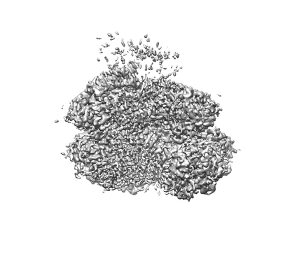
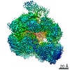
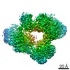
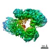
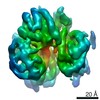
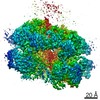
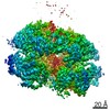
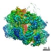
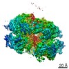
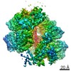
- EMDB-20004: CryoEM map of the hyperactive ClpB mutant K476C, bound to casein,... -
+ Open data

- Basic information
| Entry | Database: EMDB / ID: EMD-20004 | |||||||||
|---|---|---|---|---|---|---|---|---|---|---|
| Title | CryoEM map of the hyperactive ClpB mutant K476C, bound to casein, pre-state | |||||||||
 Map data Map data | hyperactive ClpB mutant K476C, bound to casein, state_1 | |||||||||
 Sample Sample |
| |||||||||
 Keywords Keywords | disaggregase / CLPB / AAA+ / CHAPERONE | |||||||||
| Function / homology |  Function and homology information Function and homology informationcellular response to heat / response to heat / protein refolding / ATP hydrolysis activity / ATP binding / membrane / identical protein binding / cytoplasm / cytosol Similarity search - Function | |||||||||
| Biological species |   | |||||||||
| Method | single particle reconstruction / cryo EM / Resolution: 2.9 Å | |||||||||
 Authors Authors | Rizo AR / Lin J-B | |||||||||
 Citation Citation | Journal: Nat Commun / Year: 2019 Title: Structural basis for substrate gripping and translocation by the ClpB AAA+ disaggregase. Authors: Alexandrea N Rizo / JiaBei Lin / Stephanie N Gates / Eric Tse / Stephen M Bart / Laura M Castellano / Frank DiMaio / James Shorter / Daniel R Southworth /  Abstract: Bacterial ClpB and yeast Hsp104 are homologous Hsp100 protein disaggregases that serve critical functions in proteostasis by solubilizing protein aggregates. Two AAA+ nucleotide binding domains (NBDs) ...Bacterial ClpB and yeast Hsp104 are homologous Hsp100 protein disaggregases that serve critical functions in proteostasis by solubilizing protein aggregates. Two AAA+ nucleotide binding domains (NBDs) power polypeptide translocation through a central channel comprised of a hexameric spiral of protomers that contact substrate via conserved pore-loop interactions. Here we report cryo-EM structures of a hyperactive ClpB variant bound to the model substrate, casein in the presence of slowly hydrolysable ATPγS, which reveal the translocation mechanism. Distinct substrate-gripping interactions are identified for NBD1 and NBD2 pore loops. A trimer of N-terminal domains define a channel entrance that binds the polypeptide substrate adjacent to the topmost NBD1 contact. NBD conformations at the seam interface reveal how ATP hydrolysis-driven substrate disengagement and re-binding are precisely tuned to drive a directional, stepwise translocation cycle. | |||||||||
| History |
|
- Structure visualization
| Movie |
Movie viewer |
|---|---|
| Structure viewer | EM map: SurfViewMolmilJmol/JSmol |
| Supplemental images |
- Downloads & links
-EMDB archive
| Map data | emd_20004.map.gz | 59.7 MB | EMDB map data format | |
|---|---|---|---|---|
| Header (meta data) | emd-20004-v30.xmlemd-20004.xml | 16.1 KB 16.1 KB | Display Display | EMDB header |
| Images |  emd_20004.png emd_20004.png | 39.5 KB | ||
| Filedesc metadata | emd-20004.cif.gz | 6.6 KB | ||
| Archive directory |  http://ftp.pdbj.org/pub/emdb/structures/EMD-20004ftp://ftp.pdbj.org/pub/emdb/structures/EMD-20004 http://ftp.pdbj.org/pub/emdb/structures/EMD-20004ftp://ftp.pdbj.org/pub/emdb/structures/EMD-20004 | HTTPS FTP |
-Related structure data
| Related structure data | 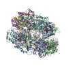 6oaxMC 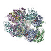 6oayC 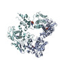 6og1C 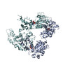 6og2C  6og3C C: citing same article ( M: atomic model generated by this map |
|---|---|
| Similar structure data |

-Links
| EMDB pages | EMDB (EBI/PDBe) / EMDataResource |
|---|---|
| Related items in Molecule of the Month |


-Map
| File | Download / File: emd_20004.map.gz / Format: CCP4 / Size: 64 MB / Type: IMAGE STORED AS FLOATING POINT NUMBER (4 BYTES) | ||||||||||||||||||||||||||||||||||||||||||||||||||||||||||||
|---|---|---|---|---|---|---|---|---|---|---|---|---|---|---|---|---|---|---|---|---|---|---|---|---|---|---|---|---|---|---|---|---|---|---|---|---|---|---|---|---|---|---|---|---|---|---|---|---|---|---|---|---|---|---|---|---|---|---|---|---|---|
| Annotation | hyperactive ClpB mutant K476C, bound to casein, state_1 | ||||||||||||||||||||||||||||||||||||||||||||||||||||||||||||
| Projections & slices | Image control
Images are generated by Spider. | ||||||||||||||||||||||||||||||||||||||||||||||||||||||||||||
| Voxel size | X=Y=Z: 1.032 Å | ||||||||||||||||||||||||||||||||||||||||||||||||||||||||||||
| Density |
| ||||||||||||||||||||||||||||||||||||||||||||||||||||||||||||
| Symmetry | Space group: 1 | ||||||||||||||||||||||||||||||||||||||||||||||||||||||||||||
| Details | EMDB XML:
CCP4 map header:
| ||||||||||||||||||||||||||||||||||||||||||||||||||||||||||||
 Z (Sec.)
 Y (Row.)
 X (Col.)


-Supplemental data
- Sample components
-Entire : hyperactive ClpB mutant K476C bound to casein
| Entire | Name: hyperactive ClpB mutant K476C bound to casein |
|---|---|
| Components |
|
-Supramolecule #1: hyperactive ClpB mutant K476C bound to casein
| Supramolecule | Name: hyperactive ClpB mutant K476C bound to casein / type: complex / ID: 1 / Parent: 0 / Macromolecule list: #1-#2 |
|---|
-Supramolecule #2: hyperactive ClpB mutant K476C
| Supramolecule | Name: hyperactive ClpB mutant K476C / type: complex / ID: 2 / Parent: 1 / Macromolecule list: #1 |
|---|---|
| Source (natural) | Organism: |
-Supramolecule #3: alpha-S1-casein
| Supramolecule | Name: alpha-S1-casein / type: complex / ID: 3 / Parent: 1 / Macromolecule list: #2 Details: Protein was purchased from Sigma-Aldrich. Purified from bovine milk. |
|---|---|
| Source (natural) | Organism: |
-Macromolecule #1: Hyperactive disaggregase ClpB
| Macromolecule | Name: Hyperactive disaggregase ClpB / type: protein_or_peptide / ID: 1 / Number of copies: 6 / Enantiomer: LEVO |
|---|---|
| Source (natural) | Organism: |
| Molecular weight | Theoretical: 97.018469 KDa |
| Recombinant expression | Organism: |
| Sequence | String: MRLDRLTNKF QLALADAQSL ALGHDNQFIE PLHLMSALLN QEGGSVSPLL TSAGINAGQL RTDINQALNR LPQVEGTGGD VQPSQDLVR VLNLCDNVAQ KRGDNFISSE LFVLAALESR GTVADILKAA GATTANITQA IEQMRGGESV NDQGAEDQRQ A LKKYTIDL ...String: MRLDRLTNKF QLALADAQSL ALGHDNQFIE PLHLMSALLN QEGGSVSPLL TSAGINAGQL RTDINQALNR LPQVEGTGGD VQPSQDLVR VLNLCDNVAQ KRGDNFISSE LFVLAALESR GTVADILKAA GATTANITQA IEQMRGGESV NDQGAEDQRQ A LKKYTIDL TERAEQGKLD PVIGRDEEIR RTIQVLQRRT KNNPVLIGEP GVGKTAIVEG LAQRIINGEV PEGLKGRRVL AL DMGALVA GAKYRGEFEE RLKGVLNDLA KQEGNVILFI DELHTMVGAG KADGAMDAGN MLKPALARGE LHCVGATTLD EYR QYIEKD AALERRFQKV FVAEPSVEDT IAILRGLKER YELHHHVQIT DPAIVAAATL SHRYIADRQL PDKAIDLIDE AASS IRMQI DSKPEELDRL DRRIIQLKLE QQALMKESDE ASKKRLDMLN EELSDKERQY SELEEEWKAE KASLSGTQTI KCELE QAKI AIEQARRVGD LARMSELQYG KIPELEKQLE AATQLEGKTM RLLRNKVTDA EIAEVLARWT GIPVSRMMES EREKLL RME QELHHRVIGQ NEAVDAVSNA IRRSRAGLAD PNRPIGSFLF LGPTGVGKTE LCKALANFMF DSDEAMVRID MSEFMEK HS VSRLVGAPPG YVGYEEGGYL TEAVRRRPYS VILLDEVEKA HPDVFNILLQ VLDDGRLTDG QGRTVDFRNT VVIMTSNL G SDLIQERFGE LDYAHMKELV LGVVSHNFRP EFINRIDEVV VFHPLGEQHI ASIAQIQLKR LYKRLEERGY EIHISDEAL KLLSENGYDP VYGARPLKRA IQQQIENPLA QQILSGELVP GKVIRLEVNE DRIVAVQRSR SHHHHHH |
-Macromolecule #2: Alpha-S1-casein
| Macromolecule | Name: Alpha-S1-casein / type: protein_or_peptide / ID: 2 / Number of copies: 1 / Enantiomer: LEVO |
|---|---|
| Source (natural) | Organism: |
| Molecular weight | Theoretical: 2.230741 KDa |
| Sequence | String: (UNK)(UNK)(UNK)(UNK)(UNK)(UNK)(UNK)(UNK)(UNK)(UNK) (UNK)(UNK)(UNK)(UNK)(UNK)(UNK) (UNK)(UNK)(UNK) (UNK)(UNK)(UNK)(UNK)(UNK)(UNK)(UNK) |
-Macromolecule #3: PHOSPHOTHIOPHOSPHORIC ACID-ADENYLATE ESTER
| Macromolecule | Name: PHOSPHOTHIOPHOSPHORIC ACID-ADENYLATE ESTER / type: ligand / ID: 3 / Number of copies: 9 / Formula: AGS |
|---|---|
| Molecular weight | Theoretical: 523.247 Da |
| Chemical component information |  ChemComp-AGS: |
-Macromolecule #4: ADENOSINE-5'-DIPHOSPHATE
| Macromolecule | Name: ADENOSINE-5'-DIPHOSPHATE / type: ligand / ID: 4 / Number of copies: 3 / Formula: ADP |
|---|---|
| Molecular weight | Theoretical: 427.201 Da |
| Chemical component information |  ChemComp-ADP: |
-Experimental details
-Structure determination
| Method | cryo EM |
|---|---|
 Processing Processing | single particle reconstruction |
| Aggregation state | particle |
-Sample preparation
| Concentration | 1.5 mg/mL |
|---|---|
| Buffer | pH: 7.5 |
| Grid | Details: unspecified |
| Vitrification | Cryogen name: ETHANE |
- Electron microscopy
| Microscope | FEI TITAN KRIOS |
|---|---|
| Image recording | Film or detector model: GATAN K2 SUMMIT (4k x 4k) / Detector mode: SUPER-RESOLUTION / Average electron dose: 56.0 e/Å2 |
| Electron beam | Acceleration voltage: 300 kV / Electron source:  FIELD EMISSION GUN FIELD EMISSION GUN |
| Electron optics | Illumination mode: FLOOD BEAM / Imaging mode: BRIGHT FIELD / Cs: 2.6 mm |
| Experimental equipment |  Model: Titan Krios / Image courtesy: FEI Company |
