 Movie
Movie Controller

[English] 日本語
 Yorodumi
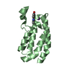
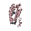
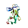
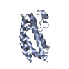
- PDB-1xwv: Structure of the house dust mite allergen Der f 2: Implications f... -
+ Open data

- Basic information
| Entry | Database: PDB / ID: 1xwv | ||||||
|---|---|---|---|---|---|---|---|
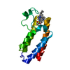
| Title | Structure of the house dust mite allergen Der f 2: Implications for function and molecular basis of IgE cross-reactivity | ||||||
 Components Components | Der f II | ||||||
 Keywords Keywords | ALLERGEN / BETA SHEETS | ||||||
| Function / homology |  Function and homology information Function and homology information | ||||||
| Biological species |  Dermatophagoides farinae (American house dust mite) Dermatophagoides farinae (American house dust mite) | ||||||
| Method |  X-RAY DIFFRACTION / MOLECULAR REPLACEMENT / Resolution: 1.83 Å X-RAY DIFFRACTION / MOLECULAR REPLACEMENT / Resolution: 1.83 Å | ||||||
 Authors Authors | Johannessen, B.R. / Skov, L.K. / Kastrup, J.S. / Kristensen, O. / Bolwig, C. / Larsen, J.N. / Spangfort, M. / Lund, K. / Gajhede, M. | ||||||
 Citation Citation | Journal: Febs Lett. / Year: 2005 Title: Structure of the house dust mite allergen Der f 2: implications for function and molecular basis of IgE cross-reactivity. Authors: Johannessen, B.R. / Skov, L.K. / Kastrup, J.S. / Kristensen, O. / Bolwig, C. / Larsen, J.N. / Spangfort, M. / Lund, K. / Gajhede, M. | ||||||
| History |
|
- Structure visualization
| Structure viewer | Molecule: MolmilJmol/JSmol |
|---|
- Downloads & links
-Download
| PDBx/mmCIF format | 1xwv.cif.gz | 71.3 KB | Display | PDBx/mmCIF format |
|---|---|---|---|---|
| PDB format | pdb1xwv.ent.gz | 51.5 KB | Display | PDB format |
| PDBx/mmJSON format | 1xwv.json.gz | Tree view | PDBx/mmJSON format | |
| Others |  Other downloads Other downloads |
-Validation report
| Arichive directory | https://data.pdbj.org/pub/pdb/validation_reports/xw/1xwvftp://data.pdbj.org/pub/pdb/validation_reports/xw/1xwv | HTTPS FTP |
|---|
-Related structure data
| Related structure data |  1ktjS S: Starting model for refinement |
|---|---|
| Similar structure data |


-Links
 PDBj
PDBj
- Assembly
| Deposited unit | 
| ||||||||
|---|---|---|---|---|---|---|---|---|---|
| 1 | 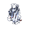
| ||||||||
| 2 | 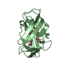
| ||||||||
| Unit cell |
|
-Components
| #1: Protein | Mass: 14054.177 Da / Num. of mol.: 2 Source method: isolated from a genetically manipulated source Source: (gene. exp.) Dermatophagoides farinae (American house dust mite)Plasmid: pGAPZalpha-A / Production host:  Pichia pastoris (fungus) / References: UniProt: Q00855 Pichia pastoris (fungus) / References: UniProt: Q00855#2: Chemical | ChemComp-PE3 / |   Mass: 634.751 Da / Num. of mol.: 1 / Source method: obtained synthetically / Formula: C28H58O15 Mass: 634.751 Da / Num. of mol.: 1 / Source method: obtained synthetically / Formula: C28H58O15#3: Chemical | ChemComp-XPE / |   Mass: 458.541 Da / Num. of mol.: 1 / Source method: obtained synthetically / Formula: C20H42O11 / Comment: precipitant*YM Mass: 458.541 Da / Num. of mol.: 1 / Source method: obtained synthetically / Formula: C20H42O11 / Comment: precipitant*YM#4: Water | ChemComp-HOH / |  Mass: 18.015 Da / Num. of mol.: 240 / Source method: isolated from a natural source / Formula: H2O Mass: 18.015 Da / Num. of mol.: 240 / Source method: isolated from a natural source / Formula: H2OHas protein modification | Y | |
|---|
-Experimental details
-Experiment
| Experiment | Method: X-RAY DIFFRACTION / Number of used crystals: 1 |
|---|
- Sample preparation
| Crystal | Density Matthews: 2 Å3/Da / Density % sol: 37.2 % |
|---|---|
| Crystal grow | Temperature: 293 K / Method: vapor diffusion, hanging drop / pH: 6 Details: PEG 4000, potassium citrate, ammonium acetate, pH 6.0, VAPOR DIFFUSION, HANGING DROP, temperature 293K |
-Data collection
| Diffraction | Mean temperature: 100 K |
|---|---|
| Diffraction source | Source: ROTATING ANODE / Type: RIGAKU RU300 / Wavelength: 1.5418 |
| Detector | Type: MAR scanner 345 mm plate / Detector: IMAGE PLATE / Date: Dec 5, 2002 / Details: mirrors |
| Radiation | Protocol: SINGLE WAVELENGTH / Monochromatic (M) / Laue (L): M / Scattering type: x-ray |
| Radiation wavelength | Wavelength: 1.5418 Å / Relative weight: 1 |
| Reflection | Resolution: 1.83→24.43 Å / Num. all: 18043 / Num. obs: 18043 / % possible obs: 93.4 % / Observed criterion σ(F): 0 / Observed criterion σ(I): -3 / Redundancy: 3.9 % / Biso Wilson estimate: 18.7 Å2 / Rmerge(I) obs: 0.049 / Rsym value: 0.049 / Net I/σ(I): 9 |
| Reflection shell | Resolution: 1.83→1.94 Å / Redundancy: 3.8 % / Rmerge(I) obs: 0.24 / Mean I/σ(I) obs: 3 / Num. unique all: 2626 / Rsym value: 0.24 / % possible all: 85.9 |
- Processing
| Software |
| |||||||||||||||||||||||||
|---|---|---|---|---|---|---|---|---|---|---|---|---|---|---|---|---|---|---|---|---|---|---|---|---|---|---|
| Refinement | Method to determine structure: MOLECULAR REPLACEMENT Starting model: PDB ENTRY 1KTJ Resolution: 1.83→24.43 Å / Isotropic thermal model: RESTRAINED / Cross valid method: THROUGHOUT / σ(F): 0 / σ(I): 0
| |||||||||||||||||||||||||
| Displacement parameters | Biso mean: 30.5 Å2
| |||||||||||||||||||||||||
| Refine analyze |
| |||||||||||||||||||||||||
| Refinement step | Cycle: LAST / Resolution: 1.83→24.43 Å
| |||||||||||||||||||||||||
| Refine LS restraints |
| |||||||||||||||||||||||||
| LS refinement shell | Resolution: 1.83→1.94 Å / Rfactor Rfree error: 0.026
|
