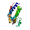
 Movie
Movie Controller


+ Open data

- Basic information
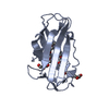
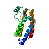
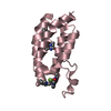
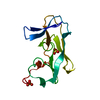
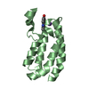
| Entry | Database: PDB / ID: 6lu6 | ||||||
|---|---|---|---|---|---|---|---|
| Title | Crystal structure of BPTF-BRD with ligand DCBPin5-2 bound | ||||||
 Components Components | Nucleosome-remodeling factor subunit BPTF | ||||||
 Keywords Keywords | ANTITUMOR PROTEIN / BPTF Bromodomain / Lysine acetylation / small-molecule inhibitor / high-throughput screening / BIOSYNTHETIC PROTEIN | ||||||
| Function / homology |  Function and homology information Function and homology informationNURF complex / regulation of transcription by RNA polymerase II / zinc ion binding Similarity search - Function | ||||||
| Biological species |  Homo sapiens (human) Homo sapiens (human) | ||||||
| Method |  X-RAY DIFFRACTION / SYNCHROTRON / MOLECULAR REPLACEMENT / Resolution: 1.97006302049 Å X-RAY DIFFRACTION / SYNCHROTRON / MOLECULAR REPLACEMENT / Resolution: 1.97006302049 Å | ||||||
 Authors Authors | Lu, T. / Lu, H.B. / Wang, J. / Lin, H. / Lu, W. / Luo, C. | ||||||
 Citation Citation | Journal: To Be Published Title: Discovery and Optimization of Small-Molecule Inhibitors for the BPTF Bromodomains Proteins Authors: Lu, T. / Lu, H.B. / Wang, J. / Han, J. / Xiao, S. / Jiang, H. / Chen, Y. / Yang, F. / Li, Q. / Jiang, H.L. / Chen, K.X. / Lu, W.C. / Lin, H. / Luo, C. | ||||||
| History |
|
- Structure visualization
| Structure viewer | Molecule: MolmilJmol/JSmol |
|---|
- Downloads & links
-Download
| PDBx/mmCIF format | 6lu6.cif.gz | 46.2 KB | Display | PDBx/mmCIF format |
|---|---|---|---|---|
| PDB format | pdb6lu6.ent.gz | 25.4 KB | Display | PDB format |
| PDBx/mmJSON format | 6lu6.json.gz | Tree view | PDBx/mmJSON format | |
| Others |  Other downloads Other downloads |
-Validation report
| Arichive directory | https://data.pdbj.org/pub/pdb/validation_reports/lu/6lu6ftp://data.pdbj.org/pub/pdb/validation_reports/lu/6lu6 | HTTPS FTP |
|---|
-Related structure data
| Related structure data |  6lu5C  7f5cC  7f5dC  7f5eC  3qztS S: Starting model for refinement C: citing same article ( |
|---|---|
| Similar structure data |

-Links
 PDBj
PDBj- Assembly
| Deposited unit | 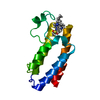
| ||||||||||||
|---|---|---|---|---|---|---|---|---|---|---|---|---|---|
| 1 |
| ||||||||||||
| Unit cell |
|
-Components
| #1: Protein | Mass: 13031.885 Da / Num. of mol.: 1 / Fragment: Bromodomain Source method: isolated from a genetically manipulated source Source: (gene. exp.) Homo sapiens (human) / Gene: BPTF / Production host:  |
|---|---|
| #2: Chemical | ChemComp-EUU / 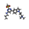  Mass: 415.552 Da / Num. of mol.: 1 / Source method: obtained synthetically / Formula: C21H29N5O2S / Feature type: SUBJECT OF INVESTIGATION Mass: 415.552 Da / Num. of mol.: 1 / Source method: obtained synthetically / Formula: C21H29N5O2S / Feature type: SUBJECT OF INVESTIGATION |
| #3: Water | ChemComp-HOH /  Mass: 18.015 Da / Num. of mol.: 26 / Source method: isolated from a natural source / Formula: H2O Mass: 18.015 Da / Num. of mol.: 26 / Source method: isolated from a natural source / Formula: H2O |
| Has ligand of interest | Y |
-Experimental details
-Experiment
| Experiment | Method: X-RAY DIFFRACTION / Number of used crystals: 1 |
|---|
- Sample preparation
| Crystal | Density Matthews: 2.16 Å3/Da / Density % sol: 43 % |
|---|---|
| Crystal grow | Temperature: 289 K / Method: vapor diffusion, sitting drop / pH: 8.5 Details: 0.2 M Lithium sulfate monohydrate, 0.1 M Tris pH 8.5, 25% PEG3350 |
-Data collection
| Diffraction | Mean temperature: 100 K / Serial crystal experiment: N |
|---|---|
| Diffraction source | Source: SYNCHROTRON / Site: SSRF  / Beamline: BL19U1 / Wavelength: 0.98 Å / Beamline: BL19U1 / Wavelength: 0.98 Å |
| Detector | Type: DECTRIS PILATUS 6M / Detector: PIXEL / Date: Dec 30, 2019 |
| Radiation | Protocol: SINGLE WAVELENGTH / Monochromatic (M) / Laue (L): M / Scattering type: x-ray |
| Radiation wavelength | Wavelength: 0.98 Å / Relative weight: 1 |
| Reflection | Resolution: 1.97→33.82 Å / Num. obs: 7299 / % possible obs: 88.7 % / Redundancy: 2.2 % / Biso Wilson estimate: 25.5461904813 Å2 / CC1/2: 0.992 / Rmerge(I) obs: 0.1108 / Rpim(I) all: 0.1108 / Rrim(I) all: 0.1567 / Net I/σ(I): 2.2 |
| Reflection shell | Resolution: 1.97→2.04 Å / Rmerge(I) obs: 0.9473 / Num. unique obs: 753 / CC1/2: 0.841 / Rpim(I) all: 0.9473 |
- Processing
| Software |
| ||||||||||||||||||||||||||||
|---|---|---|---|---|---|---|---|---|---|---|---|---|---|---|---|---|---|---|---|---|---|---|---|---|---|---|---|---|---|
| Refinement | Method to determine structure: MOLECULAR REPLACEMENT Starting model: 3QZT Resolution: 1.97006302049→33.8181389703 Å / SU ML: 0.450709209554 / Cross valid method: FREE R-VALUE / σ(F): 1.34349388682 / Phase error: 44.6521649335
| ||||||||||||||||||||||||||||
| Solvent computation | Shrinkage radii: 0.9 Å / VDW probe radii: 1.11 Å | ||||||||||||||||||||||||||||
| Displacement parameters | Biso mean: 31.121167247 Å2 | ||||||||||||||||||||||||||||
| Refinement step | Cycle: LAST / Resolution: 1.97006302049→33.8181389703 Å
| ||||||||||||||||||||||||||||
| Refine LS restraints |
| ||||||||||||||||||||||||||||
| LS refinement shell |
|
