 Movie
Movie Controller

[English] 日本語
 Yorodumi
- PDB-1wrr: Urate oxidase from aspergillus flavus complexed with 5-amino 6-ni... -
+ Open data

- Basic information
| Entry | Database: PDB / ID: 1wrr | ||||||
|---|---|---|---|---|---|---|---|
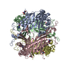
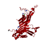
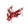
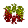
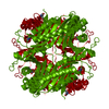
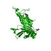
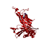
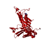
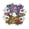
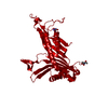
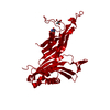
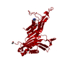
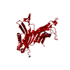
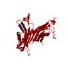
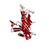
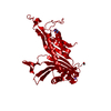
| Title | Urate oxidase from aspergillus flavus complexed with 5-amino 6-nitro uracil | ||||||
 Components Components | Uricase | ||||||
 Keywords Keywords | OXIDOREDUCTASE / uric acid degradation / dimeric barrel / tunnel-shaped protein | ||||||
| Function / homology |  Function and homology information Function and homology informationurate oxidase activity / purine nucleobase catabolic process / factor-independent urate hydroxylase / urate catabolic process / peroxisome Similarity search - Function | ||||||
| Biological species |  | ||||||
| Method |  X-RAY DIFFRACTION / SYNCHROTRON / SIR under pressure of noble gaz / Resolution: 1.64 Å X-RAY DIFFRACTION / SYNCHROTRON / SIR under pressure of noble gaz / Resolution: 1.64 Å | ||||||
 Authors Authors | Retailleau, P. / Colloc'h, N. / Vivares, D. / Bonnete, F. / Castro, B. / El Hajji, M. / Prange, T. | ||||||
 Citation Citation | Journal: Acta Crystallogr.,Sect.D / Year: 2005 Title: Urate oxidase from Aspergillus flavus: new crystal-packing contacts in relation to the content of the active site. Authors: Retailleau, P. / Colloc'h, N. / Vivares, D. / Bonnete, F. / Castro, B. / El Hajji, M. / Prange, T. #1: Journal: Nat.Struct.Biol. / Year: 1997 Title: Crystal structure of the protein drug urate oxidase-inhibitor complex at 2.05 A resolution Authors: Colloc'h, N. / El Hajji, M. / Bachet, B. / L'Hermite, G. / Schiltz, M. / Castro, B. / Mornon, J.P. | ||||||
| History |
|
- Structure visualization
| Structure viewer | Molecule: MolmilJmol/JSmol |
|---|
- Downloads & links
-Download
| PDBx/mmCIF format | 1wrr.cif.gz | 77.7 KB | Display | PDBx/mmCIF format |
|---|---|---|---|---|
| PDB format | pdb1wrr.ent.gz | 58.2 KB | Display | PDB format |
| PDBx/mmJSON format | 1wrr.json.gz | Tree view | PDBx/mmJSON format | |
| Others |  Other downloads Other downloads |
-Validation report
| Summary document | 1wrr_validation.pdf.gz | 438.8 KB | Display | wwPDB validaton report |
|---|---|---|---|---|
| Full document | 1wrr_full_validation.pdf.gz | 439.4 KB | Display | |
| Data in XML | 1wrr_validation.xml.gz | 17 KB | Display | |
| Data in CIF | 1wrr_validation.cif.gz | 23.6 KB | Display | |
| Arichive directory | https://data.pdbj.org/pub/pdb/validation_reports/wr/1wrrftp://data.pdbj.org/pub/pdb/validation_reports/wr/1wrr | HTTPS FTP |
-Related structure data


-Links
 PDBj
PDBj
- Assembly
| Deposited unit | 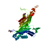
| ||||||||
|---|---|---|---|---|---|---|---|---|---|
| 1 | 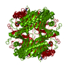
| ||||||||
| Unit cell |
|
-Components
| #1: Protein | Mass: 34199.586 Da / Num. of mol.: 1 Source method: isolated from a genetically manipulated source Source: (gene. exp.)  References: UniProt: Q00511, factor-independent urate hydroxylase |
|---|---|
| #2: Chemical | ChemComp-UNC /   Mass: 172.099 Da / Num. of mol.: 1 / Source method: obtained synthetically / Formula: C4H4N4O4 Mass: 172.099 Da / Num. of mol.: 1 / Source method: obtained synthetically / Formula: C4H4N4O4 |
| #3: Water | ChemComp-HOH /  Mass: 18.015 Da / Num. of mol.: 215 / Source method: isolated from a natural source / Formula: H2O Mass: 18.015 Da / Num. of mol.: 215 / Source method: isolated from a natural source / Formula: H2O |
| Has protein modification | Y |
-Experimental details
-Experiment
| Experiment | Method: X-RAY DIFFRACTION / Number of used crystals: 1 |
|---|
- Sample preparation
| Crystal | Density Matthews: 2.8 Å3/Da / Density % sol: 55.8 % |
|---|---|
| Crystal grow | Temperature: 291 K / Method: vapor diffusion, sitting drop / pH: 8 Details: 8.5MG/ML PROTEIN, 0.2MG/ML DIAMINOURACIL, 5-7%(W/V) PEG 8000, 100MM TRIS/HCL, pH 8.00, VAPOR DIFFUSION, SITTING DROP, temperature 291K |
-Data collection
| Diffraction | Mean temperature: 283 K |
|---|---|
| Diffraction source | Source: SYNCHROTRON / Site: LURE  / Beamline: DW32 / Wavelength: 0.972 Å / Beamline: DW32 / Wavelength: 0.972 Å |
| Detector | Type: MARRESEARCH / Detector: IMAGE PLATE / Date: Oct 25, 2003 / Details: curvated mirrors |
| Radiation | Monochromator: Si (111) / Protocol: SINGLE WAVELENGTH / Monochromatic (M) / Laue (L): M / Scattering type: x-ray |
| Radiation wavelength | Wavelength: 0.972 Å / Relative weight: 1 |
| Reflection | Resolution: 1.64→35 Å / Num. obs: 48554 / % possible obs: 99.9 % / Redundancy: 6 % / Biso Wilson estimate: 28.5 Å2 / Rsym value: 0.037 / Net I/σ(I): 17.8 |
| Reflection shell | Resolution: 1.64→1.68 Å / Mean I/σ(I) obs: 10.3 / Rsym value: 0.285 / % possible all: 100 |
- Processing
| Software |
| ||||||||||||||||||||||||||||||||||||||||||||||||||||||||||||||||||||||||||||||||||||||||||||||||||||||||||||||||||||||||||||||||||||||||||||||||||||||||||||||||
|---|---|---|---|---|---|---|---|---|---|---|---|---|---|---|---|---|---|---|---|---|---|---|---|---|---|---|---|---|---|---|---|---|---|---|---|---|---|---|---|---|---|---|---|---|---|---|---|---|---|---|---|---|---|---|---|---|---|---|---|---|---|---|---|---|---|---|---|---|---|---|---|---|---|---|---|---|---|---|---|---|---|---|---|---|---|---|---|---|---|---|---|---|---|---|---|---|---|---|---|---|---|---|---|---|---|---|---|---|---|---|---|---|---|---|---|---|---|---|---|---|---|---|---|---|---|---|---|---|---|---|---|---|---|---|---|---|---|---|---|---|---|---|---|---|---|---|---|---|---|---|---|---|---|---|---|---|---|---|---|---|---|
| Refinement | Method to determine structure: SIR under pressure of noble gaz Resolution: 1.64→28 Å / Cor.coef. Fo:Fc: 0.971 / Cor.coef. Fo:Fc free: 0.966 / SU B: 1.343 / SU ML: 0.046 / Cross valid method: THROUGHOUT / ESU R: 0.078 / ESU R Free: 0.076 Stereochemistry target values: MAXIMUM LIKELIHOOD WITH PHASES Details: The structre was refined also with buster-TNT. The final refinement statistics were derived from REFMAC 5. Major refinement was proceeded using BUSTER-TNT.
| ||||||||||||||||||||||||||||||||||||||||||||||||||||||||||||||||||||||||||||||||||||||||||||||||||||||||||||||||||||||||||||||||||||||||||||||||||||||||||||||||
| Solvent computation | Solvent model: BABINET MASK | ||||||||||||||||||||||||||||||||||||||||||||||||||||||||||||||||||||||||||||||||||||||||||||||||||||||||||||||||||||||||||||||||||||||||||||||||||||||||||||||||
| Displacement parameters | Biso mean: 21.976 Å2
| ||||||||||||||||||||||||||||||||||||||||||||||||||||||||||||||||||||||||||||||||||||||||||||||||||||||||||||||||||||||||||||||||||||||||||||||||||||||||||||||||
| Refinement step | Cycle: LAST / Resolution: 1.64→28 Å
| ||||||||||||||||||||||||||||||||||||||||||||||||||||||||||||||||||||||||||||||||||||||||||||||||||||||||||||||||||||||||||||||||||||||||||||||||||||||||||||||||
| Refine LS restraints |
| ||||||||||||||||||||||||||||||||||||||||||||||||||||||||||||||||||||||||||||||||||||||||||||||||||||||||||||||||||||||||||||||||||||||||||||||||||||||||||||||||
| LS refinement shell | Resolution: 1.64→1.682 Å / Total num. of bins used: 20
|
