- PDB-1vrn: PHOTOSYNTHETIC REACTION CENTER BLASTOCHLORIS VIRIDIS (ATCC) -
+
Open data
ID or keywords:
Loading...
-
Basic information
Entry
Database: PDB / ID: 1vrn
Title
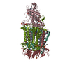
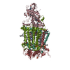
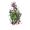
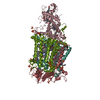
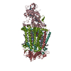
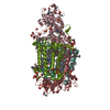
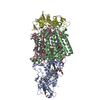
PHOTOSYNTHETIC REACTION CENTER BLASTOCHLORIS VIRIDIS (ATCC)
Components
(Reaction center protein ...) x 3
Photosynthetic reaction center cytochrome c subunit
Keywords
PHOTOSYNTHESIS / INTEGRAL MEMBRANE PROTEIN / UBIQUINONE / SECONDARY QUINONE (QB)
Function / homology
Function and homology information
plasma membrane-derived chromatophore membrane / plasma membrane light-harvesting complex / bacteriochlorophyll binding / photosynthetic electron transport in photosystem II / photosynthesis, light reaction / photosynthesis / electron transfer activity / iron ion binding / heme binding / metal ion binding Similarity search - Function
Photosynthetic Reaction Center, subunit C; domain 2 / Photosynthetic Reaction Center, subunit C, domain 2 / Photosynthetic reaction centre, cytochrome c subunit / Multihaem cytochrome, PRC subunit superfamily / Photosynthetic reaction centre cytochrome C subunit / Photosynthetic Reaction Center; Chain H, domain 1 / Photosynthetic reaction centre, H subunit, N-terminal domain / Photosynthetic Reaction Center; Chain H, domain 2 / Photosynthetic Reaction Center, subunit H, domain 2 / Photosynthetic Reaction Center, subunit M; domain 1 ...Photosynthetic Reaction Center, subunit C; domain 2 / Photosynthetic Reaction Center, subunit C, domain 2 / Photosynthetic reaction centre, cytochrome c subunit / Multihaem cytochrome, PRC subunit superfamily / Photosynthetic reaction centre cytochrome C subunit / Photosynthetic Reaction Center; Chain H, domain 1 / Photosynthetic reaction centre, H subunit, N-terminal domain / Photosynthetic Reaction Center; Chain H, domain 2 / Photosynthetic Reaction Center, subunit H, domain 2 / Photosynthetic Reaction Center, subunit M; domain 1 / Photosystem II protein D1-like / Multiheme cytochrome c family profile. / Photosynthetic reaction centre, H subunit / Bacterial photosynthetic reaction centre, H-chain, C-terminal / Photosynthetic reaction centre, H subunit, N-terminal / PRC-barrel domain / Photosynthetic reaction centre, H subunit, N-terminal domain superfamily / Photosynthetic reaction centre, H-chain N-terminal region / Photosynthetic reaction centre, M subunit / PRC-barrel domain / PRC-barrel-like superfamily / Photosynthetic reaction centre, L subunit / Multiheme cytochrome superfamily / : / Photosynthetic reaction centre, L/M / Photosystem II protein D1/D2 superfamily / Photosynthetic reaction centre protein / Photosynthetic reaction center proteins signature. / Few Secondary Structures / Irregular / Prokaryotic membrane lipoprotein lipid attachment site profile. / Alpha-Beta Complex / Up-down Bundle / Orthogonal Bundle / Mainly Alpha / Alpha Beta Similarity search - Domain/homology
BACTERIOCHLOROPHYLL B / BACTERIOPHEOPHYTIN B / : / HEME C / MENAQUINONE-9 / 15-cis-1,2-dihydroneurosporene / UBIQUINONE-7 / Reaction center protein H chain / Reaction center protein L chain / Reaction center protein M chain / Photosynthetic reaction center cytochrome c subunit Similarity search - Component
#89 - May 2007 Aconitase and Iron Regulatory Protein 1 similarity (1)
-
Assembly
Deposited unit
C: Photosynthetic reaction center cytochrome c subunit H: Reaction center protein H chain L: Reaction center protein L chain M: Reaction center protein M chain hetero molecules
C: Photosynthetic reaction center cytochrome c subunit H: Reaction center protein H chain L: Reaction center protein L chain M: Reaction center protein M chain hetero molecules
C: Photosynthetic reaction center cytochrome c subunit H: Reaction center protein H chain L: Reaction center protein L chain M: Reaction center protein M chain hetero molecules
In the structure databanks used in Yorodumi, some data are registered as the other names, "COVID-19 virus" and "2019-nCoV". Here are the details of the virus and the list of structure data.
Jan 31, 2019. EMDB accession codes are about to change! (news from PDBe EMDB page)
EMDB accession codes are about to change! (news from PDBe EMDB page)
The allocation of 4 digits for EMDB accession codes will soon come to an end. Whilst these codes will remain in use, new EMDB accession codes will include an additional digit and will expand incrementally as the available range of codes is exhausted. The current 4-digit format prefixed with “EMD-” (i.e. EMD-XXXX) will advance to a 5-digit format (i.e. EMD-XXXXX), and so on. It is currently estimated that the 4-digit codes will be depleted around Spring 2019, at which point the 5-digit format will come into force.
The EM Navigator/Yorodumi systems omit the EMD- prefix.
Related info.:Q: What is EMD? / ID/Accession-code notation in Yorodumi/EM Navigator
Yorodumi is a browser for structure data from EMDB, PDB, SASBDB, etc.
This page is also the successor to EM Navigator detail page, and also detail information page/front-end page for Omokage search.
The word "yorodu" (or yorozu) is an old Japanese word meaning "ten thousand". "mi" (miru) is to see.
Related info.:EMDB / PDB / SASBDB / Comparison of 3 databanks / Yorodumi Search / Aug 31, 2016. New EM Navigator & Yorodumi / Yorodumi Papers / Jmol/JSmol / Function and homology information / Changes in new EM Navigator and Yorodumi
 Movie
Movie Controller


 Open data
 Basic information
 Components
 Keywords
 Function and homology information
 Blastochloris viridis (bacteria)
 X-RAY DIFFRACTION /
  Authors
 Citation
 Structure visualization
 Downloads & links
 Other downloads
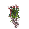


 PDBj
PDBj


 Assembly


 Mass: 618.503 Da / Num. of mol.: 4 / Source method: obtained synthetically / Formula: C34H34FeN4O4
 Mass: 96.063 Da / Num. of mol.: 7 / Source method: obtained synthetically / Formula: SO4
 Mass: 229.402 Da / Num. of mol.: 6 / Source method: obtained synthetically / Formula: C14H31NO / Comment: LDAO, detergent*YM
 Mass: 909.488 Da / Num. of mol.: 4 / Source method: obtained synthetically / Formula: C55H72MgN4O6
 Mass: 887.199 Da / Num. of mol.: 2 / Source method: obtained synthetically / Formula: C55H74N4O6
 Mass: 658.992 Da / Num. of mol.: 1 / Source method: obtained synthetically / Formula: C44H66O4
 Mass: 55.845 Da / Num. of mol.: 1 / Source method: obtained synthetically / Formula: Fe
 Mass: 785.233 Da / Num. of mol.: 1 / Source method: obtained synthetically / Formula: C56H80O2
 Mass: 540.904 Da / Num. of mol.: 1 / Source method: obtained synthetically / Formula: C40H60
 Sample preparation
 / Beamline: 14-BM-C / Wavelength: 0.9
  Processing