 Movie
Movie Controller

[English] 日本語
 Yorodumi
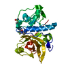
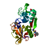
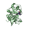
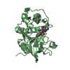
- PDB-1u9v: Crystal Structure of the Cysteine Protease Human Cathepsin K in C... -
+ Open data

- Basic information
| Entry | Database: PDB / ID: 1u9v | ||||||
|---|---|---|---|---|---|---|---|
| Title | Crystal Structure of the Cysteine Protease Human Cathepsin K in Complex with the Covalent Inhibitor NVP-ABE854 | ||||||
 Components Components | Cathepsin K | ||||||
 Keywords Keywords | HYDROLASE / SULFHYDRYL PROTEINASE | ||||||
| Function / homology |  Function and homology information Function and homology informationcathepsin K / negative regulation of cartilage development / RUNX1 regulates transcription of genes involved in differentiation of keratinocytes / endolysosome lumen / thyroid hormone generation / Trafficking and processing of endosomal TLR / proteoglycan binding / Activation of Matrix Metalloproteinases / Collagen degradation / collagen catabolic process ...cathepsin K / negative regulation of cartilage development / RUNX1 regulates transcription of genes involved in differentiation of keratinocytes / endolysosome lumen / thyroid hormone generation / Trafficking and processing of endosomal TLR / proteoglycan binding / Activation of Matrix Metalloproteinases / Collagen degradation / collagen catabolic process / fibronectin binding / extracellular matrix disassembly / bone resorption / mitophagy / collagen binding / Degradation of the extracellular matrix / cysteine-type peptidase activity / MHC class II antigen presentation / lysosomal lumen / : / lysosome / apical plasma membrane / serine-type endopeptidase activity / external side of plasma membrane / cysteine-type endopeptidase activity / proteolysis / : / extracellular region / nucleoplasm Similarity search - Function | ||||||
| Biological species |  Homo sapiens (human) Homo sapiens (human) | ||||||
| Method |  X-RAY DIFFRACTION / MOLECULAR REPLACEMENT / Resolution: 2.2 Å X-RAY DIFFRACTION / MOLECULAR REPLACEMENT / Resolution: 2.2 Å | ||||||
 Authors Authors | Cowan-Jacob, S.W. | ||||||
 Citation Citation | Journal: J.Med.Chem. / Year: 2004 Title: Novel purine nitrile derived inhibitors of the cysteine protease cathepsin K Authors: Altmann, E. / Cowan-Jacob, S.W. / Missbach, M. | ||||||
| History |
|
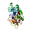
- Structure visualization
| Structure viewer | Molecule: MolmilJmol/JSmol |
|---|
- Downloads & links
-Download
| PDBx/mmCIF format | 1u9v.cif.gz | 56 KB | Display | PDBx/mmCIF format |
|---|---|---|---|---|
| PDB format | pdb1u9v.ent.gz | 39.7 KB | Display | PDB format |
| PDBx/mmJSON format | 1u9v.json.gz | Tree view | PDBx/mmJSON format | |
| Others |  Other downloads Other downloads |
-Validation report
| Arichive directory | https://data.pdbj.org/pub/pdb/validation_reports/u9/1u9vftp://data.pdbj.org/pub/pdb/validation_reports/u9/1u9v | HTTPS FTP |
|---|
-Related structure data
| Related structure data | 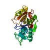 1u9wC 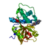 1u9xC 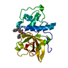 1memS S: Starting model for refinement C: citing same article ( |
|---|---|
| Similar structure data |


-Links
 PDBj
PDBj


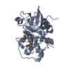
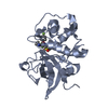
- Assembly
| Deposited unit | 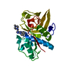
| ||||||||
|---|---|---|---|---|---|---|---|---|---|
| 1 |
| ||||||||
| Unit cell |
|
-Components
| #1: Protein | Mass: 23737.727 Da / Num. of mol.: 1 Source method: isolated from a genetically manipulated source Source: (gene. exp.) Homo sapiens (human) / Cell line (production host): SF21 / Production host:   Spodoptera frugiperda (fall armyworm) / References: UniProt: P43235, cathepsin K Spodoptera frugiperda (fall armyworm) / References: UniProt: P43235, cathepsin K |
|---|---|
| #2: Chemical | ChemComp-IHE / 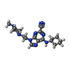  Mass: 368.479 Da / Num. of mol.: 1 / Source method: obtained synthetically / Formula: C19H28N8 Mass: 368.479 Da / Num. of mol.: 1 / Source method: obtained synthetically / Formula: C19H28N8 |
| #3: Water | ChemComp-HOH /  Mass: 18.015 Da / Num. of mol.: 81 / Source method: isolated from a natural source / Formula: H2O Mass: 18.015 Da / Num. of mol.: 81 / Source method: isolated from a natural source / Formula: H2O |
| Has protein modification | Y |
-Experimental details
-Experiment
| Experiment | Method: X-RAY DIFFRACTION / Number of used crystals: 1 |
|---|
- Sample preparation
| Crystal | Density Matthews: 2.36 Å3/Da / Density % sol: 47 % |
|---|---|
| Crystal grow | Temperature: 293 K / Method: vapor diffusion, hanging drop / pH: 8.7 Details: PEG 4000, Tris, magnesium chloride, pH 8.7, VAPOR DIFFUSION, HANGING DROP, temperature 293K |
-Data collection
| Diffraction | Mean temperature: 294 K |
|---|---|
| Diffraction source | Source: ROTATING ANODE / Type: ENRAF-NONIUS FR571 / Wavelength: 1.5418 / Wavelength: 1.5418 Å |
| Detector | Type: MARRESEARCH / Detector: IMAGE PLATE / Date: Oct 28, 1999 / Details: MONOCHROMATOR |
| Radiation | Monochromator: GRAPHITE / Protocol: SINGLE WAVELENGTH / Monochromatic (M) / Laue (L): M / Scattering type: x-ray |
| Radiation wavelength | Wavelength: 1.5418 Å / Relative weight: 1 |
| Reflection | Resolution: 2.2→35.2 Å / Num. all: 12003 / Num. obs: 12003 / % possible obs: 99 % / Observed criterion σ(F): 0 / Observed criterion σ(I): 0 / Redundancy: 9.9 % / Rmerge(I) obs: 0.0606 / Rsym value: 0.0606 / Net I/σ(I): 48.76 |
| Reflection shell | Resolution: 2.2→2.3 Å / Redundancy: 9.1 % / Rmerge(I) obs: 0.1824 / Mean I/σ(I) obs: 16.6 / Rsym value: 0.1824 / % possible all: 92.3 |
- Processing
| Software |
| |||||||||||||||||||||||||
|---|---|---|---|---|---|---|---|---|---|---|---|---|---|---|---|---|---|---|---|---|---|---|---|---|---|---|
| Refinement | Method to determine structure: MOLECULAR REPLACEMENT Starting model: PDB ENTRY 1MEM Resolution: 2.2→20 Å / Data cutoff high absF: 100000 / Data cutoff low absF: 0.1 / σ(F): 0 / Stereochemistry target values: Engh & Huber
| |||||||||||||||||||||||||
| Displacement parameters | Biso mean: 21.27 Å2 | |||||||||||||||||||||||||
| Refinement step | Cycle: LAST / Resolution: 2.2→20 Å
| |||||||||||||||||||||||||
| Refine LS restraints |
| |||||||||||||||||||||||||
| Xplor file | Serial no: 1 / Param file: PARHCSDX.PRO / Topol file: TOPHCSDX.PRO |
