 Movie
Movie Controller

[English] 日本語
 Yorodumi
- PDB-1m5k: Crystal structure of a hairpin ribozyme in the catalytically-acti... -
+ Open data

- Basic information
| Entry | Database: PDB / ID: 1m5k | |||||||||
|---|---|---|---|---|---|---|---|---|---|---|
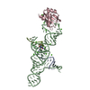
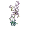
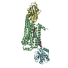
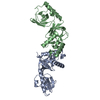
| Title | Crystal structure of a hairpin ribozyme in the catalytically-active conformation | |||||||||
 Components Components |
| |||||||||
 Keywords Keywords | TRANSLATION/RNA / HAIRPIN RIBOZYME / CATALYTIC RNA / U1A RNA BINDING PROTEIN DOCKED CONFORMATION / SUBSTRATE INHIBITOR STRAND / TRANSLATION-RNA COMPLEX | |||||||||
| Function / homology |  Function and homology information Function and homology informationU1 snRNP binding / U1 snRNP / U1 snRNA binding / U4/U6 x U5 tri-snRNP complex / mRNA Splicing - Major Pathway / spliceosomal complex / mRNA splicing, via spliceosome / DNA binding / RNA binding / nucleoplasm ...U1 snRNP binding / U1 snRNP / U1 snRNA binding / U4/U6 x U5 tri-snRNP complex / mRNA Splicing - Major Pathway / spliceosomal complex / mRNA splicing, via spliceosome / DNA binding / RNA binding / nucleoplasm / identical protein binding / nucleus Similarity search - Function | |||||||||
| Biological species |  Homo sapiens (human) Homo sapiens (human) | |||||||||
| Method |  X-RAY DIFFRACTION / SYNCHROTRON / MAD / Resolution: 2.4 Å X-RAY DIFFRACTION / SYNCHROTRON / MAD / Resolution: 2.4 Å | |||||||||
 Authors Authors | Rupert, P.B. / Ferre-D'Amare, A.R. | |||||||||
 Citation Citation | Journal: Science / Year: 2002 Title: Transition state stabilization by a catalytic RNA Authors: Rupert, P.B. / Massey, A.P. / Sigurdsson, S.T. / Ferre-D'Amare, A.R. #1: Journal: Nature / Year: 2001Title: Crystal structure of a hairpin ribozyme-inhibitor complex with implications for catalysis Authors: Rupert, P.B. / Ferre-D'Amare, A.R. | |||||||||
| History |
|
- Structure visualization
| Structure viewer | Molecule: MolmilJmol/JSmol |
|---|
- Downloads & links
-Download
| PDBx/mmCIF format | 1m5k.cif.gz | 181.2 KB | Display | PDBx/mmCIF format |
|---|---|---|---|---|
| PDB format | pdb1m5k.ent.gz | 135.7 KB | Display | PDB format |
| PDBx/mmJSON format | 1m5k.json.gz | Tree view | PDBx/mmJSON format | |
| Others |  Other downloads Other downloads |
-Validation report
| Arichive directory | https://data.pdbj.org/pub/pdb/validation_reports/m5/1m5kftp://data.pdbj.org/pub/pdb/validation_reports/m5/1m5k | HTTPS FTP |
|---|
-Related structure data
| Related structure data | |
|---|---|
| Similar structure data |


-Links
 PDBj
PDBj


- Assembly
| Deposited unit | 
| ||||||||||
|---|---|---|---|---|---|---|---|---|---|---|---|
| 1 | 
| ||||||||||
| 2 | 
| ||||||||||
| Unit cell |
|
-Components
-RNA chain , 2 types, 4 molecules ADBE
| #1: RNA chain | Mass: 6739.887 Da / Num. of mol.: 2 / Source method: obtained synthetically Details: THIS SEQUENCE OCCURS NATURALLY IN SATELLITE TOBACCO RINGSPOT VIRUS #2: RNA chain | Mass: 29969.754 Da / Num. of mol.: 2 / Source method: obtained synthetically Details: THIS SEQUENCE OCCURS NATURALLY IN SATELLITE TOBACCO RINGSPOT VIRUS |
|---|
-Protein , 1 types, 2 molecules CF
| #3: Protein | Mass: 11498.472 Da / Num. of mol.: 2 / Fragment: U1A RNA BINDING DOMAIN / Mutation: Y31H,Q36R Source method: isolated from a genetically manipulated source Source: (gene. exp.) Homo sapiens (human) / Gene: SNRPA / Production host:  |
|---|
-Non-polymers , 3 types, 138 molecules 


| #4: Chemical | ChemComp-CA /  Mass: 40.078 Da / Num. of mol.: 33 / Source method: obtained synthetically / Formula: Ca Mass: 40.078 Da / Num. of mol.: 33 / Source method: obtained synthetically / Formula: Ca#5: Chemical |  Mass: 35.453 Da / Num. of mol.: 2 / Source method: obtained synthetically / Formula: Cl Mass: 35.453 Da / Num. of mol.: 2 / Source method: obtained synthetically / Formula: Cl#6: Water | ChemComp-HOH / | Mass: 18.015 Da / Num. of mol.: 103 / Source method: isolated from a natural source / Formula: H2O |
|---|
-Experimental details
-Experiment
| Experiment | Method: X-RAY DIFFRACTION / Number of used crystals: 1 |
|---|
- Sample preparation
| Crystal | Density Matthews: 2.85 Å3/Da / Density % sol: 56.78 % | ||||||||||||||||||||||||||||||||||||||||||
|---|---|---|---|---|---|---|---|---|---|---|---|---|---|---|---|---|---|---|---|---|---|---|---|---|---|---|---|---|---|---|---|---|---|---|---|---|---|---|---|---|---|---|---|
| Crystal grow | Temperature: 300 K / Method: vapor diffusion, sitting drop / pH: 5 Details: MPD, ammonium chloride, calcium chloride, pH 5.0, VAPOR DIFFUSION, SITTING DROP at 300K, VAPOR DIFFUSION, SITTING DROP | ||||||||||||||||||||||||||||||||||||||||||
| Components of the solutions |
| ||||||||||||||||||||||||||||||||||||||||||
| Crystal grow | *PLUS Temperature: 22 ℃ / Method: vapor diffusion / Details: Rupert, P.B., (2001) Nature, 410, 780. | ||||||||||||||||||||||||||||||||||||||||||
| Components of the solutions | *PLUS
|
-Data collection
| Diffraction | Mean temperature: 150 K |
|---|---|
| Diffraction source | Source: SYNCHROTRON / Site: ALS  / Beamline: 5.0.2 / Wavelength: 1.1 Å / Beamline: 5.0.2 / Wavelength: 1.1 Å |
| Detector | Type: ADSC QUANTUM 4 / Detector: CCD / Date: Oct 5, 2000 |
| Radiation | Protocol: SINGLE WAVELENGTH / Monochromatic (M) / Laue (L): M / Scattering type: x-ray |
| Radiation wavelength | Wavelength: 1.1 Å / Relative weight: 1 |
| Reflection | Resolution: 2.4→61.07 Å / Num. all: 41156 / Num. obs: 41156 / % possible obs: 95.2 % / Observed criterion σ(F): 0 / Observed criterion σ(I): 0 / Redundancy: 3.6 % / Biso Wilson estimate: 43.2 Å2 / Rsym value: 0.067 / Net I/σ(I): 22.8 |
| Reflection shell | Resolution: 2.4→2.49 Å / Rmerge(I) obs: 0.261 / Mean I/σ(I) obs: 2.1 / Num. unique all: 3147 / % possible all: 73.9 |
| Reflection | *PLUS Highest resolution: 2.4 Å / Lowest resolution: 100 Å / Num. obs: 41290 / % possible obs: 95.6 % / Rmerge(I) obs: 0.067 |
| Reflection shell | *PLUS % possible obs: 73.9 % / Rmerge(I) obs: 0.261 |
- Processing
| Software |
| ||||||||||||||||||||||||||||||||||||
|---|---|---|---|---|---|---|---|---|---|---|---|---|---|---|---|---|---|---|---|---|---|---|---|---|---|---|---|---|---|---|---|---|---|---|---|---|---|
| Refinement | Method to determine structure: MAD / Resolution: 2.4→61.07 Å / Rfactor Rfree error: 0.004 / Data cutoff high absF: 1376780.72 / Data cutoff low absF: 0 / Isotropic thermal model: RESTRAINED / Cross valid method: THROUGHOUT / σ(F): 0 / σ(I): 0 Stereochemistry target values: Engh & Huber, Parkinson et al.
| ||||||||||||||||||||||||||||||||||||
| Solvent computation | Solvent model: FLAT MODEL / Bsol: 38.68 Å2 / ksol: 0.275 e/Å3 | ||||||||||||||||||||||||||||||||||||
| Displacement parameters | Biso mean: 86.7 Å2
| ||||||||||||||||||||||||||||||||||||
| Refine analyze |
| ||||||||||||||||||||||||||||||||||||
| Refinement step | Cycle: LAST / Resolution: 2.4→61.07 Å
| ||||||||||||||||||||||||||||||||||||
| Refine LS restraints |
| ||||||||||||||||||||||||||||||||||||
| LS refinement shell | Resolution: 2.4→2.55 Å / Rfactor Rfree error: 0.018 / Total num. of bins used: 6
| ||||||||||||||||||||||||||||||||||||
| Xplor file |
| ||||||||||||||||||||||||||||||||||||
| Refinement | *PLUS Rfactor obs: 0.23 / Rfactor Rfree: 0.285 / Rfactor Rwork: 0.229 | ||||||||||||||||||||||||||||||||||||
| Solvent computation | *PLUS | ||||||||||||||||||||||||||||||||||||
| Displacement parameters | *PLUS | ||||||||||||||||||||||||||||||||||||
| Refine LS restraints | *PLUS
| ||||||||||||||||||||||||||||||||||||
| LS refinement shell | *PLUS Rfactor Rfree: 0.41 / Rfactor Rwork: 0.376 |
