 Movie
Movie Controller

[English] 日本語
 Yorodumi
- PDB-1lc8: Crystal Structure of L-Threonine-O-3-phosphate Decarboxylase from... -
+ Open data

- Basic information
| Entry | Database: PDB / ID: 1lc8 | ||||||
|---|---|---|---|---|---|---|---|
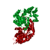
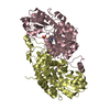
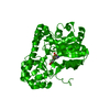
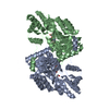
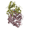
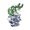
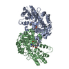
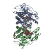
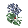
| Title | Crystal Structure of L-Threonine-O-3-phosphate Decarboxylase from S. enterica complexed with its reaction intermediate | ||||||
 Components Components | L-Threonine-O-3-Phosphate Decarboxylase | ||||||
 Keywords Keywords | LYASE / CobD / L-threonine-O-3-phosphate / PLP-dependent decarboxylase / cobalamin | ||||||
| Function / homology |  Function and homology information Function and homology informationthreonine-phosphate decarboxylase / threonine-phosphate decarboxylase activity / cobalamin biosynthetic process / pyridoxal phosphate binding / protein homodimerization activity / identical protein binding Similarity search - Function | ||||||
| Biological species |  Salmonella enterica (bacteria) Salmonella enterica (bacteria) | ||||||
| Method |  X-RAY DIFFRACTION / SYNCHROTRON / MOLECULAR REPLACEMENT / Resolution: 1.8 Å X-RAY DIFFRACTION / SYNCHROTRON / MOLECULAR REPLACEMENT / Resolution: 1.8 Å | ||||||
 Authors Authors | Cheong, C.-G. / Escalante-Semerena, J. / Rayment, I. | ||||||
 Citation Citation | Journal: Biochemistry / Year: 2002 Title: Structural studies of the L-threonine-O-3-phosphate decarboxylase (CobD) enzyme from Salmonella enterica: the apo, substrate, and product-aldimine complexes. Authors: Cheong, C.G. / Escalante-Semerena, J.C. / Rayment, I. | ||||||
| History |
| ||||||
| Remark 999 | SEQUENCE According to the authors, the GenBank entry is in error because the original DNA sequence ... SEQUENCE According to the authors, the GenBank entry is in error because the original DNA sequence had some errors. The electron density also supports it. The new sequence is Gln25, Ser30, Val42, Arg44 and Ala45. Arg44 lacks side chain density. The organism name in this GenBank entry is Salmonella typhimurium. Salmonella typhimurium has been changed to Salmonella enterica. Therefore, the two names are same. |
- Structure visualization
| Structure viewer | Molecule: MolmilJmol/JSmol |
|---|
- Downloads & links
-Download
| PDBx/mmCIF format | 1lc8.cif.gz | 88.3 KB | Display | PDBx/mmCIF format |
|---|---|---|---|---|
| PDB format | pdb1lc8.ent.gz | 65.4 KB | Display | PDB format |
| PDBx/mmJSON format | 1lc8.json.gz | Tree view | PDBx/mmJSON format | |
| Others |  Other downloads Other downloads |
-Validation report
| Summary document | 1lc8_validation.pdf.gz | 750.2 KB | Display | wwPDB validaton report |
|---|---|---|---|---|
| Full document | 1lc8_full_validation.pdf.gz | 754.5 KB | Display | |
| Data in XML | 1lc8_validation.xml.gz | 18.2 KB | Display | |
| Data in CIF | 1lc8_validation.cif.gz | 26.8 KB | Display | |
| Arichive directory | https://data.pdbj.org/pub/pdb/validation_reports/lc/1lc8ftp://data.pdbj.org/pub/pdb/validation_reports/lc/1lc8 | HTTPS FTP |
-Related structure data
| Related structure data |  1l4nC  1l5fC  1l5kC  1l5lC  1l5mC  1l5nC 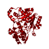 1lc5C 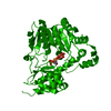 1lc7C  1kus C: citing same article ( S: Starting model for refinement |
|---|---|
| Similar structure data |


-Links
 PDBj
PDBj- Assembly
| Deposited unit | 
| ||||||||||
|---|---|---|---|---|---|---|---|---|---|---|---|
| 1 | 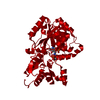
| ||||||||||
| Unit cell |
| ||||||||||
| Details | the second subunit of biological dimer can be generated by the operation of crystallographic two-fold symmetry axis |
-Components
| #1: Protein | Mass: 40849.848 Da / Num. of mol.: 1 Source method: isolated from a genetically manipulated source Source: (gene. exp.) Salmonella enterica (bacteria) / Gene: cobD / Production host: |
|---|---|
| #2: Chemical | ChemComp-33P / {  Mass: 386.232 Da / Num. of mol.: 1 / Source method: obtained synthetically / Formula: C11H20N2O9P2 Mass: 386.232 Da / Num. of mol.: 1 / Source method: obtained synthetically / Formula: C11H20N2O9P2 |
| #3: Water | ChemComp-HOH /  Mass: 18.015 Da / Num. of mol.: 271 / Source method: isolated from a natural source / Formula: H2O Mass: 18.015 Da / Num. of mol.: 271 / Source method: isolated from a natural source / Formula: H2O |
-Experimental details
-Experiment
| Experiment | Method: X-RAY DIFFRACTION / Number of used crystals: 1 |
|---|
- Sample preparation
| Crystal | Density Matthews: 2.47 Å3/Da / Density % sol: 50.12 % | ||||||||||||||||||||||||||||||||||||||||||
|---|---|---|---|---|---|---|---|---|---|---|---|---|---|---|---|---|---|---|---|---|---|---|---|---|---|---|---|---|---|---|---|---|---|---|---|---|---|---|---|---|---|---|---|
| Crystal grow | Temperature: 298 K / pH: 6 Details: PEG methyl ether 2000, pH 6.0, VAPOR DIFFUSION, HANGING DROP at 298K | ||||||||||||||||||||||||||||||||||||||||||
| Crystal grow | *PLUS Method: batch method | ||||||||||||||||||||||||||||||||||||||||||
| Components of the solutions | *PLUS
|
-Data collection
| Diffraction | Mean temperature: 100 K |
|---|---|
| Diffraction source | Source: SYNCHROTRON / Site: APS  / Beamline: 19-BM / Wavelength: 0.9763 Å / Beamline: 19-BM / Wavelength: 0.9763 Å |
| Detector | Detector: CCD |
| Radiation | Protocol: SINGLE WAVELENGTH / Monochromatic (M) / Laue (L): M / Scattering type: x-ray |
| Radiation wavelength | Wavelength: 0.9763 Å / Relative weight: 1 |
| Reflection | Resolution: 1.8→500 Å / Num. obs: 37471 / % possible obs: 99.4 % / Redundancy: 7.6 % / Rmerge(I) obs: 0.078 / Net I/σ(I): 31.9 |
| Reflection shell | Resolution: 1.8→1.86 Å / Rmerge(I) obs: 0.331 / Mean I/σ(I) obs: 5.8 / % possible all: 99.2 |
| Reflection | *PLUS Highest resolution: 1.8 Å / Lowest resolution: 500 Å / Rmerge(I) obs: 0.078 |
| Reflection shell | *PLUS % possible obs: 99.2 % / Rmerge(I) obs: 0.331 |
- Processing
| Software |
| ||||||||||||||||||||
|---|---|---|---|---|---|---|---|---|---|---|---|---|---|---|---|---|---|---|---|---|---|
| Refinement | Method to determine structure: MOLECULAR REPLACEMENT Starting model: PDB entry 1KUS 1kus Resolution: 1.8→500 Å / Cross valid method: THROUGHOUT / σ(F): 0
| ||||||||||||||||||||
| Refinement step | Cycle: LAST / Resolution: 1.8→500 Å
| ||||||||||||||||||||
| Refine LS restraints |
| ||||||||||||||||||||
| Refinement | *PLUS Highest resolution: 1.8 Å / Lowest resolution: 500 Å / Num. reflection obs: 35608 / Rfactor Rfree: 0.229 / Rfactor Rwork: 0.196 | ||||||||||||||||||||
| Solvent computation | *PLUS | ||||||||||||||||||||
| Displacement parameters | *PLUS |
