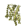
 Movie
Movie Controller


+ Open data

- Basic information
| Entry | Database: PDB / ID: 1gu3 | |||||||||
|---|---|---|---|---|---|---|---|---|---|---|
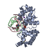
| Title | CBM4 structure and function | |||||||||
 Components Components | ENDOGLUCANASE C | |||||||||
 Keywords Keywords | CARBOHYDRATE-BINDING MODULE / CARBOHYDRATE BINDING MODULE / CBM / GLUCAN / CELLULOSE | |||||||||
| Function / homology |  Function and homology information Function and homology information | |||||||||
| Biological species |  CELLULOMONAS FIMI (bacteria) CELLULOMONAS FIMI (bacteria) | |||||||||
| Method |  X-RAY DIFFRACTION / MOLECULAR REPLACEMENT / Resolution: 2.3 Å X-RAY DIFFRACTION / MOLECULAR REPLACEMENT / Resolution: 2.3 Å | |||||||||
 Authors Authors | Nurizzo, D. / Notenboom, V. / Davies, G.J. | |||||||||
 Citation Citation | Journal: J.Mol.Biol. / Year: 2002 Title: Differential Oligosaccharide Recognition by Evolutionarily-Related Beta-1,4 and Beta-1,3 Glucan-Binding Modules Authors: Boraston, A.B. / Nurizzo, D. / Notenboom, V. / Ducros, V. / Rose, D.R. / Kilburn, D.G. / Davies, G.J. | |||||||||
| History |
|
- Structure visualization
| Structure viewer | Molecule: MolmilJmol/JSmol |
|---|
- Downloads & links
-Download
| PDBx/mmCIF format | 1gu3.cif.gz | 41.9 KB | Display | PDBx/mmCIF format |
|---|---|---|---|---|
| PDB format | pdb1gu3.ent.gz | 29.5 KB | Display | PDB format |
| PDBx/mmJSON format | 1gu3.json.gz | Tree view | PDBx/mmJSON format | |
| Others |  Other downloads Other downloads |
-Validation report
| Arichive directory | https://data.pdbj.org/pub/pdb/validation_reports/gu/1gu3ftp://data.pdbj.org/pub/pdb/validation_reports/gu/1gu3 | HTTPS FTP |
|---|
-Related structure data
| Related structure data |  1guiC  1uloS S: Starting model for refinement C: citing same article ( |
|---|---|
| Similar structure data |


-Links
 PDBj
PDBj


- Assembly
| Deposited unit | 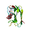
| ||||||||||||
|---|---|---|---|---|---|---|---|---|---|---|---|---|---|
| 1 | 
| ||||||||||||
| Unit cell |
| ||||||||||||
| Components on special symmetry positions |
|
-Components
| #1: Protein | Mass: 15088.256 Da / Num. of mol.: 1 Fragment: CARBOHYDRATE BINDING MODULE FAMILY 4, RESIDUES 1-149 Source method: isolated from a genetically manipulated source Source: (gene. exp.) CELLULOMONAS FIMI (bacteria)Description: CARBOHYDRATE BINDING MODULE OF CELLULASE 9B FROM CELLULOMONAS FIMI Plasmid: PTUG / Production host: |
|---|---|
| #2: Polysaccharide | beta-D-glucopyranose-(1-4)-beta-D-glucopyranose-(1-4)-beta-D-glucopyranose-(1-4)-beta-D- ...beta-D-glucopyranose-(1-4)-beta-D-glucopyranose-(1-4)-beta-D-glucopyranose-(1-4)-beta-D-glucopyranose-(1-4)-beta-D-glucopyranose / beta-cellopentaose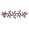  Source method: isolated from a genetically manipulated source Details: oligosaccharide / References: beta-cellopentaose |
| #3: Water | ChemComp-HOH /  Mass: 18.015 Da / Num. of mol.: 89 / Source method: isolated from a natural source / Formula: H2O Mass: 18.015 Da / Num. of mol.: 89 / Source method: isolated from a natural source / Formula: H2O |
| Has protein modification | Y |
-Experimental details
-Experiment
| Experiment | Method: X-RAY DIFFRACTION / Number of used crystals: 1 |
|---|
- Sample preparation
| Crystal | Density Matthews: 3 Å3/Da / Density % sol: 59 % | |||||||||||||||
|---|---|---|---|---|---|---|---|---|---|---|---|---|---|---|---|---|
| Crystal grow | pH: 5 / Details: 1.8M AMMONIUM SULFATE, 3% ISOPROPANOL, pH 5.00 | |||||||||||||||
| Crystal grow | *PLUS Method: vapor diffusion, hanging drop | |||||||||||||||
| Components of the solutions | *PLUS
|
-Data collection
| Diffraction | Mean temperature: 100 K |
|---|---|
| Diffraction source | Source: ROTATING ANODE / Type: RIGAKU / Wavelength: 1.5418 |
| Detector | Type: MARRESEARCH / Detector: IMAGE PLATE / Details: OSMICS MIRRORS |
| Radiation | Protocol: SINGLE WAVELENGTH / Monochromatic (M) / Laue (L): M / Scattering type: x-ray |
| Radiation wavelength | Wavelength: 1.5418 Å / Relative weight: 1 |
| Reflection | Resolution: 2.3→20 Å / Num. obs: 8313 / % possible obs: 93.6 % / Redundancy: 4.3 % / Rmerge(I) obs: 0.082 / Net I/σ(I): 18 |
| Reflection shell | Resolution: 2.3→2.38 Å / Redundancy: 2.4 % / Rmerge(I) obs: 0.26 / Mean I/σ(I) obs: 3.6 / % possible all: 61 |
| Reflection | *PLUS Lowest resolution: 20 Å |
| Reflection shell | *PLUS Highest resolution: 2.3 Å / % possible obs: 61 % / Rmerge(I) obs: 0.26 |
- Processing
| Software |
| ||||||||||||||||||||||||||||||||||||||||||||||||||||||||||||||||||||||||||||||||||||||||||||||||||||||||||||||||||||||||||||||||||||||||||||||||||||||||||||||||||||||||||||||||||||||
|---|---|---|---|---|---|---|---|---|---|---|---|---|---|---|---|---|---|---|---|---|---|---|---|---|---|---|---|---|---|---|---|---|---|---|---|---|---|---|---|---|---|---|---|---|---|---|---|---|---|---|---|---|---|---|---|---|---|---|---|---|---|---|---|---|---|---|---|---|---|---|---|---|---|---|---|---|---|---|---|---|---|---|---|---|---|---|---|---|---|---|---|---|---|---|---|---|---|---|---|---|---|---|---|---|---|---|---|---|---|---|---|---|---|---|---|---|---|---|---|---|---|---|---|---|---|---|---|---|---|---|---|---|---|---|---|---|---|---|---|---|---|---|---|---|---|---|---|---|---|---|---|---|---|---|---|---|---|---|---|---|---|---|---|---|---|---|---|---|---|---|---|---|---|---|---|---|---|---|---|---|---|---|---|
| Refinement | Method to determine structure: MOLECULAR REPLACEMENT Starting model: PDB ENTRY 1ULO Resolution: 2.3→20 Å / Cor.coef. Fo:Fc: 0.926 / Cor.coef. Fo:Fc free: 0.885 / SU B: 7.612 / SU ML: 0.181 / Cross valid method: THROUGHOUT / ESU R: 0.377 / ESU R Free: 0.264 / Stereochemistry target values: MAXIMUM LIKELIHOOD
| ||||||||||||||||||||||||||||||||||||||||||||||||||||||||||||||||||||||||||||||||||||||||||||||||||||||||||||||||||||||||||||||||||||||||||||||||||||||||||||||||||||||||||||||||||||||
| Solvent computation | Ion probe radii: 0.8 Å / Shrinkage radii: 0.8 Å / VDW probe radii: 1.4 Å / Solvent model: BABINET MODEL WITH MASK | ||||||||||||||||||||||||||||||||||||||||||||||||||||||||||||||||||||||||||||||||||||||||||||||||||||||||||||||||||||||||||||||||||||||||||||||||||||||||||||||||||||||||||||||||||||||
| Displacement parameters | Biso mean: 25.32 Å2
| ||||||||||||||||||||||||||||||||||||||||||||||||||||||||||||||||||||||||||||||||||||||||||||||||||||||||||||||||||||||||||||||||||||||||||||||||||||||||||||||||||||||||||||||||||||||
| Refinement step | Cycle: LAST / Resolution: 2.3→20 Å
| ||||||||||||||||||||||||||||||||||||||||||||||||||||||||||||||||||||||||||||||||||||||||||||||||||||||||||||||||||||||||||||||||||||||||||||||||||||||||||||||||||||||||||||||||||||||
| Refine LS restraints |
|