 Movie
Movie Controller

[English] 日本語
 Yorodumi
- PDB-1gag: CRYSTAL STRUCTURE OF THE INSULIN RECEPTOR KINASE IN COMPLEX WITH ... -
+ Open data

- Basic information
| Entry | Database: PDB / ID: 1gag | ||||||
|---|---|---|---|---|---|---|---|
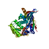
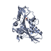
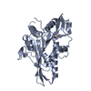
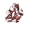
| Title | CRYSTAL STRUCTURE OF THE INSULIN RECEPTOR KINASE IN COMPLEX WITH A BISUBSTRATE INHIBITOR | ||||||
 Components Components |
| ||||||
 Keywords Keywords | TRANSFERASE/TRANSFERASE INHIBITOR / PROTEIN KINASE INHIBITOR / TYROSINE KINASE / transferase / signaling protein / transferase-transferase inhibitor complex | ||||||
| Function / homology |  Function and homology information Function and homology informationregulation of female gonad development / positive regulation of meiotic cell cycle / insulin-like growth factor II binding / positive regulation of developmental growth / male sex determination / insulin receptor complex / insulin-like growth factor I binding / insulin receptor activity / positive regulation of protein-containing complex disassembly / exocrine pancreas development ...regulation of female gonad development / positive regulation of meiotic cell cycle / insulin-like growth factor II binding / positive regulation of developmental growth / male sex determination / insulin receptor complex / insulin-like growth factor I binding / insulin receptor activity / positive regulation of protein-containing complex disassembly / exocrine pancreas development / dendritic spine maintenance / adrenal gland development / insulin binding / cargo receptor activity / PTB domain binding / Signaling by Insulin receptor / IRS activation / neuronal cell body membrane / positive regulation of respiratory burst / amyloid-beta clearance / regulation of embryonic development / insulin receptor substrate binding / positive regulation of receptor internalization / epidermis development / positive regulation of glycogen biosynthetic process / Signal attenuation / protein kinase activator activity / transport across blood-brain barrier / phosphatidylinositol 3-kinase binding / heart morphogenesis / Insulin receptor recycling / insulin-like growth factor receptor binding / neuron projection maintenance / positive regulation of mitotic nuclear division / receptor-mediated endocytosis / Insulin receptor signalling cascade / dendrite membrane / positive regulation of glycolytic process / positive regulation of D-glucose import across plasma membrane / learning / receptor protein-tyrosine kinase / caveola / receptor internalization / cellular response to growth factor stimulus / male gonad development / cellular response to insulin stimulus / memory / positive regulation of nitric oxide biosynthetic process / insulin receptor signaling pathway / protein autophosphorylation / late endosome / glucose homeostasis / amyloid-beta binding / PI5P, PP2A and IER3 Regulate PI3K/AKT Signaling / protein tyrosine kinase activity / positive regulation of MAPK cascade / positive regulation of canonical NF-kappaB signal transduction / positive regulation of phosphatidylinositol 3-kinase/protein kinase B signal transduction / lysosome / signaling receptor complex / endosome membrane / positive regulation of cell migration / G protein-coupled receptor signaling pathway / external side of plasma membrane / protein domain specific binding / axon / positive regulation of cell population proliferation / symbiont entry into host cell / regulation of DNA-templated transcription / positive regulation of DNA-templated transcription / GTP binding / protein-containing complex binding / extracellular exosome / ATP binding / membrane / identical protein binding / plasma membrane Similarity search - Function | ||||||
| Biological species |  Homo sapiens (human) Homo sapiens (human) | ||||||
| Method |  X-RAY DIFFRACTION / FOURIER SYNTHESIS / Resolution: 2.7 Å X-RAY DIFFRACTION / FOURIER SYNTHESIS / Resolution: 2.7 Å | ||||||
 Authors Authors | Parang, K. / Till, J.H. / Ablooglu, A.J. / Kohanski, R.A. / Hubbard, S.R. / Cole, P.A. | ||||||
 Citation Citation | Journal: Nat.Struct.Biol. / Year: 2001 Title: Mechanism-based design of a protein kinase inhibitor. Authors: Parang, K. / Till, J.H. / Ablooglu, A.J. / Kohanski, R.A. / Hubbard, S.R. / Cole, P.A. #1: Journal: Embo J. / Year: 1997Title: Crystal structure of the activated insulin receptor tyrosine kinase in complex with peptide substrate and ATP analog Authors: Hubbard, S.R. | ||||||
| History |
|
- Structure visualization
| Structure viewer | Molecule: MolmilJmol/JSmol |
|---|
- Downloads & links
-Download
| PDBx/mmCIF format | 1gag.cif.gz | 78.8 KB | Display | PDBx/mmCIF format |
|---|---|---|---|---|
| PDB format | pdb1gag.ent.gz | 56.8 KB | Display | PDB format |
| PDBx/mmJSON format | 1gag.json.gz | Tree view | PDBx/mmJSON format | |
| Others |  Other downloads Other downloads |
-Validation report
| Arichive directory | https://data.pdbj.org/pub/pdb/validation_reports/ga/1gagftp://data.pdbj.org/pub/pdb/validation_reports/ga/1gag | HTTPS FTP |
|---|
-Related structure data
| Related structure data |  1ir3S S: Starting model for refinement |
|---|---|
| Similar structure data |


-Links
 PDBj
PDBj


- Assembly
| Deposited unit | 
| ||||||||||
|---|---|---|---|---|---|---|---|---|---|---|---|
| 1 |
| ||||||||||
| 2 | 
| ||||||||||
| Unit cell |
|
-Components
| #1: Protein | Mass: 35032.738 Da / Num. of mol.: 1 / Fragment: TYROSINE KINASE DOMAIN / Mutation: C981S, Y984F Source method: isolated from a genetically manipulated source Source: (gene. exp.) Homo sapiens (human) / Production host:   Spodoptera frugiperda (fall armyworm) / References: UniProt: P06213, EC: 2.7.1.112 Spodoptera frugiperda (fall armyworm) / References: UniProt: P06213, EC: 2.7.1.112 | ||||||
|---|---|---|---|---|---|---|---|
| #2: Protein/peptide | Mass: 1323.495 Da / Num. of mol.: 1 / Source method: obtained synthetically Details: PHE107 OF PEPTIDE 13-MER MODIFIED BY ATP GAMMA S PLUS FOUR-ATOM LINKER, RESIDUE 112 | ||||||
| #3: Chemical |   Mass: 24.305 Da / Num. of mol.: 2 / Source method: obtained synthetically / Formula: Mg Mass: 24.305 Da / Num. of mol.: 2 / Source method: obtained synthetically / Formula: Mg#4: Chemical | ChemComp-112 / |   Mass: 580.298 Da / Num. of mol.: 1 / Source method: obtained synthetically / Formula: C12H19N6O13P3S Mass: 580.298 Da / Num. of mol.: 1 / Source method: obtained synthetically / Formula: C12H19N6O13P3S#5: Water | ChemComp-HOH / |  Mass: 18.015 Da / Num. of mol.: 23 / Source method: isolated from a natural source / Formula: H2O Mass: 18.015 Da / Num. of mol.: 23 / Source method: isolated from a natural source / Formula: H2OHas protein modification | Y | |
-Experimental details
-Experiment
| Experiment | Method: X-RAY DIFFRACTION / Number of used crystals: 1 |
|---|
- Sample preparation
| Crystal | Density Matthews: 2.4 Å3/Da / Density % sol: 50 % | ||||||||||||||||||||||||||||||||||||||||||
|---|---|---|---|---|---|---|---|---|---|---|---|---|---|---|---|---|---|---|---|---|---|---|---|---|---|---|---|---|---|---|---|---|---|---|---|---|---|---|---|---|---|---|---|
| Crystal grow | Temperature: 277 K / Method: vapor diffusion, hanging drop / pH: 7.5 Details: PEG 8000, Tris-HCl, pH 7.5, VAPOR DIFFUSION, HANGING DROP, temperature 277K | ||||||||||||||||||||||||||||||||||||||||||
| Crystal grow | *PLUS Temperature: 4 ℃ | ||||||||||||||||||||||||||||||||||||||||||
| Components of the solutions | *PLUS
|
-Data collection
| Diffraction | Mean temperature: 110 K |
|---|---|
| Diffraction source | Source: ROTATING ANODE / Type: RIGAKU RU200 / Wavelength: 1.5418 Å |
| Detector | Type: RIGAKU RAXIS II / Detector: IMAGE PLATE / Date: Jul 3, 2000 |
| Radiation | Protocol: SINGLE WAVELENGTH / Monochromatic (M) / Laue (L): M / Scattering type: x-ray |
| Radiation wavelength | Wavelength: 1.5418 Å / Relative weight: 1 |
| Reflection | Resolution: 2.7→30 Å / Num. all: 42217 / Num. obs: 42217 / % possible obs: 96.5 % / Observed criterion σ(F): 0 / Observed criterion σ(I): 0 / Redundancy: 4.4 % / Rmerge(I) obs: 0.114 / Net I/σ(I): 7.6 |
| Reflection shell | Resolution: 2.7→2.8 Å / Rmerge(I) obs: 0.339 / % possible all: 91.9 |
| Reflection | *PLUS Lowest resolution: 30 Å / Num. obs: 9633 / Num. measured all: 42217 |
| Reflection shell | *PLUS % possible obs: 91.9 % |
- Processing
| Software |
| ||||||||||||||||||||||||||||||||||||||||
|---|---|---|---|---|---|---|---|---|---|---|---|---|---|---|---|---|---|---|---|---|---|---|---|---|---|---|---|---|---|---|---|---|---|---|---|---|---|---|---|---|---|
| Refinement | Method to determine structure: FOURIER SYNTHESIS Starting model: PDB ENTRY 1IR3 Resolution: 2.7→30 Å / σ(F): 0 / σ(I): 0 / Stereochemistry target values: Engh & Huber
| ||||||||||||||||||||||||||||||||||||||||
| Solvent computation | Solvent model: CNS / Bsol: 10.14 Å2 / ksol: 0.312 e/Å3 | ||||||||||||||||||||||||||||||||||||||||
| Displacement parameters | Biso mean: 20.6 Å2
| ||||||||||||||||||||||||||||||||||||||||
| Refinement step | Cycle: LAST / Resolution: 2.7→30 Å
| ||||||||||||||||||||||||||||||||||||||||
| Refine LS restraints |
| ||||||||||||||||||||||||||||||||||||||||
| Software | *PLUS Name: CNS / Classification: refinement | ||||||||||||||||||||||||||||||||||||||||
| Refinement | *PLUS Highest resolution: 2.7 Å / Lowest resolution: 30 Å / σ(F): 0 / % reflection Rfree: 5.4 % | ||||||||||||||||||||||||||||||||||||||||
| Solvent computation | *PLUS | ||||||||||||||||||||||||||||||||||||||||
| Displacement parameters | *PLUS Biso mean: 20.6 Å2 | ||||||||||||||||||||||||||||||||||||||||
| Refine LS restraints | *PLUS
|
