 Movie
Movie Controller

[English] 日本語
 Yorodumi
- PDB-1ftm: CRYSTAL STRUCTURE OF THE GLUR2 LIGAND BINDING CORE (S1S2J) IN COM... -
+ Open data

- Basic information
| Entry | Database: PDB / ID: 1ftm | ||||||
|---|---|---|---|---|---|---|---|
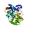
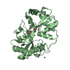
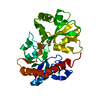
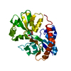
| Title | CRYSTAL STRUCTURE OF THE GLUR2 LIGAND BINDING CORE (S1S2J) IN COMPLEX WITH AMPA AT 1.7 RESOLUTION | ||||||
 Components Components | GLUTAMATE RECEPTOR SUBUNIT 2 | ||||||
 Keywords Keywords | MEMBRANE PROTEIN / AMPA Receptor / GluR2 / S1S2 / ligand-binding core / agonist | ||||||
| Function / homology |  Function and homology information Function and homology informationspine synapse / dendritic spine neck / dendritic spine head / cellular response to amine stimulus / Activation of AMPA receptors / perisynaptic space / ligand-gated monoatomic cation channel activity / AMPA glutamate receptor activity / response to lithium ion / Trafficking of GluR2-containing AMPA receptors ...spine synapse / dendritic spine neck / dendritic spine head / cellular response to amine stimulus / Activation of AMPA receptors / perisynaptic space / ligand-gated monoatomic cation channel activity / AMPA glutamate receptor activity / response to lithium ion / Trafficking of GluR2-containing AMPA receptors / kainate selective glutamate receptor activity / AMPA glutamate receptor complex / cellular response to glycine / extracellularly glutamate-gated ion channel activity / ionotropic glutamate receptor complex / immunoglobulin binding / asymmetric synapse / conditioned place preference / regulation of receptor recycling / glutamate receptor binding / Unblocking of NMDA receptors, glutamate binding and activation / positive regulation of synaptic transmission / regulation of synaptic transmission, glutamatergic / response to fungicide / glutamate-gated receptor activity / cytoskeletal protein binding / regulation of long-term synaptic depression / extracellular ligand-gated monoatomic ion channel activity / cellular response to brain-derived neurotrophic factor stimulus / glutamate-gated calcium ion channel activity / presynaptic active zone membrane / somatodendritic compartment / ionotropic glutamate receptor binding / dendrite membrane / ligand-gated monoatomic ion channel activity involved in regulation of presynaptic membrane potential / ionotropic glutamate receptor signaling pathway / dendrite cytoplasm / synaptic membrane / SNARE binding / dendritic shaft / transmitter-gated monoatomic ion channel activity involved in regulation of postsynaptic membrane potential / synaptic transmission, glutamatergic / PDZ domain binding / protein tetramerization / establishment of protein localization / postsynaptic density membrane / modulation of chemical synaptic transmission / cerebral cortex development / receptor internalization / Schaffer collateral - CA1 synapse / terminal bouton / synaptic vesicle / synaptic vesicle membrane / presynapse / signaling receptor activity / amyloid-beta binding / growth cone / presynaptic membrane / scaffold protein binding / perikaryon / chemical synaptic transmission / dendritic spine / postsynaptic membrane / neuron projection / postsynaptic density / axon / external side of plasma membrane / neuronal cell body / dendrite / synapse / protein kinase binding / protein-containing complex binding / glutamatergic synapse / cell surface / endoplasmic reticulum / protein-containing complex / identical protein binding / membrane / plasma membrane Similarity search - Function | ||||||
| Biological species |  | ||||||
| Method |  X-RAY DIFFRACTION / SYNCHROTRON / Resolution: 1.7 Å X-RAY DIFFRACTION / SYNCHROTRON / Resolution: 1.7 Å | ||||||
 Authors Authors | Gouaux, E. / Armstrong, N. | ||||||
 Citation Citation | Journal: Neuron / Year: 2000 Title: Mechanisms for activation and antagonism of an AMPA-sensitive glutamate receptor: crystal structures of the GluR2 ligand binding core. Authors: Armstrong, N. / Gouaux, E. #1: Journal: Protein Sci. / Year: 1998Title: Probing the ligand binding domain of the GluR2 receptor by proteolysis and deletion mutagenesis defines domain boundaries and yields a crystallizable construct Authors: Chen, G.Q. / Sun, Y. / Jin, R. / Gouaux, E. | ||||||
| History |
|
- Structure visualization
| Structure viewer | Molecule: MolmilJmol/JSmol |
|---|
- Downloads & links
-Download
| PDBx/mmCIF format | 1ftm.cif.gz | 166.1 KB | Display | PDBx/mmCIF format |
|---|---|---|---|---|
| PDB format | pdb1ftm.ent.gz | 130.6 KB | Display | PDB format |
| PDBx/mmJSON format | 1ftm.json.gz | Tree view | PDBx/mmJSON format | |
| Others |  Other downloads Other downloads |
-Validation report
| Summary document | 1ftm_validation.pdf.gz | 395.9 KB | Display | wwPDB validaton report |
|---|---|---|---|---|
| Full document | 1ftm_full_validation.pdf.gz | 401.9 KB | Display | |
| Data in XML | 1ftm_validation.xml.gz | 16.5 KB | Display | |
| Data in CIF | 1ftm_validation.cif.gz | 27.3 KB | Display | |
| Arichive directory | https://data.pdbj.org/pub/pdb/validation_reports/ft/1ftmftp://data.pdbj.org/pub/pdb/validation_reports/ft/1ftm | HTTPS FTP |
-Related structure data


-Links
 PDBj
PDBj


- Assembly
| Deposited unit | 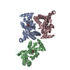
| ||||||||
|---|---|---|---|---|---|---|---|---|---|
| 1 | 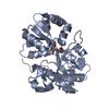
| ||||||||
| 2 | 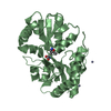
| ||||||||
| 3 | 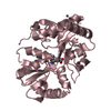
| ||||||||
| 4 | 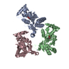
| ||||||||
| 5 | 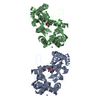
| ||||||||
| Unit cell |
|
-Components
| #1: Protein | Mass: 29221.682 Da / Num. of mol.: 3 / Fragment: GLUR2-FLOP LIGAND BINDING CORE (S1S2J) Source method: isolated from a genetically manipulated source Source: (gene. exp.)  #2: Chemical | ChemComp-ZN /   Mass: 65.409 Da / Num. of mol.: 5 / Source method: obtained synthetically / Formula: Zn Mass: 65.409 Da / Num. of mol.: 5 / Source method: obtained synthetically / Formula: Zn#3: Chemical |   Mass: 186.165 Da / Num. of mol.: 3 / Source method: obtained synthetically / Formula: C7H10N2O4 Mass: 186.165 Da / Num. of mol.: 3 / Source method: obtained synthetically / Formula: C7H10N2O4#4: Water | ChemComp-HOH / |  Mass: 18.015 Da / Num. of mol.: 420 / Source method: isolated from a natural source / Formula: H2O Mass: 18.015 Da / Num. of mol.: 420 / Source method: isolated from a natural source / Formula: H2OHas protein modification | Y | |
|---|
-Experimental details
-Experiment
| Experiment | Method: X-RAY DIFFRACTION / Number of used crystals: 1 |
|---|
- Sample preparation
| Crystal | Density Matthews: 2.5 Å3/Da / Density % sol: 50.76 % | ||||||||||||||||||||||||||||||||||||||||||||||||
|---|---|---|---|---|---|---|---|---|---|---|---|---|---|---|---|---|---|---|---|---|---|---|---|---|---|---|---|---|---|---|---|---|---|---|---|---|---|---|---|---|---|---|---|---|---|---|---|---|---|
| Crystal grow | Temperature: 277 K / Method: vapor diffusion, hanging drop / pH: 6.5 Details: 15% PEG 8000 0.2 M Zinc Acetate 0.1 M Cacodylate, pH 6.5, VAPOR DIFFUSION, HANGING DROP, temperature 277K | ||||||||||||||||||||||||||||||||||||||||||||||||
| Crystal grow | *PLUS Temperature: 4 ℃ / pH: 7 | ||||||||||||||||||||||||||||||||||||||||||||||||
| Components of the solutions | *PLUS
|
-Data collection
| Diffraction | Mean temperature: 110 K |
|---|---|
| Diffraction source | Source: SYNCHROTRON / Site: NSLS  / Beamline: X4A / Wavelength: 0.979 / Beamline: X4A / Wavelength: 0.979 |
| Detector | Type: ADSC QUANTUM 4 / Detector: CCD / Date: Oct 19, 1999 |
| Radiation | Protocol: SINGLE WAVELENGTH / Monochromatic (M) / Laue (L): M / Scattering type: x-ray |
| Radiation wavelength | Wavelength: 0.979 Å / Relative weight: 1 |
| Reflection | Resolution: 1.7→20 Å / Num. all: 97543 / Num. obs: 91073 / % possible obs: 93.5 % / Observed criterion σ(F): 0 / Observed criterion σ(I): 0 / Redundancy: 5.53 % / Biso Wilson estimate: 16.617 Å2 / Rmerge(I) obs: 0.068 / Net I/σ(I): 29.2 |
| Reflection shell | Resolution: 1.7→1.76 Å / Redundancy: 2.03 % / Rmerge(I) obs: 0.108 / Num. unique all: 6518 / % possible all: 67.7 |
| Reflection | *PLUS Num. measured all: 503581 |
| Reflection shell | *PLUS % possible obs: 67.7 % |
- Processing
| Software |
| ||||||||||||||||||||
|---|---|---|---|---|---|---|---|---|---|---|---|---|---|---|---|---|---|---|---|---|---|
| Refinement | Resolution: 1.7→20 Å / σ(F): 2 / σ(I): 0 / Stereochemistry target values: Engh & Huber
| ||||||||||||||||||||
| Refinement step | Cycle: LAST / Resolution: 1.7→20 Å
| ||||||||||||||||||||
| Refine LS restraints |
| ||||||||||||||||||||
| Software | *PLUS Name: X-PLOR / Version: 3.851 / Classification: refinement | ||||||||||||||||||||
| Refinement | *PLUS Highest resolution: 1.7 Å / Lowest resolution: 20 Å / σ(F): 2 / % reflection Rfree: 5 % / Rfactor obs: 0.209 / Rfactor Rwork: 0.21 | ||||||||||||||||||||
| Solvent computation | *PLUS | ||||||||||||||||||||
| Displacement parameters | *PLUS |
