 Movie
Movie Controller


+ Open data

- Basic information
| Entry | Database: PDB / ID: 1fq7 | |||||||||
|---|---|---|---|---|---|---|---|---|---|---|
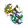
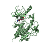
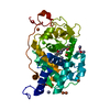
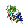
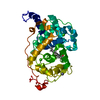
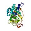
| Title | X-RAY STRUCTURE OF INHIBITOR CP-72,647 BOUND TO SACCHAROPEPSIN | |||||||||
 Components Components | SACCHAROPEPSIN | |||||||||
 Keywords Keywords | HYDROLASE/HYDROLASE INHIBITOR / Hydrophobic inhibitor / HYDROLASE-HYDROLASE INHIBITOR complex | |||||||||
| Function / homology |  Function and homology information Function and homology informationsaccharopepsin / MHC class II antigen presentation / microautophagy / cytoplasm to vacuole targeting by the Cvt pathway / oligosaccharide binding / fungal-type vacuole / pexophagy / Neutrophil degranulation / : / macroautophagy ...saccharopepsin / MHC class II antigen presentation / microautophagy / cytoplasm to vacuole targeting by the Cvt pathway / oligosaccharide binding / fungal-type vacuole / pexophagy / Neutrophil degranulation / : / macroautophagy / autophagy / disordered domain specific binding / peptidase activity / aspartic-type endopeptidase activity / endoplasmic reticulum / protein-containing complex / mitochondrion Similarity search - Function | |||||||||
| Biological species |  | |||||||||
| Method |  X-RAY DIFFRACTION / SYNCHROTRON / Resolution: 2.8 Å X-RAY DIFFRACTION / SYNCHROTRON / Resolution: 2.8 Å | |||||||||
 Authors Authors | Cronin, N.B. / Badasso, M.O. / Tickle, I.J. / Dreyer, T. / Hoover, D.J. / Rosati, R.L. / Humblet, C.C. / Lunney, E.A. / Cooper, J.B. | |||||||||
 Citation Citation | Journal: J.Mol.Biol. / Year: 2000 Title: X-ray structures of five renin inhibitors bound to saccharopepsin: exploration of active-site specificity. Authors: Cronin, N.B. / Badasso, M.O. / J Tickle, I. / Dreyer, T. / Hoover, D.J. / Rosati, R.L. / Humblet, C.C. / Lunney, E.A. / Cooper, J.B. #1: Journal: J.Mol.Biol. / Year: 1997Title: The Three-dimensional Structure at 2.4 A Resolution of Glycosylated Proteinase A from the Lysosome-like Vacuole of Saccharomyces cerevisiae Authors: Aguilar, C.F. / Cronin, N.B. / Badasso, M. / Dreyer, T. / Newman, M.P. / Cooper, J.B. / Hoover, D.J. / Wood, S.P. / Johnson, M.S. / Blundell, T.L. | |||||||||
| History |
|
- Structure visualization
| Structure viewer | Molecule: MolmilJmol/JSmol |
|---|
- Downloads & links
-Download
| PDBx/mmCIF format | 1fq7.cif.gz | 86.3 KB | Display | PDBx/mmCIF format |
|---|---|---|---|---|
| PDB format | pdb1fq7.ent.gz | 62.3 KB | Display | PDB format |
| PDBx/mmJSON format | 1fq7.json.gz | Tree view | PDBx/mmJSON format | |
| Others |  Other downloads Other downloads |
-Validation report
| Arichive directory | https://data.pdbj.org/pub/pdb/validation_reports/fq/1fq7ftp://data.pdbj.org/pub/pdb/validation_reports/fq/1fq7 | HTTPS FTP |
|---|
-Related structure data


-Links
 PDBj
PDBj


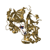
- Assembly
| Deposited unit | 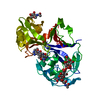
| ||||||||
|---|---|---|---|---|---|---|---|---|---|
| 1 |
| ||||||||
| 2 | 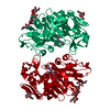
| ||||||||
| Unit cell |
|
-Components
| #1: Protein | Mass: 35774.551 Da / Num. of mol.: 1 / Source method: isolated from a natural source / Source: (natural) |
|---|---|
| #2: Polysaccharide | beta-D-arabino-hexopyranos-2-ulose-(1-2)-beta-D-mannopyranose-(1-3)-beta-D-mannopyranose-(1-4)-2- ...beta-D-arabino-hexopyranos-2-ulose-(1-2)-beta-D-mannopyranose-(1-3)-beta-D-mannopyranose-(1-4)-2-acetamido-2-deoxy-beta-D-glucopyranose-(1-4)-2-acetamido-2-deoxy-beta-D-glucopyranose Type: oligosaccharide / Mass: 908.807 Da / Num. of mol.: 1 Source method: isolated from a genetically manipulated source |
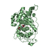
| #3: Chemical | ChemComp-2Y3 /   Type: peptide-like, Peptide-like / Class: Enzyme inhibitor / Mass: 682.893 Da / Num. of mol.: 1 / Source method: obtained synthetically / Formula: C37H58N6O6 / References: CP-72,647 Type: peptide-like, Peptide-like / Class: Enzyme inhibitor / Mass: 682.893 Da / Num. of mol.: 1 / Source method: obtained synthetically / Formula: C37H58N6O6 / References: CP-72,647 |
| #4: Sugar | ChemComp-NAG /   Type: D-saccharide, beta linking / Mass: 221.208 Da / Num. of mol.: 1 Type: D-saccharide, beta linking / Mass: 221.208 Da / Num. of mol.: 1Source method: isolated from a genetically manipulated source Formula: C8H15NO6 |
| #5: Water | ChemComp-HOH /  Mass: 18.015 Da / Num. of mol.: 65 / Source method: isolated from a natural source / Formula: H2O Mass: 18.015 Da / Num. of mol.: 65 / Source method: isolated from a natural source / Formula: H2O |
| Has protein modification | Y |
-Experimental details
-Experiment
| Experiment | Method: X-RAY DIFFRACTION / Number of used crystals: 1 |
|---|
- Sample preparation
| Crystal | Density Matthews: 3.31 Å3/Da / Density % sol: 62.87 % | ||||||||||||||||||||||||
|---|---|---|---|---|---|---|---|---|---|---|---|---|---|---|---|---|---|---|---|---|---|---|---|---|---|
| Crystal grow | Temperature: 298 K / Method: vapor diffusion, hanging drop / pH: 5.5 Details: PEG 6000, sodium acetate, pH 5.5, VAPOR DIFFUSION, HANGING DROP, temperature 298K | ||||||||||||||||||||||||
| Crystal | *PLUS Density % sol: 56 % | ||||||||||||||||||||||||
| Crystal grow | *PLUS Method: vapor diffusion | ||||||||||||||||||||||||
| Components of the solutions | *PLUS
|
-Data collection
| Diffraction | Mean temperature: 298 K |
|---|---|
| Diffraction source | Source: SYNCHROTRON / Site: EMBL/DESY, HAMBURG  / Beamline: X11 / Wavelength: 0.928 / Beamline: X11 / Wavelength: 0.928 |
| Detector | Type: MARRESEARCH / Detector: IMAGE PLATE / Date: Jun 15, 1994 |
| Radiation | Protocol: SINGLE WAVELENGTH / Monochromatic (M) / Laue (L): M / Scattering type: x-ray |
| Radiation wavelength | Wavelength: 0.928 Å / Relative weight: 1 |
| Reflection | Resolution: 2.8→28.3 Å / Num. all: 11434 / % possible obs: 95.9 % / Observed criterion σ(F): 3 / Observed criterion σ(I): 3 / Redundancy: 4.4 % / Rmerge(I) obs: 0.16 |
| Reflection | *PLUS Num. obs: 11457 / Num. measured all: 49475 / Rmerge(I) obs: 0.104 |
| Reflection shell | *PLUS % possible obs: 95.3 % / Rmerge(I) obs: 0.45 |
- Processing
| Software |
| |||||||||||||||||||||
|---|---|---|---|---|---|---|---|---|---|---|---|---|---|---|---|---|---|---|---|---|---|---|
| Refinement | Resolution: 2.8→28.3 Å / σ(F): 0 / σ(I): 0 / Stereochemistry target values: Engh & Huber
| |||||||||||||||||||||
| Refinement step | Cycle: LAST / Resolution: 2.8→28.3 Å
| |||||||||||||||||||||
| Software | *PLUS Name: RESTRAIN / Classification: refinement | |||||||||||||||||||||
| Refinement | *PLUS Highest resolution: 2.8 Å / Lowest resolution: 28.3 Å / σ(F): 0 / Rfactor Rfree: 0.27 / Rfactor Rwork: 0.19 | |||||||||||||||||||||
| Solvent computation | *PLUS | |||||||||||||||||||||
| Displacement parameters | *PLUS | |||||||||||||||||||||
| Refine LS restraints | *PLUS
|
