 Movie
Movie Controller


+ Open data

- Basic information
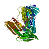
| Entry | Database: PDB / ID: 1f5a | ||||||
|---|---|---|---|---|---|---|---|
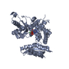
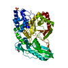
| Title | CRYSTAL STRUCTURE OF MAMMALIAN POLY(A) POLYMERASE | ||||||
 Components Components | POLY(A) POLYMERASE | ||||||
 Keywords Keywords | TRANSFERASE / mRNA processing / transcription / RNA-binding / phosphorylation / nuclear protein / alternative splicing helical turn motif / nucleotidyl transferase catalytic domain | ||||||
| Function / homology |  Function and homology information Function and homology informationmRNA 3'-end processing / RNA Polymerase II Transcription Termination / Processing of Intronless Pre-mRNAs / Processing of Capped Intron-Containing Pre-mRNA / cytosolic mRNA polyadenylation / co-transcriptional mRNA 3'-end processing, cleavage and polyadenylation pathway / polynucleotide adenylyltransferase / poly(A) RNA polymerase activity / manganese ion binding / magnesium ion binding ...mRNA 3'-end processing / RNA Polymerase II Transcription Termination / Processing of Intronless Pre-mRNAs / Processing of Capped Intron-Containing Pre-mRNA / cytosolic mRNA polyadenylation / co-transcriptional mRNA 3'-end processing, cleavage and polyadenylation pathway / polynucleotide adenylyltransferase / poly(A) RNA polymerase activity / manganese ion binding / magnesium ion binding / RNA binding / nucleoplasm / ATP binding / nucleus Similarity search - Function | ||||||
| Biological species |  | ||||||
| Method |  X-RAY DIFFRACTION / SYNCHROTRON / Resolution: 2.5 Å X-RAY DIFFRACTION / SYNCHROTRON / Resolution: 2.5 Å | ||||||
 Authors Authors | Martin, G. / Keller, W. / Doublie, S. | ||||||
 Citation Citation | Journal: EMBO J. / Year: 2000 Title: Crystal structure of mammalian poly(A) polymerase in complex with an analog of ATP. Authors: Martin, G. / Keller, W. / Doublie, S. #1: Journal: Embo J. / Year: 1996Title: Mutational analysis of mammalian poly(A) polymerase identifies a region for primer binding and catalytic domain, homologous to the family X polymerases, and to other nucleotidyltransferases. Authors: Martin, G. / Keller, W. #2: Journal: Protein Sci. / Year: 1999Title: Mapping of ATP binding regions in poly(A) polymerases by photoaffinity labeling and by mutational analysis identifies a domain conserved in many nucleotidyltransferases. Authors: Martin, G. / Jeno, P. / Keller, W. | ||||||
| History |
|
- Structure visualization
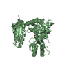
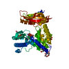
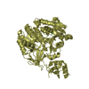
| Structure viewer | Molecule: MolmilJmol/JSmol |
|---|
- Downloads & links
-Download
| PDBx/mmCIF format | 1f5a.cif.gz | 113.3 KB | Display | PDBx/mmCIF format |
|---|---|---|---|---|
| PDB format | pdb1f5a.ent.gz | 86.2 KB | Display | PDB format |
| PDBx/mmJSON format | 1f5a.json.gz | Tree view | PDBx/mmJSON format | |
| Others |  Other downloads Other downloads |
-Validation report
| Arichive directory | https://data.pdbj.org/pub/pdb/validation_reports/f5/1f5aftp://data.pdbj.org/pub/pdb/validation_reports/f5/1f5a | HTTPS FTP |
|---|
-Related structure data
| Similar structure data |
|---|

-Links
 PDBj
PDBj


- Assembly
| Deposited unit | 
| ||||||||
|---|---|---|---|---|---|---|---|---|---|
| 1 |
| ||||||||
| Unit cell |
| ||||||||
| Details | The biological assembly is a monomer. |
-Components
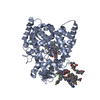
| #1: Protein | Mass: 59444.172 Da / Num. of mol.: 1 Fragment: C-TERMINAL DELETION MUTANT MISSING RESIDUES 514-738 Source method: isolated from a genetically manipulated source Source: (gene. exp.)  References: UniProt: P25500, polynucleotide adenylyltransferase | ||||||||
|---|---|---|---|---|---|---|---|---|---|
| #2: Chemical |   Mass: 54.938 Da / Num. of mol.: 3 / Source method: obtained synthetically / Formula: Mn Mass: 54.938 Da / Num. of mol.: 3 / Source method: obtained synthetically / Formula: Mn#3: Chemical | ChemComp-3AT / | 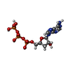  Mass: 491.182 Da / Num. of mol.: 1 / Source method: obtained synthetically / Formula: C10H16N5O12P3 Mass: 491.182 Da / Num. of mol.: 1 / Source method: obtained synthetically / Formula: C10H16N5O12P3#4: Chemical | ChemComp-3PO / |   Mass: 257.955 Da / Num. of mol.: 1 / Source method: obtained synthetically / Formula: H5O10P3 Mass: 257.955 Da / Num. of mol.: 1 / Source method: obtained synthetically / Formula: H5O10P3#5: Water | ChemComp-HOH / |  Mass: 18.015 Da / Num. of mol.: 170 / Source method: isolated from a natural source / Formula: H2O Mass: 18.015 Da / Num. of mol.: 170 / Source method: isolated from a natural source / Formula: H2OHas protein modification | Y | |
-Experimental details
-Experiment
| Experiment | Method: X-RAY DIFFRACTION / Number of used crystals: 2 |
|---|
- Sample preparation
| Crystal | Density Matthews: 2.73 Å3/Da / Density % sol: 55 % | ||||||||||||||||||||||||||||||||||||||||||
|---|---|---|---|---|---|---|---|---|---|---|---|---|---|---|---|---|---|---|---|---|---|---|---|---|---|---|---|---|---|---|---|---|---|---|---|---|---|---|---|---|---|---|---|
| Crystal grow | Temperature: 297 K / Method: vapor diffusion, hanging drop / pH: 6 Details: PEG8000, Ammonium sulfate, MES buffer, Calcium chloride, manganese chloride, pH 6, VAPOR DIFFUSION, HANGING DROP, temperature 297K | ||||||||||||||||||||||||||||||||||||||||||
| Crystal | *PLUS Density % sol: 55 % | ||||||||||||||||||||||||||||||||||||||||||
| Crystal grow | *PLUS Temperature: 24 ℃ / Method: vapor diffusion / Details: used microseeding | ||||||||||||||||||||||||||||||||||||||||||
| Components of the solutions | *PLUS
|
-Data collection
| Diffraction |
| ||||||||||||||||||
|---|---|---|---|---|---|---|---|---|---|---|---|---|---|---|---|---|---|---|---|
| Diffraction source |
| ||||||||||||||||||
| Detector |
| ||||||||||||||||||
| Radiation | Protocol: SINGLE WAVELENGTH / Monochromatic (M) / Laue (L): M / Scattering type: x-ray | ||||||||||||||||||
| Radiation wavelength |
| ||||||||||||||||||
| Reflection | Resolution: 2.5→20 Å / Num. obs: 19890 / % possible obs: 87 % / Observed criterion σ(F): 0 / Observed criterion σ(I): 0 / Redundancy: 4.2 % / Biso Wilson estimate: 19.3 Å2 / Rmerge(I) obs: 0.085 / Net I/σ(I): 16.7 | ||||||||||||||||||
| Reflection shell | Resolution: 2.5→2.67 Å / Rmerge(I) obs: 0.267 / % possible all: 65.1 |

- Processing
| Software |
| ||||||||||||||||||||||||||||||||||||
|---|---|---|---|---|---|---|---|---|---|---|---|---|---|---|---|---|---|---|---|---|---|---|---|---|---|---|---|---|---|---|---|---|---|---|---|---|---|
| Refinement | Resolution: 2.5→20 Å / Rfactor Rfree error: 0.007 / Cross valid method: THROUGHOUT / σ(F): 0 / σ(I): 0 / Stereochemistry target values: MHLH
| ||||||||||||||||||||||||||||||||||||
| Solvent computation | Solvent model: FLAT MODEL / Bsol: 30.0537 Å2 / ksol: 0.352814 e/Å3 | ||||||||||||||||||||||||||||||||||||
| Displacement parameters | Biso mean: 29 Å2 | ||||||||||||||||||||||||||||||||||||
| Refine analyze |
| ||||||||||||||||||||||||||||||||||||
| Refinement step | Cycle: LAST / Resolution: 2.5→20 Å
| ||||||||||||||||||||||||||||||||||||
| Refine LS restraints |
| ||||||||||||||||||||||||||||||||||||
| LS refinement shell | Resolution: 2.5→2.67 Å / Rfactor Rfree: 0.31 / Rfactor Rwork: 0.254 | ||||||||||||||||||||||||||||||||||||
| Xplor file | Serial no: 1 / Param file: protein_rep.param / Topol file: protein.top | ||||||||||||||||||||||||||||||||||||
| Software | *PLUS Name: CNS / Version: 1 / Classification: refinement | ||||||||||||||||||||||||||||||||||||
| Refinement | *PLUS Highest resolution: 2.5 Å / Lowest resolution: 20 Å / σ(F): 0 / % reflection Rfree: 7 % / Rfactor obs: 0.22 | ||||||||||||||||||||||||||||||||||||
| Solvent computation | *PLUS | ||||||||||||||||||||||||||||||||||||
| Displacement parameters | *PLUS Biso mean: 29 Å2 | ||||||||||||||||||||||||||||||||||||
| Refine LS restraints | *PLUS
| ||||||||||||||||||||||||||||||||||||
| LS refinement shell | *PLUS Highest resolution: 2.5 Å / Rfactor Rfree: 0.31 / Rfactor Rwork: 0.254 |
