 Movie
Movie Controller

[English] 日本語
 Yorodumi
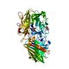
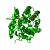
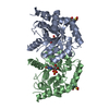
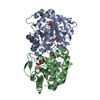
- PDB-1dqx: CRYSTAL STRUCTURE OF OROTIDINE 5'-PHOSPHATE DECARBOXYLASE COMPLEX... -
+ Open data

- Basic information
| Entry | Database: PDB / ID: 1dqx | ||||||
|---|---|---|---|---|---|---|---|
| Title | CRYSTAL STRUCTURE OF OROTIDINE 5'-PHOSPHATE DECARBOXYLASE COMPLEXED TO 6-HYDROXYURIDINE 5'-PHOSPHATE (BMP) | ||||||
 Components Components | OROTIDINE 5'-PHOSPHATE DECARBOXYLASE | ||||||
 Keywords Keywords | LYASE / orotidine 5'phosphate decarboxylase / uridine 5'phosphate / UMP / OMP / 6-hydroxyuridine 5'-phosphate / BMP / TIM barrel | ||||||
| Function / homology |  Function and homology information Function and homology informationUMP biosynthetic process / orotidine-5'-phosphate decarboxylase / orotidine-5'-phosphate decarboxylase activity / 'de novo' UMP biosynthetic process / 'de novo' pyrimidine nucleobase biosynthetic process / cytosol Similarity search - Function | ||||||
| Biological species |  | ||||||
| Method |  X-RAY DIFFRACTION / Resolution: 2.4 Å X-RAY DIFFRACTION / Resolution: 2.4 Å | ||||||
 Authors Authors | Milburn, M.V. / Miller, B.G. / Hassell, A.M. / Wolfenden, R. / Short, S.A. | ||||||
 Citation Citation | Journal: Proc.Natl.Acad.Sci.USA / Year: 2000 Title: Anatomy of a proficient enzyme: the structure of orotidine 5'-monophosphate decarboxylase in the presence and absence of a potential transition state analog. Authors: Miller, B.G. / Hassell, A.M. / Wolfenden, R. / Milburn, M.V. / Short, S.A. | ||||||
| History |
|
- Structure visualization
| Structure viewer | Molecule: MolmilJmol/JSmol |
|---|
- Downloads & links
-Download
| PDBx/mmCIF format | 1dqx.cif.gz | 224 KB | Display | PDBx/mmCIF format |
|---|---|---|---|---|
| PDB format | pdb1dqx.ent.gz | 180.1 KB | Display | PDB format |
| PDBx/mmJSON format | 1dqx.json.gz | Tree view | PDBx/mmJSON format | |
| Others |  Other downloads Other downloads |
-Validation report
| Arichive directory | https://data.pdbj.org/pub/pdb/validation_reports/dq/1dqxftp://data.pdbj.org/pub/pdb/validation_reports/dq/1dqx | HTTPS FTP |
|---|
-Related structure data


-Links
 PDBj
PDBj
- Assembly
| Deposited unit | 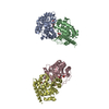
| ||||||||
|---|---|---|---|---|---|---|---|---|---|
| 1 | 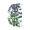
| ||||||||
| 2 | 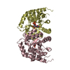
| ||||||||
| Unit cell |
| ||||||||
| Details | The biological assembly is a dimer from molecule A (E BMP molecule) two-fold related to molecule B (F BMP molecule), or molecule C (G BMP molecule) two-fold related to molecule D (H BMP molecule) |
-Components
| #1: Protein | Mass: 29347.586 Da / Num. of mol.: 4 / Mutation: S2H, N267D Source method: isolated from a genetically manipulated source Source: (gene. exp.) Plasmid: PCDA6022 / Production host:  References: UniProt: P03962, orotidine-5'-phosphate decarboxylase #2: Chemical | ChemComp-BMP /   Type: RNA linking / Mass: 340.181 Da / Num. of mol.: 4 / Source method: obtained synthetically / Formula: C9H13N2O10P Type: RNA linking / Mass: 340.181 Da / Num. of mol.: 4 / Source method: obtained synthetically / Formula: C9H13N2O10P#3: Water | ChemComp-HOH / |  Mass: 18.015 Da / Num. of mol.: 501 / Source method: isolated from a natural source / Formula: H2O Mass: 18.015 Da / Num. of mol.: 501 / Source method: isolated from a natural source / Formula: H2ONonpolymer details | BMP is a proposed transition state analog | |
|---|
-Experimental details
-Experiment
| Experiment | Method: X-RAY DIFFRACTION / Number of used crystals: 1 |
|---|
- Sample preparation
| Crystal | Density Matthews: 2.53 Å3/Da / Density % sol: 51.42 % | |||||||||||||||
|---|---|---|---|---|---|---|---|---|---|---|---|---|---|---|---|---|
| Crystal grow | Temperature: 295 K / Method: vapor diffusion, hanging drop / pH: 4.7 Details: sodium phosphate, polyethylene glycol 3350, pH 4.7, VAPOR DIFFUSION, HANGING DROP, temperature 295K | |||||||||||||||
| Crystal grow | *PLUS Temperature: 22 ℃ | |||||||||||||||
| Components of the solutions | *PLUS
|
-Data collection
| Diffraction | Mean temperature: 93 K |
|---|---|
| Diffraction source | Source: ROTATING ANODE / Type: RIGAKU RU200 / Wavelength: 1.5418 |
| Detector | Type: RIGAKU RAXIS IV / Detector: IMAGE PLATE / Date: Jun 5, 1999 |
| Radiation | Protocol: SINGLE WAVELENGTH / Monochromatic (M) / Laue (L): M / Scattering type: x-ray |
| Radiation wavelength | Wavelength: 1.5418 Å / Relative weight: 1 |
| Reflection | Resolution: 2.4→50 Å / Num. all: 46031 / Num. obs: 42013 / % possible obs: 91.3 % / Observed criterion σ(F): 0 / Observed criterion σ(I): 0 / Redundancy: 6.5 % / Biso Wilson estimate: 17.2 Å2 / Rmerge(I) obs: 0.061 / Net I/σ(I): 11.1 |
| Reflection shell | Resolution: 2.4→2.5 Å / Redundancy: 3.4 % / Rmerge(I) obs: 0.122 / % possible all: 78.4 |
- Processing
| Software |
| ||||||||||||||||||||||||||||||||||||||||
|---|---|---|---|---|---|---|---|---|---|---|---|---|---|---|---|---|---|---|---|---|---|---|---|---|---|---|---|---|---|---|---|---|---|---|---|---|---|---|---|---|---|
| Refinement | Resolution: 2.4→50 Å / Rfactor Rfree error: 0.005 / Data cutoff high absF: 2388386.14 / Data cutoff low absF: 0 / Isotropic thermal model: RESTRAINED / Cross valid method: THROUGHOUT / σ(F): 0 / σ(I): 0 / Stereochemistry target values: Engh & Huber
| ||||||||||||||||||||||||||||||||||||||||
| Solvent computation | Solvent model: FLAT MODEL / Bsol: 87.85 Å2 / ksol: 0.38 e/Å3 | ||||||||||||||||||||||||||||||||||||||||
| Displacement parameters | Biso mean: 35 Å2
| ||||||||||||||||||||||||||||||||||||||||
| Refine analyze |
| ||||||||||||||||||||||||||||||||||||||||
| Refinement step | Cycle: LAST / Resolution: 2.4→50 Å
| ||||||||||||||||||||||||||||||||||||||||
| Refine LS restraints |
| ||||||||||||||||||||||||||||||||||||||||
| Refine LS restraints NCS | NCS model details: CONSTRAINED | ||||||||||||||||||||||||||||||||||||||||
| LS refinement shell | Resolution: 2.4→2.49 Å / Rfactor Rfree error: 0.024 / Total num. of bins used: 10
| ||||||||||||||||||||||||||||||||||||||||
| Xplor file |
| ||||||||||||||||||||||||||||||||||||||||
| Software | *PLUS Name: CNS / Version: 0.5 / Classification: refinement | ||||||||||||||||||||||||||||||||||||||||
| Refinement | *PLUS σ(F): 0 / % reflection Rfree: 6.1 % | ||||||||||||||||||||||||||||||||||||||||
| Solvent computation | *PLUS | ||||||||||||||||||||||||||||||||||||||||
| Displacement parameters | *PLUS Biso mean: 35 Å2 | ||||||||||||||||||||||||||||||||||||||||
| Refine LS restraints | *PLUS
| ||||||||||||||||||||||||||||||||||||||||
| LS refinement shell | *PLUS Rfactor Rfree: 0.302 / % reflection Rfree: 6.6 % / Rfactor Rwork: 0.304 / Rfactor obs: 0.231 |
