 Movie
Movie Controller

[English] 日本語
 Yorodumi
- PDB-1dae: DETHIOBIOTIN SYNTHETASE COMPLEXED WITH 3-(1-AMINOETHYL) NONANEDIO... -
+ Open data

- Basic information
| Entry | Database: PDB / ID: 1dae | ||||||
|---|---|---|---|---|---|---|---|
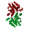
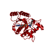
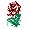
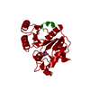
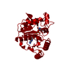
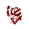
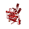
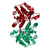
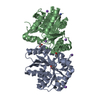
| Title | DETHIOBIOTIN SYNTHETASE COMPLEXED WITH 3-(1-AMINOETHYL) NONANEDIOIC ACID | ||||||
 Components Components | DETHIOBIOTIN SYNTHETASE | ||||||
 Keywords Keywords | LIGASE / BIOTIN BIOSYNTHESIS / MAGNESIUM / ATP-BINDING | ||||||
| Function / homology |  Function and homology information Function and homology informationdethiobiotin synthase / dethiobiotin synthase activity / biotin biosynthetic process / magnesium ion binding / protein homodimerization activity / ATP binding / cytosol Similarity search - Function | ||||||
| Biological species |  | ||||||
| Method |  X-RAY DIFFRACTION / Resolution: 1.7 Å X-RAY DIFFRACTION / Resolution: 1.7 Å | ||||||
 Authors Authors | Huang, W. / Jia, J. / Schneider, G. / Lindqvist, Y. | ||||||
 Citation Citation | Journal: Biochemistry / Year: 1995 Title: Mechanism of an ATP-dependent carboxylase, dethiobiotin synthetase, based on crystallographic studies of complexes with substrates and a reaction intermediate. Authors: Huang, W. / Jia, J. / Gibson, K.J. / Taylor, W.S. / Rendina, A.R. / Schneider, G. / Lindqvist, Y. #1: Journal: Structure / Year: 1994Title: Crystal Structure of an ATP-Dependent Carboxylase, Dethiobiotin Synthetase, at 1.65 A Resolution Authors: Huang, W. / Lindqvist, Y. / Schneider, G. / Gibson, K.J. / Flint, D. / Lorimer, G. | ||||||
| History |
|
- Structure visualization
| Structure viewer | Molecule: MolmilJmol/JSmol |
|---|
- Downloads & links
-Download
| PDBx/mmCIF format | 1dae.cif.gz | 57.4 KB | Display | PDBx/mmCIF format |
|---|---|---|---|---|
| PDB format | pdb1dae.ent.gz | 40.9 KB | Display | PDB format |
| PDBx/mmJSON format | 1dae.json.gz | Tree view | PDBx/mmJSON format | |
| Others |  Other downloads Other downloads |
-Validation report
| Summary document | 1dae_validation.pdf.gz | 374.4 KB | Display | wwPDB validaton report |
|---|---|---|---|---|
| Full document | 1dae_full_validation.pdf.gz | 376.8 KB | Display | |
| Data in XML | 1dae_validation.xml.gz | 6.3 KB | Display | |
| Data in CIF | 1dae_validation.cif.gz | 9.9 KB | Display | |
| Arichive directory | https://data.pdbj.org/pub/pdb/validation_reports/da/1daeftp://data.pdbj.org/pub/pdb/validation_reports/da/1dae | HTTPS FTP |
-Related structure data


-Links
 PDBj
PDBj- Assembly
| Deposited unit | 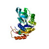
| ||||||||
|---|---|---|---|---|---|---|---|---|---|
| 1 | 
| ||||||||
| Unit cell |
|
-Components
| #1: Protein | Mass: 24028.289 Da / Num. of mol.: 1 Source method: isolated from a genetically manipulated source Source: (gene. exp.) |
|---|---|
| #2: Chemical | ChemComp-IKT /   Mass: 231.289 Da / Num. of mol.: 1 / Source method: obtained synthetically / Formula: C11H21NO4 Mass: 231.289 Da / Num. of mol.: 1 / Source method: obtained synthetically / Formula: C11H21NO4 |
| #3: Water | ChemComp-HOH /  Mass: 18.015 Da / Num. of mol.: 167 / Source method: isolated from a natural source / Formula: H2O Mass: 18.015 Da / Num. of mol.: 167 / Source method: isolated from a natural source / Formula: H2O |
-Experimental details
-Experiment
| Experiment | Method: X-RAY DIFFRACTION |
|---|
- Sample preparation
| Crystal | Density Matthews: 2.21 Å3/Da / Density % sol: 34 % | |||||||||||||||||||||
|---|---|---|---|---|---|---|---|---|---|---|---|---|---|---|---|---|---|---|---|---|---|---|
| Crystal grow | *PLUS Method: other / Details: cocrystallization | |||||||||||||||||||||
| Components of the solutions | *PLUS
|
-Data collection
| Diffraction source | Wavelength: 1.5418 |
|---|---|
| Detector | Type: RIGAKU RAXIS II / Detector: IMAGE PLATE / Date: Mar 6, 1994 |
| Radiation | Monochromatic (M) / Laue (L): M / Scattering type: x-ray |
| Radiation wavelength | Wavelength: 1.5418 Å / Relative weight: 1 |
| Reflection | Num. obs: 20347 / % possible obs: 88 % / Observed criterion σ(I): 1 / Redundancy: 2.6 % / Rmerge(I) obs: 0.05 |
| Reflection | *PLUS Highest resolution: 1.7 Å / Num. obs: 23576 / % possible obs: 95 % / Num. measured all: 60324 |
- Processing
| Software |
| ||||||||||||||||||||||||||||||||||||||||||||||||||||||||||||
|---|---|---|---|---|---|---|---|---|---|---|---|---|---|---|---|---|---|---|---|---|---|---|---|---|---|---|---|---|---|---|---|---|---|---|---|---|---|---|---|---|---|---|---|---|---|---|---|---|---|---|---|---|---|---|---|---|---|---|---|---|---|
| Refinement | Resolution: 1.7→6 Å / σ(F): 1 /
| ||||||||||||||||||||||||||||||||||||||||||||||||||||||||||||
| Displacement parameters | Biso mean: 22.7 Å2 | ||||||||||||||||||||||||||||||||||||||||||||||||||||||||||||
| Refinement step | Cycle: LAST / Resolution: 1.7→6 Å
| ||||||||||||||||||||||||||||||||||||||||||||||||||||||||||||
| Refine LS restraints |
| ||||||||||||||||||||||||||||||||||||||||||||||||||||||||||||
| Software | *PLUS Name: X-PLOR / Classification: refinement | ||||||||||||||||||||||||||||||||||||||||||||||||||||||||||||
| Refinement | *PLUS Rfactor obs: 0.173 | ||||||||||||||||||||||||||||||||||||||||||||||||||||||||||||
| Solvent computation | *PLUS | ||||||||||||||||||||||||||||||||||||||||||||||||||||||||||||
| Displacement parameters | *PLUS | ||||||||||||||||||||||||||||||||||||||||||||||||||||||||||||
| Refine LS restraints | *PLUS
|
