 Movie
Movie Controller

[English] 日本語
 Yorodumi
- PDB-1czp: ANABAENA PCC7119 [2FE-2S] FERREDOXIN IN THE REDUCED AND OXIXIZED ... -
+ Open data

- Basic information
| Entry | Database: PDB / ID: 1czp | ||||||
|---|---|---|---|---|---|---|---|
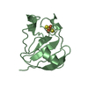
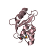
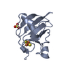
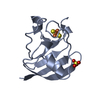
| Title | ANABAENA PCC7119 [2FE-2S] FERREDOXIN IN THE REDUCED AND OXIXIZED STATE AT 1.17 A | ||||||
 Components Components | FERREDOXIN I | ||||||
 Keywords Keywords | ELECTRON TRANSPORT / [2FE-2S] PROTEIN / CRYSTAL REDUCED WITH DITHIONITE | ||||||
| Function / homology |  Function and homology information Function and homology informationelectron transport chain / 2 iron, 2 sulfur cluster binding / electron transfer activity / metal ion binding Similarity search - Function | ||||||
| Biological species |  Nostoc sp. (bacteria) Nostoc sp. (bacteria) | ||||||
| Method |  X-RAY DIFFRACTION / SYNCHROTRON / MOLECULAR REPLACEMENT / Resolution: 1.17 Å X-RAY DIFFRACTION / SYNCHROTRON / MOLECULAR REPLACEMENT / Resolution: 1.17 Å | ||||||
 Authors Authors | Morales, R. / Charon, M.H. / Frey, M. | ||||||
 Citation Citation | Journal: Biochemistry / Year: 1999 Title: Refined X-ray structures of the oxidized, at 1.3 A, and reduced, at 1.17 A, [2Fe-2S] ferredoxin from the cyanobacterium Anabaena PCC7119 show redox-linked conformational changes. Authors: Morales, R. / Charon, M.H. / Hudry-Clergeon, G. / Petillot, Y. / Norager, S. / Medina, M. / Frey, M. | ||||||
| History |
|
- Structure visualization
| Structure viewer | Molecule: MolmilJmol/JSmol |
|---|
- Downloads & links
-Download
| PDBx/mmCIF format | 1czp.cif.gz | 99.5 KB | Display | PDBx/mmCIF format |
|---|---|---|---|---|
| PDB format | pdb1czp.ent.gz | 75.7 KB | Display | PDB format |
| PDBx/mmJSON format | 1czp.json.gz | Tree view | PDBx/mmJSON format | |
| Others |  Other downloads Other downloads |
-Validation report
| Arichive directory | https://data.pdbj.org/pub/pdb/validation_reports/cz/1czpftp://data.pdbj.org/pub/pdb/validation_reports/cz/1czp | HTTPS FTP |
|---|
-Related structure data
| Related structure data | 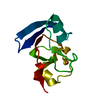 1qt9C  1fxaS C: citing same article ( S: Starting model for refinement |
|---|---|
| Similar structure data |
-Links
 PDBj
PDBj


- Assembly
| Deposited unit | 
| ||||||||||
|---|---|---|---|---|---|---|---|---|---|---|---|
| 1 | 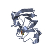
| ||||||||||
| 2 | 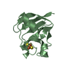
| ||||||||||
| Unit cell |
|
-Components
| #1: Protein | Mass: 10705.624 Da / Num. of mol.: 2 / Source method: isolated from a natural source / Source: (natural) Nostoc sp. (bacteria) / Strain: PCC 7119 / References: UniProt: P0A3C8#2: Chemical |   Mass: 175.820 Da / Num. of mol.: 2 / Source method: obtained synthetically / Formula: Fe2S2 Mass: 175.820 Da / Num. of mol.: 2 / Source method: obtained synthetically / Formula: Fe2S2#3: Water | ChemComp-HOH / |  Mass: 18.015 Da / Num. of mol.: 369 / Source method: isolated from a natural source / Formula: H2O Mass: 18.015 Da / Num. of mol.: 369 / Source method: isolated from a natural source / Formula: H2O |
|---|
-Experimental details
-Experiment
| Experiment | Method: X-RAY DIFFRACTION / Number of used crystals: 1 |
|---|
- Sample preparation
| Crystal | Density Matthews: 2 Å3/Da / Density % sol: 48.32 % Description: REDUCED CRYSTAL WITH SODIUM DITHIONITE IN ANAEROBIC ATMOSPHERE | |||||||||||||||
|---|---|---|---|---|---|---|---|---|---|---|---|---|---|---|---|---|
| Crystal grow | Temperature: 277 K / Method: vapor diffusion, hanging drop / pH: 5.5 Details: 50 MM SODIUM SUCCINATE PH 5.5, 2.6 M AMMONIUM SULFATE, 1% MPD, VAPOR DIFFUSION, HANGING DROP, temperature 277K | |||||||||||||||
| Crystal grow | *PLUS Temperature: 4 ℃ | |||||||||||||||
| Components of the solutions | *PLUS
|
-Data collection
| Diffraction | Mean temperature: 100 K |
|---|---|
| Diffraction source | Source: SYNCHROTRON / Site: ESRF  / Beamline: BM14 / Wavelength: 0.75 / Beamline: BM14 / Wavelength: 0.75 |
| Detector | Type: MARRESEARCH / Detector: IMAGE PLATE / Date: 1998 |
| Radiation | Protocol: SINGLE WAVELENGTH / Monochromatic (M) / Laue (L): M / Scattering type: x-ray |
| Radiation wavelength | Wavelength: 0.75 Å / Relative weight: 1 |
| Reflection | Resolution: 1.17→30 Å / Num. all: 67690 / Num. obs: 67690 / % possible obs: 95.5 % / Observed criterion σ(F): 0 / Observed criterion σ(I): 0 / Redundancy: 4.3 % / Biso Wilson estimate: 12.7 Å2 / Rmerge(I) obs: 0.033 / Net I/σ(I): 12 |
| Reflection shell | Resolution: 1.17→1.22 Å / Redundancy: 2.8 % / Rmerge(I) obs: 0.111 / Mean I/σ(I) obs: 6.1 / % possible all: 89.3 |
| Reflection | *PLUS Num. obs: 67611 / Rmerge(I) obs: 0.037 |
| Reflection shell | *PLUS % possible obs: 89.3 % |
- Processing
| Software |
| |||||||||||||||||||||||||||||||||
|---|---|---|---|---|---|---|---|---|---|---|---|---|---|---|---|---|---|---|---|---|---|---|---|---|---|---|---|---|---|---|---|---|---|---|
| Refinement | Method to determine structure: MOLECULAR REPLACEMENT Starting model: PDB ENTRY 1FXA Resolution: 1.17→15 Å / Num. parameters: 1756 / Num. restraintsaints: 2142 / Cross valid method: FREE R / σ(F): 0 / σ(I): 0 StereochEM target val spec case: [2FE-2S] CLUSTERS NOT RESTRAINED Stereochemistry target values: ENGH & HUBER Details: ANISOTROPIC SCALING APPLIED BY THE METHOD OF PARKIN, MOEZZI & HOPE, J.APPL.CRYST. 28 (1995) 53-56. ANISOTROPIC REFINEMENT REDUCED R-FREE (NO CUTOFF) BY 4 %
| |||||||||||||||||||||||||||||||||
| Solvent computation | Solvent model: MOEWS & KRETSINGER, J.MOL.BIOL. 91 (1973) 201-228 | |||||||||||||||||||||||||||||||||
| Refine analyze | Num. disordered residues: 1 / Occupancy sum hydrogen: 0 / Occupancy sum non hydrogen: 1878 | |||||||||||||||||||||||||||||||||
| Refinement step | Cycle: LAST / Resolution: 1.17→15 Å
| |||||||||||||||||||||||||||||||||
| Refine LS restraints |
| |||||||||||||||||||||||||||||||||
| Software | *PLUS Name: SHELXL-97 / Classification: refinement | |||||||||||||||||||||||||||||||||
| Refinement | *PLUS | |||||||||||||||||||||||||||||||||
| Solvent computation | *PLUS | |||||||||||||||||||||||||||||||||
| Displacement parameters | *PLUS | |||||||||||||||||||||||||||||||||
| Refine LS restraints | *PLUS
|
