 Movie
Movie Controller

[English] 日本語
 Yorodumi
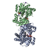
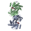
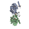
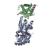
- PDB-1c1d: L-PHENYLALANINE DEHYDROGENASE STRUCTURE IN TERNARY COMPLEX WITH N... -
+ Open data

- Basic information
| Entry | Database: PDB / ID: 1c1d | ||||||
|---|---|---|---|---|---|---|---|
| Title | L-PHENYLALANINE DEHYDROGENASE STRUCTURE IN TERNARY COMPLEX WITH NADH AND L-PHENYLALANINE | ||||||
 Components Components | (L-PHENYLALANINE ...) x 2 | ||||||
 Keywords Keywords | OXIDOREDUCTASE / AMINO ACID DEHYDROGENASE / OXIDATIVE DEAMINATION MECHANISM | ||||||
| Function / homology |  Function and homology information Function and homology informationphenylalanine dehydrogenase / L-phenylalanine dehydrogenase (NAD+) activity / L-phenylalanine biosynthetic process / L-phenylalanine catabolic process / nucleotide binding Similarity search - Function | ||||||
| Biological species |  Rhodococcus sp. (bacteria) Rhodococcus sp. (bacteria) | ||||||
| Method |  X-RAY DIFFRACTION / SYNCHROTRON / Resolution: 1.25 Å X-RAY DIFFRACTION / SYNCHROTRON / Resolution: 1.25 Å | ||||||
 Authors Authors | Vanhooke, J.L. / Thoden, J.B. | ||||||
 Citation Citation | Journal: Biochemistry / Year: 2000 Title: Rhodococcus L-phenylalanine dehydrogenase: kinetics, mechanism, and structural basis for catalytic specificity. Authors: Brunhuber, N.M. / Thoden, J.B. / Blanchard, J.S. / Vanhooke, J.L. | ||||||
| History |
|
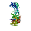
- Structure visualization
| Structure viewer | Molecule: MolmilJmol/JSmol |
|---|
- Downloads & links
-Download
| PDBx/mmCIF format | 1c1d.cif.gz | 166.7 KB | Display | PDBx/mmCIF format |
|---|---|---|---|---|
| PDB format | pdb1c1d.ent.gz | 128.6 KB | Display | PDB format |
| PDBx/mmJSON format | 1c1d.json.gz | Tree view | PDBx/mmJSON format | |
| Others |  Other downloads Other downloads |
-Validation report
| Arichive directory | https://data.pdbj.org/pub/pdb/validation_reports/c1/1c1dftp://data.pdbj.org/pub/pdb/validation_reports/c1/1c1d | HTTPS FTP |
|---|
-Related structure data


-Links
 PDBj
PDBj


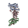
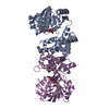
- Assembly
| Deposited unit | 
| ||||||||
|---|---|---|---|---|---|---|---|---|---|
| 1 | 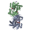
| ||||||||
| Unit cell |
|
-Components
-L-PHENYLALANINE ... , 2 types, 2 molecules AB
| #1: Protein | Mass: 36412.629 Da / Num. of mol.: 1 Source method: isolated from a genetically manipulated source Source: (gene. exp.) Rhodococcus sp. (bacteria) / Plasmid: PBL-1B / Species (production host): Escherichia coli / Production host: |
|---|---|
| #2: Protein | Mass: 36442.652 Da / Num. of mol.: 1 Source method: isolated from a genetically manipulated source Source: (gene. exp.) Rhodococcus sp. (bacteria) / Plasmid: PBL-1B / Species (production host): Escherichia coli / Production host: |
-Non-polymers , 7 types, 940 molecules 


| #3: Chemical | ChemComp-K /  Mass: 39.098 Da / Num. of mol.: 4 / Source method: obtained synthetically / Formula: K Mass: 39.098 Da / Num. of mol.: 4 / Source method: obtained synthetically / Formula: K#4: Chemical |  Type: L-peptide linking / Mass: 165.189 Da / Num. of mol.: 2 / Source method: obtained synthetically / Formula: C9H11NO2 Type: L-peptide linking / Mass: 165.189 Da / Num. of mol.: 2 / Source method: obtained synthetically / Formula: C9H11NO2#5: Chemical |  Mass: 665.441 Da / Num. of mol.: 2 / Source method: obtained synthetically / Formula: C21H29N7O14P2 Mass: 665.441 Da / Num. of mol.: 2 / Source method: obtained synthetically / Formula: C21H29N7O14P2#6: Chemical |  Mass: 22.990 Da / Num. of mol.: 2 / Source method: obtained synthetically / Formula: Na Mass: 22.990 Da / Num. of mol.: 2 / Source method: obtained synthetically / Formula: Na#7: Chemical | ChemComp-PO4 / |  Mass: 94.971 Da / Num. of mol.: 1 / Source method: obtained synthetically / Formula: PO4 Mass: 94.971 Da / Num. of mol.: 1 / Source method: obtained synthetically / Formula: PO4#8: Chemical | ChemComp-IPA / |  Mass: 60.095 Da / Num. of mol.: 1 / Source method: obtained synthetically / Formula: C3H8O Mass: 60.095 Da / Num. of mol.: 1 / Source method: obtained synthetically / Formula: C3H8O#9: Water | ChemComp-HOH / | Mass: 18.015 Da / Num. of mol.: 928 / Source method: isolated from a natural source / Formula: H2O |
|---|
-Experimental details
-Experiment
| Experiment | Method: X-RAY DIFFRACTION / Number of used crystals: 1 |
|---|
- Sample preparation
| Crystal | Density Matthews: 2.79 Å3/Da / Density % sol: 55.84 % | |||||||||||||||||||||||||||||||||||||||||||||||||||||||||||||||
|---|---|---|---|---|---|---|---|---|---|---|---|---|---|---|---|---|---|---|---|---|---|---|---|---|---|---|---|---|---|---|---|---|---|---|---|---|---|---|---|---|---|---|---|---|---|---|---|---|---|---|---|---|---|---|---|---|---|---|---|---|---|---|---|---|
| Crystal grow | pH: 8.7 / Details: pH 8.70 | |||||||||||||||||||||||||||||||||||||||||||||||||||||||||||||||
| Crystal grow | *PLUS pH: 7.8 / Method: batch method / Details: used macroseeding | |||||||||||||||||||||||||||||||||||||||||||||||||||||||||||||||
| Components of the solutions | *PLUS
|
-Data collection
| Diffraction | Mean temperature: 113 K |
|---|---|
| Diffraction source | Source: SYNCHROTRON / Site: APS  / Beamline: 19-ID / Wavelength: 0.7433 / Beamline: 19-ID / Wavelength: 0.7433 |
| Detector | Type: CUSTOM-MADE / Detector: CCD / Date: Nov 13, 1998 |
| Radiation | Protocol: SINGLE WAVELENGTH / Monochromatic (M) / Laue (L): M / Scattering type: x-ray |
| Radiation wavelength | Wavelength: 0.7433 Å / Relative weight: 1 |
| Reflection | Resolution: 1.25→50 Å / Num. obs: 216285 / % possible obs: 96.5 % / Observed criterion σ(I): 0 / Redundancy: 5.9 % / Rmerge(I) obs: 0.061 / Net I/σ(I): 24.4 |
| Reflection shell | Resolution: 1.25→1.29 Å / Redundancy: 5 % / Rmerge(I) obs: 0.255 / % possible all: 92.1 |
| Reflection | *PLUS Highest resolution: 1.25 Å / Lowest resolution: 50 Å / Observed criterion σ(I): 0 / Redundancy: 5.9 % / Num. measured all: 1286363 / Biso Wilson estimate: 0 Å2 |
| Reflection shell | *PLUS % possible obs: 92.1 % / Num. unique obs: 20510 / Num. measured obs: 103853 / Mean I/σ(I) obs: 3.4 |
- Processing
| Software |
| ||||||||||||||||||||||||||||||
|---|---|---|---|---|---|---|---|---|---|---|---|---|---|---|---|---|---|---|---|---|---|---|---|---|---|---|---|---|---|---|---|
| Refinement | Resolution: 1.25→30 Å / σ(F): 0 / Stereochemistry target values: TNT PROTGEO
| ||||||||||||||||||||||||||||||
| Refinement step | Cycle: LAST / Resolution: 1.25→30 Å
| ||||||||||||||||||||||||||||||
| Refine LS restraints |
| ||||||||||||||||||||||||||||||
| Software | *PLUS Name: TNT / Version: 5E / Classification: refinement | ||||||||||||||||||||||||||||||
| Refinement | *PLUS % reflection Rfree: 10 % / Rfactor all: 0.195 | ||||||||||||||||||||||||||||||
| Solvent computation | *PLUS | ||||||||||||||||||||||||||||||
| Displacement parameters | *PLUS | ||||||||||||||||||||||||||||||
| Refine LS restraints | *PLUS
|
