 Movie
Movie Controller


+ Open data

- Basic information
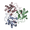
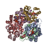
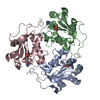
| Entry | Database: PDB / ID: 1bhn | ||||||
|---|---|---|---|---|---|---|---|
| Title | NUCLEOSIDE DIPHOSPHATE KINASE ISOFORM A FROM BOVINE RETINA | ||||||
 Components Components | NUCLEOSIDE DIPHOSPHATE TRANSFERASE | ||||||
 Keywords Keywords | PHOSPHOTRANSFERASE | ||||||
| Function / homology |  Function and homology information Function and homology informationInterconversion of nucleotide di- and triphosphates / farnesyl-diphosphate kinase / farnesyl diphosphate kinase activity / succinyl-CoA binding / ITP biosynthetic process / Ribavirin ADME / isoprenoid metabolic process / Azathioprine ADME / Hydrolases; Acting on ester bonds; Site specific endodeoxyribonucleases: cleavage is not sequence specific (deleted sub-subclass) / phosphotransferase activity, phosphate group as acceptor ...Interconversion of nucleotide di- and triphosphates / farnesyl-diphosphate kinase / farnesyl diphosphate kinase activity / succinyl-CoA binding / ITP biosynthetic process / Ribavirin ADME / isoprenoid metabolic process / Azathioprine ADME / Hydrolases; Acting on ester bonds; Site specific endodeoxyribonucleases: cleavage is not sequence specific (deleted sub-subclass) / phosphotransferase activity, phosphate group as acceptor / acetyl-CoA catabolic process / acetyl-CoA binding / nucleoside diphosphate metabolic process / coenzyme A binding / protein histidine kinase activity / regulation of fatty acid biosynthetic process / apoptotic DNA fragmentation / nucleoside-diphosphate kinase / 3'-5'-DNA exonuclease activity / UTP biosynthetic process / CTP biosynthetic process / protein hexamerization / DNA catabolic process / nucleoside diphosphate kinase activity / GTP biosynthetic process / Hydrolases; Acting on ester bonds; Exodeoxyribonucleases producing 5'-phosphomonoesters / histidine kinase / ribosomal small subunit binding / positive regulation of epithelial cell proliferation / DNA endonuclease activity / ADP binding / ruffle membrane / endocytosis / kinase activity / GDP binding / nervous system development / early endosome / cell differentiation / non-specific serine/threonine protein kinase / protein serine kinase activity / protein serine/threonine kinase activity / magnesium ion binding / DNA binding / ATP binding / identical protein binding / nucleus / cytosol / cytoplasm Similarity search - Function | ||||||
| Biological species |  | ||||||
| Method |  X-RAY DIFFRACTION / MOLECULAR REPLACEMENT / Resolution: 2.4 Å X-RAY DIFFRACTION / MOLECULAR REPLACEMENT / Resolution: 2.4 Å | ||||||
 Authors Authors | Ladner, J.E. / Abdulaev, N.G. / Kakuev, D.L. / Karaschuk, G.N. / Tordova, M. / Eisenstein, E. / Fujiwara, J.H. / Ridge, K.D. / Gilliland, G.L. | ||||||
 Citation Citation | Journal: Acta Crystallogr.,Sect.D / Year: 1999 Title: The three-dimensional structures of two isoforms of nucleoside diphosphate kinase from bovine retina. Authors: Ladner, J.E. / Abdulaev, N.G. / Kakuev, D.L. / Tordova, M. / Ridge, K.D. / Gilliland, G.L. | ||||||
| History |
|
- Structure visualization
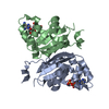
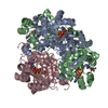
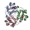
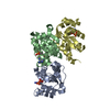
| Structure viewer | Molecule: MolmilJmol/JSmol |
|---|
- Downloads & links
-Download
| PDBx/mmCIF format | 1bhn.cif.gz | 213.3 KB | Display | PDBx/mmCIF format |
|---|---|---|---|---|
| PDB format | pdb1bhn.ent.gz | 166.5 KB | Display | PDB format |
| PDBx/mmJSON format | 1bhn.json.gz | Tree view | PDBx/mmJSON format | |
| Others |  Other downloads Other downloads |
-Validation report
| Arichive directory | https://data.pdbj.org/pub/pdb/validation_reports/bh/1bhnftp://data.pdbj.org/pub/pdb/validation_reports/bh/1bhn | HTTPS FTP |
|---|
-Related structure data
| Similar structure data |
|---|


-Links
 PDBj
PDBj

- Assembly
| Deposited unit | 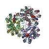
| ||||||||
|---|---|---|---|---|---|---|---|---|---|
| 1 |
| ||||||||
| Unit cell |
|
-Components
| #1: Protein | Mass: 17284.955 Da / Num. of mol.: 6 Source method: isolated from a genetically manipulated source Source: (gene. exp.)  #2: Chemical | ChemComp-35G / 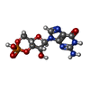  Mass: 345.205 Da / Num. of mol.: 6 / Source method: obtained synthetically / Formula: C10H12N5O7P Mass: 345.205 Da / Num. of mol.: 6 / Source method: obtained synthetically / Formula: C10H12N5O7P#3: Chemical | ChemComp-GDP /   Type: RNA linking / Mass: 443.201 Da / Num. of mol.: 6 / Source method: obtained synthetically / Formula: C10H15N5O11P2 / Comment: GDP, energy-carrying molecule*YM Type: RNA linking / Mass: 443.201 Da / Num. of mol.: 6 / Source method: obtained synthetically / Formula: C10H15N5O11P2 / Comment: GDP, energy-carrying molecule*YM#4: Water | ChemComp-HOH / |  Mass: 18.015 Da / Num. of mol.: 306 / Source method: isolated from a natural source / Formula: H2O Mass: 18.015 Da / Num. of mol.: 306 / Source method: isolated from a natural source / Formula: H2O |
|---|
-Experimental details
-Experiment
| Experiment | Method: X-RAY DIFFRACTION / Number of used crystals: 1 |
|---|
- Sample preparation
| Crystal | Density Matthews: 2.7 Å3/Da / Density % sol: 53.16 % | |||||||||||||||||||||||||||||||||||
|---|---|---|---|---|---|---|---|---|---|---|---|---|---|---|---|---|---|---|---|---|---|---|---|---|---|---|---|---|---|---|---|---|---|---|---|---|
| Crystal grow | pH: 5 / Details: pH 5 | |||||||||||||||||||||||||||||||||||
| Crystal grow | *PLUS Method: vapor diffusion, hanging dropDetails: drop contains equal volume of the reservoir solution | |||||||||||||||||||||||||||||||||||
| Components of the solutions | *PLUS
|
-Data collection
| Diffraction | Mean temperature: 100 K |
|---|---|
| Diffraction source | Source: ROTATING ANODE / Type: SIEMENS / Wavelength: 1.5418 |
| Detector | Type: SIEMENS HI-STAR / Detector: AREA DETECTOR / Date: Jan 15, 1997 / Details: COLLIMATOR |
| Radiation | Monochromator: GRAPHITE(002) / Monochromatic (M) / Laue (L): M / Scattering type: x-ray |
| Radiation wavelength | Wavelength: 1.5418 Å / Relative weight: 1 |
| Reflection | Resolution: 2.4→20 Å / Num. obs: 41872 / % possible obs: 79 % / Redundancy: 4 % / Rsym value: 0.122 / Net I/σ(I): 6.8 |
| Reflection shell | Resolution: 2.4→2.51 Å / Mean I/σ(I) obs: 1.4 / % possible all: 60 |
| Reflection | *PLUS Num. measured all: 165667 / Rmerge(I) obs: 0.122 |
| Reflection shell | *PLUS Lowest resolution: 2.54 Å / % possible obs: 60 % / Rmerge(I) obs: 0.384 |
- Processing
| Software |
| ||||||||||||||||||||||||||||||||||||||||||||||||||
|---|---|---|---|---|---|---|---|---|---|---|---|---|---|---|---|---|---|---|---|---|---|---|---|---|---|---|---|---|---|---|---|---|---|---|---|---|---|---|---|---|---|---|---|---|---|---|---|---|---|---|---|
| Refinement | Method to determine structure: MOLECULAR REPLACEMENT Starting model: PARTIALLY REFINED STRUCTURE OF THE B ISOFORM Resolution: 2.4→20 Å / Isotropic thermal model: TNT BCORREL
| ||||||||||||||||||||||||||||||||||||||||||||||||||
| Solvent computation | Bsol: 231.6 Å2 / ksol: 0.672 e/Å3 | ||||||||||||||||||||||||||||||||||||||||||||||||||
| Refinement step | Cycle: LAST / Resolution: 2.4→20 Å
| ||||||||||||||||||||||||||||||||||||||||||||||||||
| Refine LS restraints |
| ||||||||||||||||||||||||||||||||||||||||||||||||||
| Software | *PLUS Name: TNT / Version: 5E / Classification: refinement | ||||||||||||||||||||||||||||||||||||||||||||||||||
| Refinement | *PLUS Rfactor obs: 0.2 | ||||||||||||||||||||||||||||||||||||||||||||||||||
| Solvent computation | *PLUS | ||||||||||||||||||||||||||||||||||||||||||||||||||
| Displacement parameters | *PLUS | ||||||||||||||||||||||||||||||||||||||||||||||||||
| Refine LS restraints | *PLUS
|
