 Movie
Movie Controller


+ Open data

- Basic information
| Entry | Database: PDB / ID: 1ak0 | |||||||||
|---|---|---|---|---|---|---|---|---|---|---|
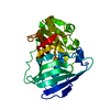
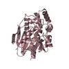
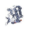
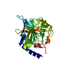
| Title | P1 NUCLEASE IN COMPLEX WITH A SUBSTRATE ANALOG | |||||||||
 Components Components | P1 NUCLEASE | |||||||||
 Keywords Keywords | ENDONUCLEASE / P1 NUCLEASE / REACTION MECHANISM / THIOPHOSPHORYLATED OLIGONUCLEOTIDES / GLYCOSYLATED PROTEIN | |||||||||
| Function / homology |  Function and homology information Function and homology informationAspergillus nuclease S1 / DNA catabolic process / hydrolase activity, acting on ester bonds / endonuclease activity / nucleic acid binding / extracellular region / metal ion binding Similarity search - Function | |||||||||
| Biological species |  Penicillium citrinum (fungus) Penicillium citrinum (fungus) | |||||||||
| Method |  X-RAY DIFFRACTION / MOLECULAR REPLACEMENT / Resolution: 1.8 Å X-RAY DIFFRACTION / MOLECULAR REPLACEMENT / Resolution: 1.8 Å | |||||||||
 Authors Authors | Romier, C. / Suck, D. | |||||||||
 Citation Citation | Journal: Proteins / Year: 1998 Title: Recognition of single-stranded DNA by nuclease P1: high resolution crystal structures of complexes with substrate analogs. Authors: Romier, C. / Dominguez, R. / Lahm, A. / Dahl, O. / Suck, D. #1: Journal: Embo J. / Year: 1991Title: Crystal Structure of Penicillium Citrinum P1 Nuclease at 2.8 A Resolution Authors: Volbeda, A. / Lahm, A. / Sakiyama, F. / Suck, D. | |||||||||
| History |
|
- Structure visualization
| Structure viewer | Molecule: MolmilJmol/JSmol |
|---|
- Downloads & links
-Download
| PDBx/mmCIF format | 1ak0.cif.gz | 72.8 KB | Display | PDBx/mmCIF format |
|---|---|---|---|---|
| PDB format | pdb1ak0.ent.gz | 52.5 KB | Display | PDB format |
| PDBx/mmJSON format | 1ak0.json.gz | Tree view | PDBx/mmJSON format | |
| Others |  Other downloads Other downloads |
-Validation report
| Arichive directory | https://data.pdbj.org/pub/pdb/validation_reports/ak/1ak0ftp://data.pdbj.org/pub/pdb/validation_reports/ak/1ak0 | HTTPS FTP |
|---|
-Related structure data
| Similar structure data |
|---|


-Links
 PDBj
PDBj- Assembly
| Deposited unit | 
| ||||||||
|---|---|---|---|---|---|---|---|---|---|
| 1 |
| ||||||||
| Unit cell |
|
-Components
-Protein , 1 types, 1 molecules A
| #1: Protein | Mass: 29251.021 Da / Num. of mol.: 1 / Source method: isolated from a natural source / Source: (natural) Penicillium citrinum (fungus) / References: UniProt: P24289, Aspergillus nuclease S1 |
|---|
-Sugars , 2 types, 3 molecules 
| #2: Polysaccharide | 2-acetamido-2-deoxy-beta-D-glucopyranose-(1-4)-2-acetamido-2-deoxy-beta-D-glucopyranose Source method: isolated from a genetically manipulated source |
|---|---|
| #3: Sugar |  Type: D-saccharide, beta linking / Mass: 221.208 Da / Num. of mol.: 2 Type: D-saccharide, beta linking / Mass: 221.208 Da / Num. of mol.: 2Source method: isolated from a genetically manipulated source Formula: C8H15NO6 |
-Non-polymers , 4 types, 162 molecules 


| #4: Chemical | ChemComp-ZN /  Mass: 65.409 Da / Num. of mol.: 4 / Source method: obtained synthetically / Formula: Zn Mass: 65.409 Da / Num. of mol.: 4 / Source method: obtained synthetically / Formula: Zn#5: Chemical | ChemComp-ADS / |  Mass: 379.352 Da / Num. of mol.: 1 / Source method: obtained synthetically / Formula: C10H14N5O5PS2 Mass: 379.352 Da / Num. of mol.: 1 / Source method: obtained synthetically / Formula: C10H14N5O5PS2#6: Chemical |  Mass: 354.340 Da / Num. of mol.: 3 / Source method: obtained synthetically / Formula: C10H15N2O6PS2 Mass: 354.340 Da / Num. of mol.: 3 / Source method: obtained synthetically / Formula: C10H15N2O6PS2#7: Water | ChemComp-HOH / | Mass: 18.015 Da / Num. of mol.: 154 / Source method: isolated from a natural source / Formula: H2O |
|---|
-Details
| Has protein modification | Y |
|---|
-Experimental details
-Experiment
| Experiment | Method: X-RAY DIFFRACTION / Number of used crystals: 1 |
|---|
- Sample preparation
| Crystal | Density Matthews: 2.71 Å3/Da / Density % sol: 54.65 % | |||||||||||||||||||||||||||||||||||
|---|---|---|---|---|---|---|---|---|---|---|---|---|---|---|---|---|---|---|---|---|---|---|---|---|---|---|---|---|---|---|---|---|---|---|---|---|
| Crystal grow | pH: 5.3 / Details: PEG 6000 12 - 20% 25 MM NA ACETATE PH 5.3 | |||||||||||||||||||||||||||||||||||
| Crystal grow | *PLUS Method: vapor diffusion, hanging drop | |||||||||||||||||||||||||||||||||||
| Components of the solutions | *PLUS
|
-Data collection
| Diffraction | Mean temperature: 293 K |
|---|---|
| Diffraction source | Type: ENRAF-NONIUS / Wavelength: 1.5418 |
| Detector | Type: MARRESEARCH / Detector: IMAGE PLATE / Date: Jan 1, 1994 |
| Radiation | Monochromator: GRAPHITE(002) / Monochromatic (M) / Laue (L): M / Scattering type: x-ray |
| Radiation wavelength | Wavelength: 1.5418 Å / Relative weight: 1 |
| Reflection | Resolution: 1.8→15 Å / Num. obs: 27854 / % possible obs: 93.2 % / Observed criterion σ(I): 2 / Redundancy: 4.3 % / Rsym value: 0.068 |
| Reflection shell | Resolution: 1.8→1.95 Å / Rsym value: 0.247 / % possible all: 89.9 |
| Reflection | *PLUS Num. measured all: 119199 / Rmerge(I) obs: 0.068 |
| Reflection shell | *PLUS % possible obs: 89.9 % / Rmerge(I) obs: 0.247 |
- Processing
| Software |
| ||||||||||||||||||||||||||||||||||||||||||||||||||||||||||||
|---|---|---|---|---|---|---|---|---|---|---|---|---|---|---|---|---|---|---|---|---|---|---|---|---|---|---|---|---|---|---|---|---|---|---|---|---|---|---|---|---|---|---|---|---|---|---|---|---|---|---|---|---|---|---|---|---|---|---|---|---|---|
| Refinement | Method to determine structure: MOLECULAR REPLACEMENT / Resolution: 1.8→8 Å / Data cutoff high absF: 10000000 / Data cutoff low absF: 0.001 / σ(F): 2
| ||||||||||||||||||||||||||||||||||||||||||||||||||||||||||||
| Displacement parameters | Biso mean: 22.8 Å2 | ||||||||||||||||||||||||||||||||||||||||||||||||||||||||||||
| Refine analyze | Luzzati d res low obs: 8 Å | ||||||||||||||||||||||||||||||||||||||||||||||||||||||||||||
| Refinement step | Cycle: LAST / Resolution: 1.8→8 Å
| ||||||||||||||||||||||||||||||||||||||||||||||||||||||||||||
| Refine LS restraints |
| ||||||||||||||||||||||||||||||||||||||||||||||||||||||||||||
| Xplor file |
| ||||||||||||||||||||||||||||||||||||||||||||||||||||||||||||
| Software | *PLUS Name: X-PLOR / Version: 3.1 / Classification: refinement | ||||||||||||||||||||||||||||||||||||||||||||||||||||||||||||
| Refinement | *PLUS | ||||||||||||||||||||||||||||||||||||||||||||||||||||||||||||
| Solvent computation | *PLUS | ||||||||||||||||||||||||||||||||||||||||||||||||||||||||||||
| Displacement parameters | *PLUS Biso mean: 21.8 Å2 |
