 Movie
Movie Controller

[English] 日本語
 Yorodumi
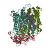
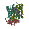
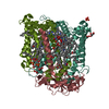
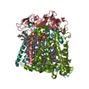
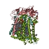
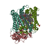
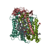
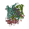
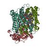
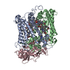
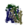
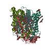
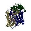
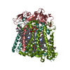
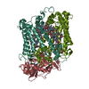
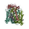
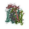
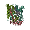
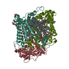
- PDB-1aig: PHOTOSYNTHETIC REACTION CENTER FROM RHODOBACTER SPHAEROIDES IN TH... -
+ Open data

- Basic information
| Entry | Database: PDB / ID: 1aig | ||||||
|---|---|---|---|---|---|---|---|
| Title | PHOTOSYNTHETIC REACTION CENTER FROM RHODOBACTER SPHAEROIDES IN THE D+QB-CHARGE SEPARATED STATE | ||||||
 Components Components | (PHOTOSYNTHETIC REACTION CENTER ...) x 3 | ||||||
 Keywords Keywords | PHOTOSYNTHETIC REACTION CENTER / INTEGRAL MEMBRANE PROTEIN / CHARGE SEPARATED | ||||||
| Function / homology |  Function and homology information Function and homology information: / : / plasma membrane-derived chromatophore membrane / plasma membrane light-harvesting complex / bacteriochlorophyll binding / photosynthetic electron transport in photosystem II / photosynthesis, light reaction / membrane => GO:0016020 / metal ion binding Similarity search - Function | ||||||
| Biological species |  Rhodobacter sphaeroides (bacteria) Rhodobacter sphaeroides (bacteria) | ||||||
| Method |  X-RAY DIFFRACTION / SYNCHROTRON / MOLECULAR REPLACEMENT / Resolution: 2.6 Å X-RAY DIFFRACTION / SYNCHROTRON / MOLECULAR REPLACEMENT / Resolution: 2.6 Å | ||||||
 Authors Authors | Stowell, M.H.B. / Mcphillips, T.M. / Soltis, S.M. / Rees, D.C. / Abresch, E. / Feher, G. | ||||||
 Citation Citation | Journal: Science / Year: 1997 Title: Light-induced structural changes in photosynthetic reaction center: implications for mechanism of electron-proton transfer. Authors: Stowell, M.H. / McPhillips, T.M. / Rees, D.C. / Soltis, S.M. / Abresch, E. / Feher, G. #1: Journal: Annu.Rev.Biochem. / Year: 1989Title: The Bacterial Photosynthetic Reaction Center as a Model for Membrane Proteins Authors: Rees, D.C. / Komiya, H. / Yeates, T.O. / Allen, J.P. / Feher, G. #2: Journal: Nature / Year: 1989Title: Structure and Function of Bacterial Photosynthetic Reaction Centres Authors: Feher, G. / Allen, J.P. / Okamura, M.Y. / Rees, D.C. #3: Journal: Proc.Natl.Acad.Sci.USA / Year: 1988Title: Structure of the Reaction Center from Rhodobacter Sphaeroides R-26: Protein-Cofactor (Quinones and Fe2+) Interactions Authors: Allen, J.P. / Feher, G. / Yeates, T.O. / Komiya, H. / Rees, D.C. #4: Journal: Proc.Natl.Acad.Sci.USA / Year: 1988Title: Structure of the Reaction Center from Rhodobacter Sphaeroides R-26 and 2.4.1: Symmetry Relations and Sequence Comparisons between Different Species Authors: Komiya, H. / Yeates, T.O. / Rees, D.C. / Allen, J.P. / Feher, G. #5: Journal: Proc.Natl.Acad.Sci.USA / Year: 1988Title: Structure of the Reaction Center from Rhodobacter Sphaeroides R-26 and 2.4.1: Protein-Cofactor (Bacteriochlorophyll, Bacteriopheophytin, and Carotenoid) Interactions Authors: Yeates, T.O. / Komiya, H. / Chirino, A. / Rees, D.C. / Allen, J.P. / Feher, G. #6: Journal: Proc.Natl.Acad.Sci.USA / Year: 1987Title: Structure of the Reaction Center from Rhodobacter Sphaeroides R-26: Membrane-Protein Interactions Authors: Yeates, T.O. / Komiya, H. / Rees, D.C. / Allen, J.P. / Feher, G. #7: Journal: Proc.Natl.Acad.Sci.USA / Year: 1987Title: Structure of the Reaction Center from Rhodobacter Sphaeroides R-26: The Cofactors Authors: Allen, J.P. / Feher, G. / Yeates, T.O. / Komiya, H. / Rees, D.C. #8: Journal: Proc.Natl.Acad.Sci.USA / Year: 1987Title: Structure of the Reaction Center from Rhodobacter Sphaeroides R-26: The Protein Subunits Authors: Allen, J.P. / Feher, G. / Yeates, T.O. / Komiya, H. / Rees, D.C. #9: Journal: Proc.Natl.Acad.Sci.USA / Year: 1986Title: Structural Homology of Reaction Centers from Rhodopseudomonas Sphaeroides and Rhodopseudomonas Viridis as Determined by X-Ray Diffraction Authors: Allen, J.P. / Feher, G. / Yeates, T.O. / Rees, D.C. / Deisenhofer, J. / Michel, H. / Huber, R. #10: Journal: Proc.Natl.Acad.Sci.USA / Year: 1984Title: Crystallization of Reaction Center from Rhodopseudomonas Sphaeroides: Preliminary Characterization Authors: Allen, J.P. / Feher, G. | ||||||
| History |
|
- Structure visualization
| Structure viewer | Molecule: MolmilJmol/JSmol |
|---|
- Downloads & links
-Download
| PDBx/mmCIF format | 1aig.cif.gz | 362.5 KB | Display | PDBx/mmCIF format |
|---|---|---|---|---|
| PDB format | pdb1aig.ent.gz | 293.4 KB | Display | PDB format |
| PDBx/mmJSON format | 1aig.json.gz | Tree view | PDBx/mmJSON format | |
| Others |  Other downloads Other downloads |
-Validation report
| Arichive directory | https://data.pdbj.org/pub/pdb/validation_reports/ai/1aigftp://data.pdbj.org/pub/pdb/validation_reports/ai/1aig | HTTPS FTP |
|---|
-Related structure data


-Links
 PDBj
PDBj


- Assembly
| Deposited unit | 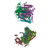
| ||||||||
|---|---|---|---|---|---|---|---|---|---|
| 1 | 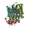
| ||||||||
| 2 | 
| ||||||||
| Unit cell |
|
-Components
-PHOTOSYNTHETIC REACTION CENTER ... , 3 types, 6 molecules LNMOHP
| #1: Protein | Mass: 31346.389 Da / Num. of mol.: 2 / Source method: isolated from a natural source / Source: (natural) Rhodobacter sphaeroides (bacteria) / Strain: R26 / References: UniProt: P02954, UniProt: P0C0Y8*PLUS#2: Protein | Mass: 34398.543 Da / Num. of mol.: 2 / Source method: isolated from a natural source / Source: (natural) Rhodobacter sphaeroides (bacteria) / Strain: R26 / References: UniProt: P02953, UniProt: P0C0Y9*PLUS#3: Protein | Mass: 28137.398 Da / Num. of mol.: 2 / Source method: isolated from a natural source / Source: (natural) Rhodobacter sphaeroides (bacteria) / Strain: R26 / References: UniProt: P11846, UniProt: P0C0Y7*PLUS |
|---|
-Non-polymers , 5 types, 104 molecules 


| #4: Chemical | ChemComp-BCL /  Mass: 911.504 Da / Num. of mol.: 8 / Source method: obtained synthetically / Formula: C55H74MgN4O6 Mass: 911.504 Da / Num. of mol.: 8 / Source method: obtained synthetically / Formula: C55H74MgN4O6#5: Chemical | ChemComp-BPH /  Mass: 889.215 Da / Num. of mol.: 4 / Source method: obtained synthetically / Formula: C55H76N4O6 Mass: 889.215 Da / Num. of mol.: 4 / Source method: obtained synthetically / Formula: C55H76N4O6#6: Chemical | ChemComp-U10 /  Mass: 863.343 Da / Num. of mol.: 4 / Source method: obtained synthetically / Formula: C59H90O4 Mass: 863.343 Da / Num. of mol.: 4 / Source method: obtained synthetically / Formula: C59H90O4#7: Chemical |  Mass: 55.845 Da / Num. of mol.: 2 / Source method: obtained synthetically / Formula: Fe Mass: 55.845 Da / Num. of mol.: 2 / Source method: obtained synthetically / Formula: Fe#8: Water | ChemComp-HOH / | Mass: 18.015 Da / Num. of mol.: 86 / Source method: isolated from a natural source / Formula: H2O |
|---|
-Details
| Nonpolymer details | ELECTRON DENSITY FOR THE UBIQUINONE-10 MOLECULES IS WEAK BEYOND THE C16 CARBON OF QB IN MOLECULE 1 ...ELECTRON DENSITY FOR THE UBIQUINONE |
|---|
-Experimental details
-Experiment
| Experiment | Method: X-RAY DIFFRACTION / Number of used crystals: 1 |
|---|
- Sample preparation
| Crystal | Density Matthews: 3.64 Å3/Da / Density % sol: 66 % | ||||||||||||||||||||||||||||||||||||||||
|---|---|---|---|---|---|---|---|---|---|---|---|---|---|---|---|---|---|---|---|---|---|---|---|---|---|---|---|---|---|---|---|---|---|---|---|---|---|---|---|---|---|
| Crystal grow | *PLUS pH: 8 / Method: vapor diffusion, sitting dropDetails: Allen, J.P., (1994) Proteins. Struct. Funct. Genet., 20, 283. | ||||||||||||||||||||||||||||||||||||||||
| Components of the solutions | *PLUS
|
-Data collection
| Diffraction | Mean temperature: 77 K |
|---|---|
| Diffraction source | Source: SYNCHROTRON / Site: SSRL  / Beamline: BL7-1 / Wavelength: 1.08 / Beamline: BL7-1 / Wavelength: 1.08 |
| Detector | Type: MARRESEARCH / Detector: IMAGE PLATE / Date: Mar 1, 1996 / Details: BENT |
| Radiation | Monochromator: SI(111) / Monochromatic (M) / Laue (L): M / Scattering type: x-ray |
| Radiation wavelength | Wavelength: 1.08 Å / Relative weight: 1 |
| Reflection | Resolution: 2.6→30 Å / Num. obs: 74104 / % possible obs: 88 % / Observed criterion σ(I): 0 / Redundancy: 3.1 % / Rsym value: 0.085 / Net I/σ(I): 14.3 |
| Reflection shell | Resolution: 2.6→2.72 Å / Mean I/σ(I) obs: 2.6 / Rsym value: 0.16 / % possible all: 86 |
| Reflection | *PLUS Num. measured all: 203621 / Rmerge(I) obs: 0.085 |
| Reflection shell | *PLUS Rmerge(I) obs: 0.16 |
- Processing
| Software |
| ||||||||||||||||||||||||||||||||||||||||||||||||||||||||||||
|---|---|---|---|---|---|---|---|---|---|---|---|---|---|---|---|---|---|---|---|---|---|---|---|---|---|---|---|---|---|---|---|---|---|---|---|---|---|---|---|---|---|---|---|---|---|---|---|---|---|---|---|---|---|---|---|---|---|---|---|---|---|
| Refinement | Method to determine structure: MOLECULAR REPLACEMENT / Resolution: 2.6→30 Å / Rfactor Rfree error: 0.005 / Data cutoff high absF: 100000 / Cross valid method: THROUGHOUT
| ||||||||||||||||||||||||||||||||||||||||||||||||||||||||||||
| Refinement step | Cycle: LAST / Resolution: 2.6→30 Å
| ||||||||||||||||||||||||||||||||||||||||||||||||||||||||||||
| Refine LS restraints |
| ||||||||||||||||||||||||||||||||||||||||||||||||||||||||||||
| LS refinement shell | Resolution: 2.6→2.72 Å / Rfactor Rfree error: 0.016 / Total num. of bins used: 8
| ||||||||||||||||||||||||||||||||||||||||||||||||||||||||||||
| Xplor file |
| ||||||||||||||||||||||||||||||||||||||||||||||||||||||||||||
| Software | *PLUS Name: X-PLOR / Version: 3.89 / Classification: refinement | ||||||||||||||||||||||||||||||||||||||||||||||||||||||||||||
| Refine LS restraints | *PLUS
|
