 Movie
Movie Controller

[English] 日本語
 Yorodumi
- PDB-1dv3: PHOTOSYNTHETIC REACTION CENTER FROM RHODOBACTER SPHAEROIDES IN TH... -
+ Open data

- Basic information
| Entry | Database: PDB / ID: 1dv3 | ||||||
|---|---|---|---|---|---|---|---|
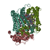
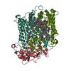
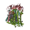
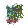
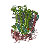
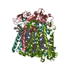
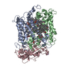
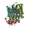
| Title | PHOTOSYNTHETIC REACTION CENTER FROM RHODOBACTER SPHAEROIDES IN THE CHARGE-SEPARATED D+QAQB-STATE WITH THE PROTON TRANSFER INHIBITOR CD2+ | ||||||
 Components Components | (PHOTOSYNTHETIC REACTION CENTER REACTION ...) x 3 | ||||||
 Keywords Keywords | PHOTOSYNTHESIS / Bacterial Photosynthesis / Rhodobacter sphaeroides / Metal Ion Binding / Cation Binding / Proton Transfer / Integral Membrane Protein | ||||||
| Function / homology |  Function and homology information Function and homology information: / : / plasma membrane-derived chromatophore membrane / plasma membrane light-harvesting complex / bacteriochlorophyll binding / photosynthetic electron transport in photosystem II / photosynthesis, light reaction / membrane => GO:0016020 / metal ion binding Similarity search - Function | ||||||
| Biological species |  Rhodobacter sphaeroides (bacteria) Rhodobacter sphaeroides (bacteria) | ||||||
| Method |  X-RAY DIFFRACTION / SYNCHROTRON / Resolution: 2.5 Å X-RAY DIFFRACTION / SYNCHROTRON / Resolution: 2.5 Å | ||||||
 Authors Authors | Axelrod, H.L. / Abresch, E.C. / Paddock, M.L. / Okamura, M.Y. / Feher, G. | ||||||
 Citation Citation | Journal: Proc.Natl.Acad.Sci.USA / Year: 2000 Title: Determination of the binding sites of the proton transfer inhibitors Cd2+ and Zn2+ in bacterial reaction centers. Authors: Axelrod, H.L. / Abresch, E.C. / Paddock, M.L. / Okamura, M.Y. / Feher, G. #1: Journal: Proc.Natl.Acad.Sci.USA / Year: 1999Title: Identification of the Proton Pathway in Bacterial Reaction Centers: Inhibition of Proton Transfer by Binding of Zn2+ or Cd2+ Authors: Paddock, M.L. / Graige, M.S. / Feher, G. / Okamura, M.Y. #2: Journal: Photosynth.Res. / Year: 1998Title: Identification of proton transfer pathways in the X-ray crystal structure of the bacterial reaction center from Rhodobacter sphaeroides Authors: Abresch, E.C. / Paddock, M.L. / Stowell, M.H.B. / McPhillips, T.M. / Axelrod, H.L. / Soltis, S.M. / Rees, D.C. / Okamura, M.Y. / Feher, G. #3: Journal: Science / Year: 1997Title: Light-Induced Structural Changes in Photosynthetic Reaction Center: Implications for Mechanism of Electron-Proton Transfer Authors: Stowell, M.H. / McPhillips, T.M. / Rees, D.C. / Soltis, S.M. / Abresch, E. / Feher, G. | ||||||
| History |
|
- Structure visualization
| Structure viewer | Molecule: MolmilJmol/JSmol |
|---|
- Downloads & links
-Download
| PDBx/mmCIF format | 1dv3.cif.gz | 365.5 KB | Display | PDBx/mmCIF format |
|---|---|---|---|---|
| PDB format | pdb1dv3.ent.gz | 294.2 KB | Display | PDB format |
| PDBx/mmJSON format | 1dv3.json.gz | Tree view | PDBx/mmJSON format | |
| Others |  Other downloads Other downloads |
-Validation report
| Arichive directory | https://data.pdbj.org/pub/pdb/validation_reports/dv/1dv3ftp://data.pdbj.org/pub/pdb/validation_reports/dv/1dv3 | HTTPS FTP |
|---|
-Related structure data


-Links
 PDBj
PDBj


- Assembly
| Deposited unit | 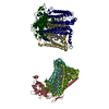
| ||||||||
|---|---|---|---|---|---|---|---|---|---|
| 1 | 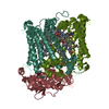
| ||||||||
| 2 | 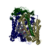
| ||||||||
| Unit cell |
| ||||||||
| Details | The biological assembly is a monomer composed of chain L, chain M, chain H, and bound cofactors |
-Components
-PHOTOSYNTHETIC REACTION CENTER REACTION ... , 3 types, 6 molecules LRMSHT
| #1: Protein | Mass: 31346.389 Da / Num. of mol.: 2 / Fragment: L CHAIN / Source method: isolated from a natural source / Source: (natural) Rhodobacter sphaeroides (bacteria) / References: UniProt: P02954, UniProt: P0C0Y8*PLUS#2: Protein | Mass: 34355.520 Da / Num. of mol.: 2 / Fragment: M CHAIN / Source method: isolated from a natural source / Source: (natural) Rhodobacter sphaeroides (bacteria) / References: UniProt: P02953, UniProt: P0C0Y9*PLUS#3: Protein | Mass: 28137.398 Da / Num. of mol.: 2 / Fragment: H CHAIN / Source method: isolated from a natural source / Source: (natural) Rhodobacter sphaeroides (bacteria) / References: UniProt: P11846, UniProt: P0C0Y7*PLUS |
|---|
-Non-polymers , 8 types, 430 molecules 


| #4: Chemical | ChemComp-BCL /  Mass: 911.504 Da / Num. of mol.: 8 / Source method: obtained synthetically / Formula: C55H74MgN4O6 Mass: 911.504 Da / Num. of mol.: 8 / Source method: obtained synthetically / Formula: C55H74MgN4O6#5: Chemical | ChemComp-BPH /  Mass: 889.215 Da / Num. of mol.: 4 / Source method: obtained synthetically / Formula: C55H76N4O6 Mass: 889.215 Da / Num. of mol.: 4 / Source method: obtained synthetically / Formula: C55H76N4O6#6: Chemical | ChemComp-U10 /  Mass: 863.343 Da / Num. of mol.: 4 / Source method: obtained synthetically / Formula: C59H90O4 Mass: 863.343 Da / Num. of mol.: 4 / Source method: obtained synthetically / Formula: C59H90O4#7: Chemical |  Mass: 55.845 Da / Num. of mol.: 2 / Source method: obtained synthetically / Formula: Fe Mass: 55.845 Da / Num. of mol.: 2 / Source method: obtained synthetically / Formula: Fe#8: Chemical |  Mass: 35.453 Da / Num. of mol.: 2 / Source method: obtained synthetically / Formula: Cl Mass: 35.453 Da / Num. of mol.: 2 / Source method: obtained synthetically / Formula: Cl#9: Chemical |  Mass: 229.402 Da / Num. of mol.: 2 / Source method: obtained synthetically / Formula: C14H31NO / Comment: LDAO, detergent*YM Mass: 229.402 Da / Num. of mol.: 2 / Source method: obtained synthetically / Formula: C14H31NO / Comment: LDAO, detergent*YM#10: Chemical |  Mass: 112.411 Da / Num. of mol.: 2 / Source method: obtained synthetically / Formula: Cd Mass: 112.411 Da / Num. of mol.: 2 / Source method: obtained synthetically / Formula: Cd#11: Water | ChemComp-HOH / | Mass: 18.015 Da / Num. of mol.: 406 / Source method: isolated from a natural source / Formula: H2O |
|---|
-Experimental details
-Experiment
| Experiment | Method: X-RAY DIFFRACTION / Number of used crystals: 1 |
|---|
- Sample preparation
| Crystal | Density Matthews: 3.66 Å3/Da / Density % sol: 66.36 % | |||||||||||||||||||||||||||||||||||
|---|---|---|---|---|---|---|---|---|---|---|---|---|---|---|---|---|---|---|---|---|---|---|---|---|---|---|---|---|---|---|---|---|---|---|---|---|
| Crystal grow | Temperature: 292 K / Method: vapor diffusion, sitting drop / pH: 8.5 Details: PEG 4000, heptanetriol, TRIS-HCL, LDAO detergent, sodium chloride, pH 8.5, VAPOR DIFFUSION, SITTING DROP, temperature 292K | |||||||||||||||||||||||||||||||||||
| Crystal grow | *PLUS Temperature: 25 ℃ / pH: 8 | |||||||||||||||||||||||||||||||||||
| Components of the solutions | *PLUS
|
-Data collection
| Diffraction | Mean temperature: 90 K |
|---|---|
| Diffraction source | Source: SYNCHROTRON / Site: SSRL  / Beamline: BL9-1 / Wavelength: 0.98 / Beamline: BL9-1 / Wavelength: 0.98 |
| Detector | Type: MARRESEARCH / Detector: IMAGE PLATE / Date: May 5, 1999 |
| Radiation | Protocol: SINGLE WAVELENGTH / Monochromatic (M) / Laue (L): M / Scattering type: x-ray |
| Radiation wavelength | Wavelength: 0.98 Å / Relative weight: 1 |
| Reflection | Resolution: 2.49→27.8 Å / Num. all: 94618 / Num. obs: 91189 / % possible obs: 94.5 % / Observed criterion σ(F): 0 / Observed criterion σ(I): 0 / Redundancy: 3.8 % / Biso Wilson estimate: 49.14 Å2 / Rmerge(I) obs: 0.086 / Net I/σ(I): 7.2 |
| Reflection shell | Resolution: 2.49→2.56 Å / Redundancy: 3.7 % / Rmerge(I) obs: 0.33 / Num. unique all: 5527 / % possible all: 78.3 |
| Reflection | *PLUS Num. obs: 12363 / Num. measured all: 91189 |
| Reflection shell | *PLUS % possible obs: 78.3 % |
- Processing
| Software |
| |||||||||||||||||||||||||
|---|---|---|---|---|---|---|---|---|---|---|---|---|---|---|---|---|---|---|---|---|---|---|---|---|---|---|
| Refinement | Resolution: 2.5→27.8 Å / σ(F): 0 / σ(I): 0 / Stereochemistry target values: Engh and Huber Details: Bulk solvent correction applied during the refinement. Refinement was carried out with a maximum likelihood target function and non-crystallographic symmetry restraints.
| |||||||||||||||||||||||||
| Refinement step | Cycle: LAST / Resolution: 2.5→27.8 Å
| |||||||||||||||||||||||||
| Refine LS restraints |
| |||||||||||||||||||||||||
| Software | *PLUS Name: CNS / Version: 0.9 / Classification: refinement | |||||||||||||||||||||||||
| Refine LS restraints | *PLUS
|
