ジャーナル: Science / 年: 2024 タイトル: Molecular mechanism of actin filament elongation by formins. 著者: Wout Oosterheert / Micaela Boiero Sanders / Johanna Funk / Daniel Prumbaum / Stefan Raunser / Peter Bieling / 要旨: Formins control the assembly of actin filaments (F-actin) that drive cell morphogenesis and motility in eukaryotes. However, their molecular interaction with F-actin and their mechanism of action ...Formins control the assembly of actin filaments (F-actin) that drive cell morphogenesis and motility in eukaryotes. However, their molecular interaction with F-actin and their mechanism of action remain unclear. In this work, we present high-resolution cryo-electron microscopy structures of F-actin barbed ends bound by three distinct formins, revealing a common asymmetric formin conformation imposed by the filament. Formation of new intersubunit contacts during actin polymerization sterically displaces formin and triggers its translocation. This "undock-and-lock" mechanism explains how actin-filament growth is coordinated with formin movement. Filament elongation speeds are controlled by the positioning and stability of actin-formin interfaces, which distinguish fast and slow formins. Furthermore, we provide a structure of the actin-formin-profilin ring complex, which resolves how profilin is rapidly released from the barbed end during filament elongation.
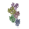
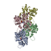
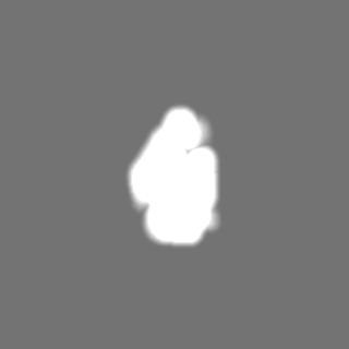
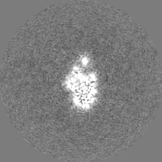
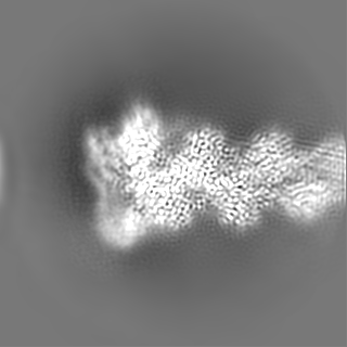
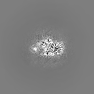
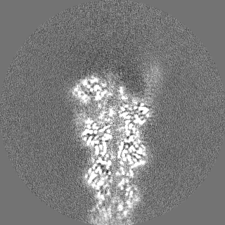
Unsharpened cryo-EM density map of actin-Cdc12. This reconstruction was computed with more particles, but displays weaker density for the FH2L domain of Cdc12 due to flexibility.
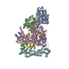
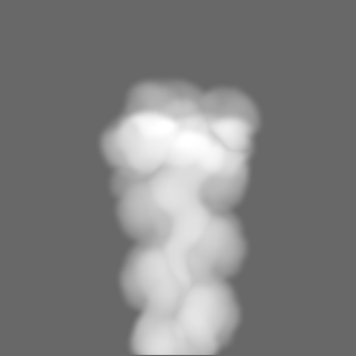
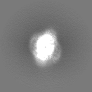
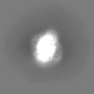
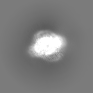
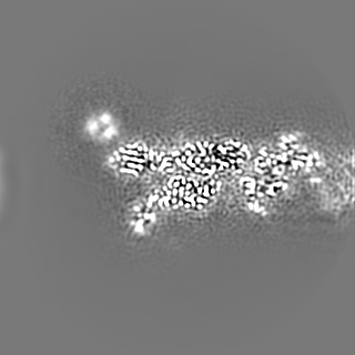
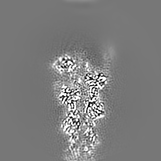
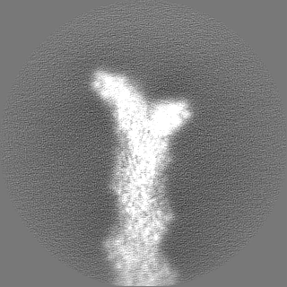
Composite map of two reconstructions of the formin Cdc12 bound to the barbed end of phalloidin-stabilized F-actin. This map was created using phenix and was used for visualization purposes.
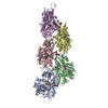
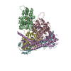
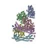
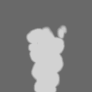
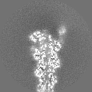
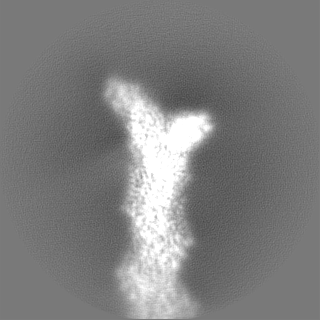
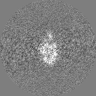
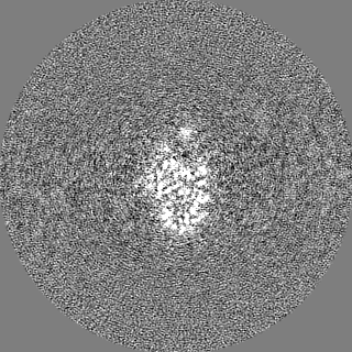
Unfiltered half map 1 of actin-Cdc12. This reconstruction was computed with more particles, but displays weaker density for the FH2L domain of Cdc12 due to flexibility.
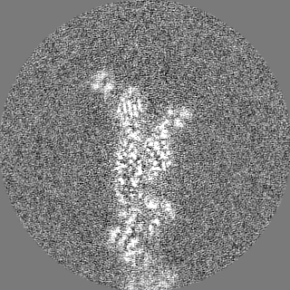
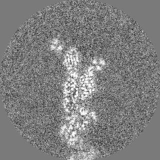
Unfiltered half map 2 of actin-Cdc12. This reconstruction was computed with more particles, but displays weaker density for the FH2L domain of Cdc12 due to flexibility.
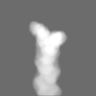
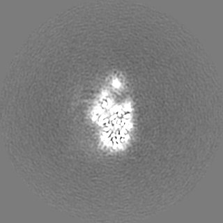
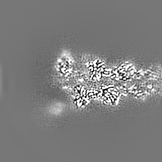
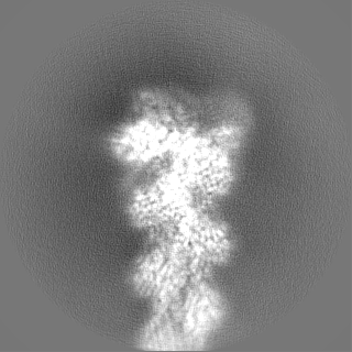
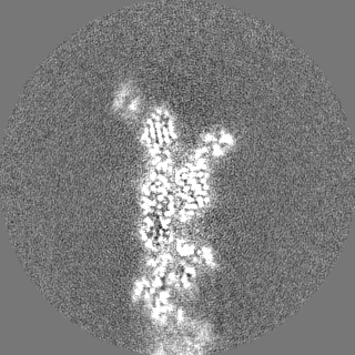
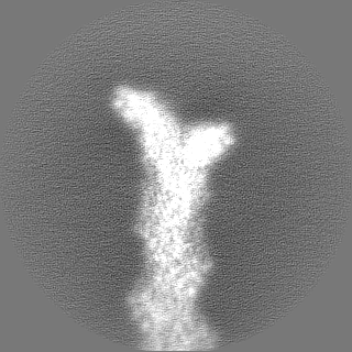
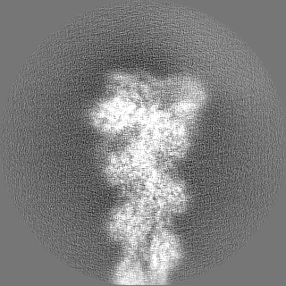
Sharpened density map of actin-Cdc12. This reconstruction was computed with more particles, but displays weaker density for the FH2L domain of Cdc12 due to flexibility.
超分子 #1: Complex of the dimeric FH2 domain of S. Pombe Cdc12 bound to the ...
超分子
名称: Complex of the dimeric FH2 domain of S. Pombe Cdc12 bound to the barbed end of phalloidin stabilized F-actin. タイプ: complex / ID: 1 / 親要素: 0 / 含まれる分子: #1-#3 詳細: Human beta-actin and S. Pombe Cdc12 were purified separately, phalloidin (from Amanita phalloides) was bought from sigma. All components were mixed to assemble the complex prior to cryo-EM grid preparation.
名称: Cell division control protein 12 / タイプ: protein_or_peptide / ID: 2 詳細: FH2 domain of S. pombe Cdc12, purified from E. coli cells. コピー数: 2 / 光学異性体: LEVO
 ムービー
ムービー コントローラー


 データを開く
 基本情報
基本情報
 マップデータ
 試料
試料 キーワード
キーワード 機能・相同性情報
 Homo sapiens (ヒト) /
 Amanita phalloides (タマゴテングタケ)
 データ登録者
 ドイツ, European Union, 3件
  引用
引用 構造の表示
構造の表示
 ダウンロードとリンク
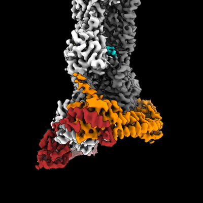 emd_19496.png
 http://ftp.pdbj.org/pub/emdb/structures/EMD-19496
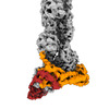


 Z
Z Y
Y X
X


 試料の構成要素
 Trichoplusia ni (イラクサキンウワバ)


 解析
解析 電子顕微鏡法
 FIELD EMISSION GUN