Movie
Movie Controller


+ Open data

- Basic information
| Entry | 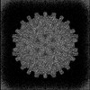 | ||||||||||||||||||
|---|---|---|---|---|---|---|---|---|---|---|---|---|---|---|---|---|---|---|---|
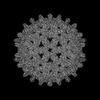
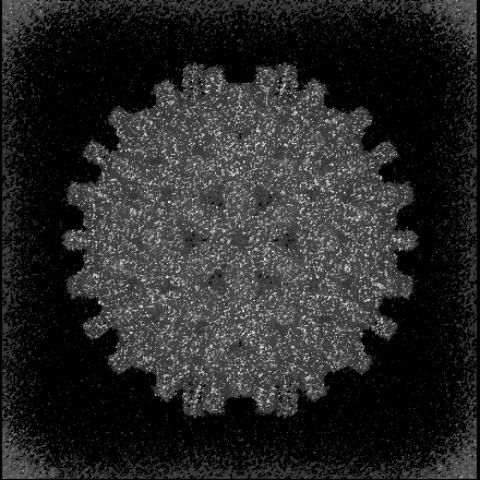
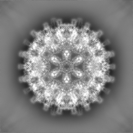
| Title | Hepatitis B core protein with bound SLLGRM-dimer | ||||||||||||||||||
 Map data Map data | |||||||||||||||||||
 Sample Sample |
| ||||||||||||||||||
 Keywords Keywords | Hepatitis B core protein / Spikes SLLGRM-dimer aggregator Hepatitis B virus Capsid LIKE PARTICLE / VIRUS LIKE PARTICLE | ||||||||||||||||||
| Function / homology | Hepatitis B virus, capsid N-terminal / Hepatitis core protein, putative zinc finger / Hepatitis core antigen / Viral capsid core domain supefamily, Hepatitis B virus / Hepatitis core antigen / virus-mediated perturbation of host defense response / structural molecule activity / extracellular region / External core antigen Function and homology information Function and homology information | ||||||||||||||||||
| Biological species |   Hepatitis B virus / synthetic construct (others) Hepatitis B virus / synthetic construct (others) | ||||||||||||||||||
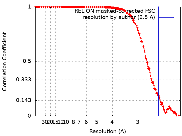
| Method | single particle reconstruction / cryo EM / Resolution: 2.5 Å | ||||||||||||||||||
 Authors Authors | Makbul C / Khayenko V / Maric MH / Bottcher B | ||||||||||||||||||
| Funding support |  Germany, 5 items Germany, 5 items
| ||||||||||||||||||
 Citation Citation | Journal: To Be Published Title: Peptide dimers aggregate Hepatitis B core proteins in live cells Authors: Khayenko V / Makbul C / Schulte C / Hemmelmann N / Bottcher B / Maric HM | ||||||||||||||||||
| History |
|
- Structure visualization
| Supplemental images |
|---|
- Downloads & links
-EMDB archive
| Map data | emd_18001.map.gz | 301.3 MB | EMDB map data format | |
|---|---|---|---|---|
| Header (meta data) | emd-18001-v30.xmlemd-18001.xml | 19.4 KB 19.4 KB | Display Display | EMDB header |
| FSC (resolution estimation) | emd_18001_fsc.xml | 15.4 KB | Display | FSC data file |
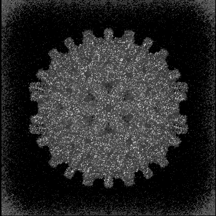
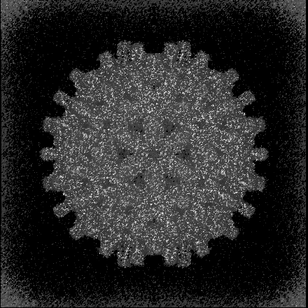
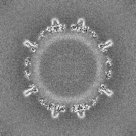
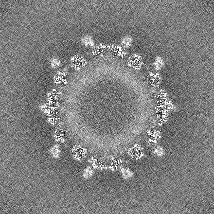
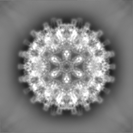
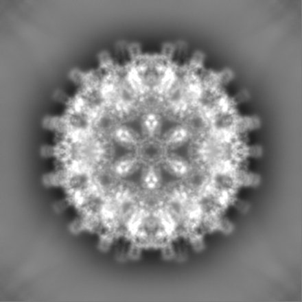
| Images |  emd_18001.png emd_18001.png | 259.1 KB | ||
| Masks | emd_18001_msk_1.map | 325 MB | Mask map | |
| Filedesc metadata | emd-18001.cif.gz | 6.2 KB | ||
| Others | emd_18001_half_map_1.map.gzemd_18001_half_map_2.map.gz | 300 MB 300 MB | ||
| Archive directory |  http://ftp.pdbj.org/pub/emdb/structures/EMD-18001ftp://ftp.pdbj.org/pub/emdb/structures/EMD-18001 http://ftp.pdbj.org/pub/emdb/structures/EMD-18001ftp://ftp.pdbj.org/pub/emdb/structures/EMD-18001 | HTTPS FTP |
-Validation report
| Summary document | emd_18001_validation.pdf.gz | 1.2 MB | Display | EMDB validaton report |
|---|---|---|---|---|
| Full document | emd_18001_full_validation.pdf.gz | 1.2 MB | Display | |
| Data in XML | emd_18001_validation.xml.gz | 23.5 KB | Display | |
| Data in CIF | emd_18001_validation.cif.gz | 31.1 KB | Display | |
| Arichive directory | https://ftp.pdbj.org/pub/emdb/validation_reports/EMD-18001ftp://ftp.pdbj.org/pub/emdb/validation_reports/EMD-18001 | HTTPS FTP |
-Related structure data
| Related structure data | 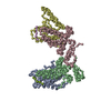 8px6MC 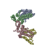 8pwoC 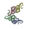 8px3C C: citing same article ( M: atomic model generated by this map |
|---|---|
| Similar structure data |

-Links
| EMDB pages | EMDB (EBI/PDBe) / EMDataResource |
|---|---|
| Related items in Molecule of the Month |

-Map
| File | Download / File: emd_18001.map.gz / Format: CCP4 / Size: 325 MB / Type: IMAGE STORED AS FLOATING POINT NUMBER (4 BYTES) | ||||||||||||||||||||||||||||||||||||
|---|---|---|---|---|---|---|---|---|---|---|---|---|---|---|---|---|---|---|---|---|---|---|---|---|---|---|---|---|---|---|---|---|---|---|---|---|---|
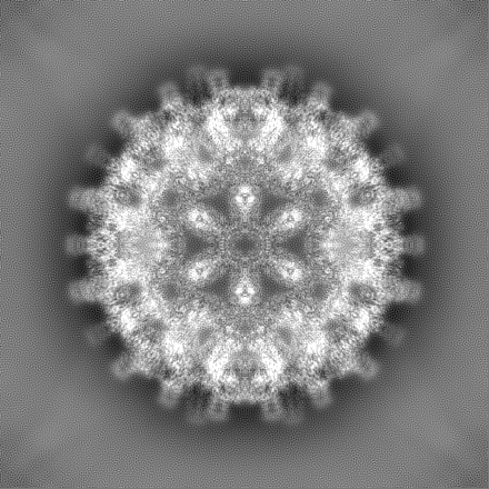
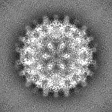
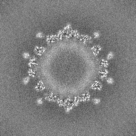
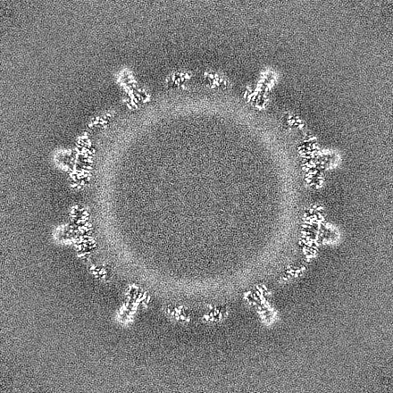
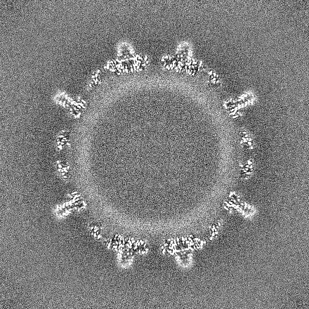
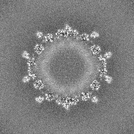
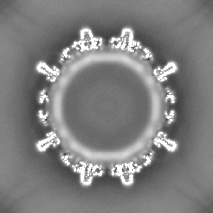
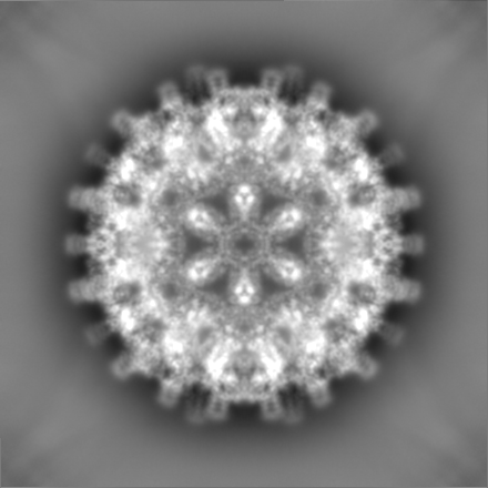
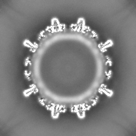
| Projections & slices | Image control
Images are generated by Spider. | ||||||||||||||||||||||||||||||||||||
| Voxel size | X=Y=Z: 1.0635 Å | ||||||||||||||||||||||||||||||||||||
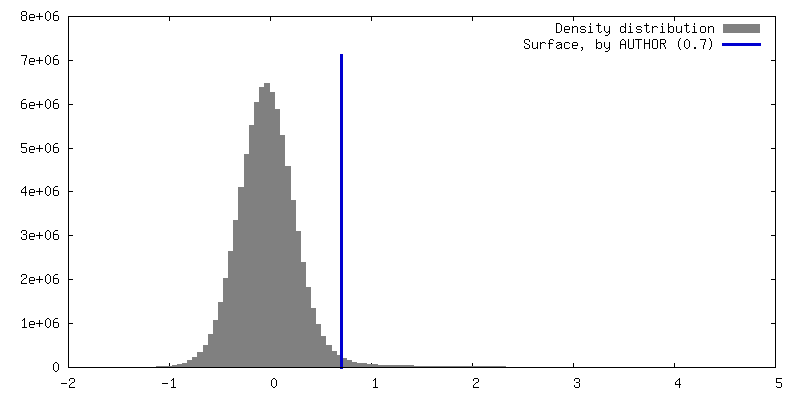
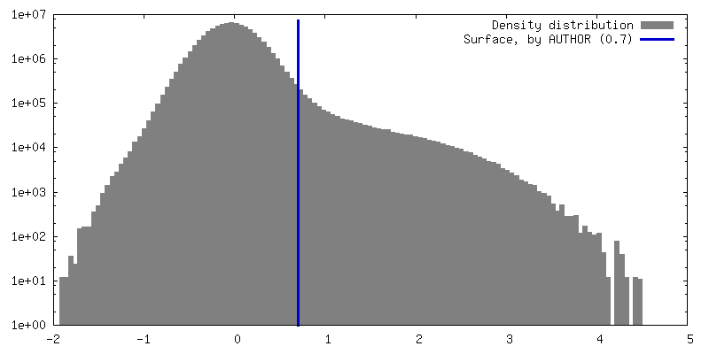
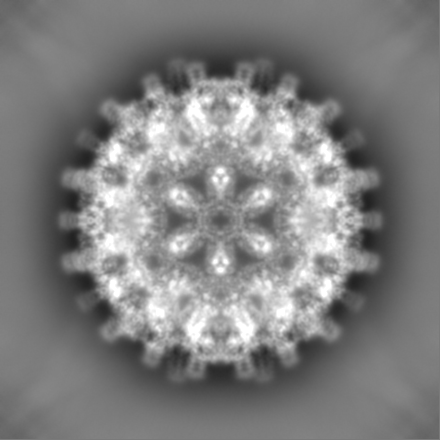
| Density |
| ||||||||||||||||||||||||||||||||||||
| Symmetry | Space group: 1 | ||||||||||||||||||||||||||||||||||||
| Details | EMDB XML:
|
 Z (Sec.)
 Y (Row.)
 X (Col.)


-Supplemental data
-Mask #1
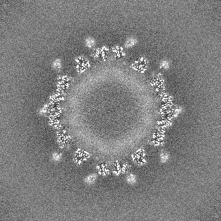
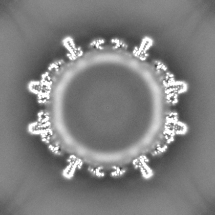
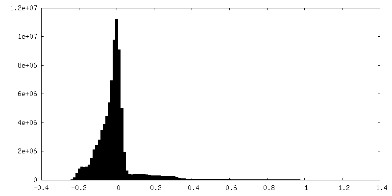
| File | emd_18001_msk_1.map | ||||||||||||
|---|---|---|---|---|---|---|---|---|---|---|---|---|---|
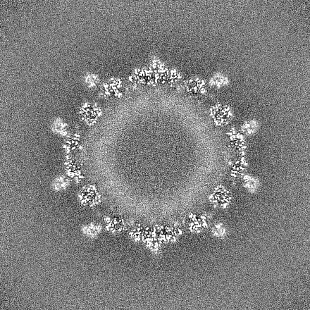
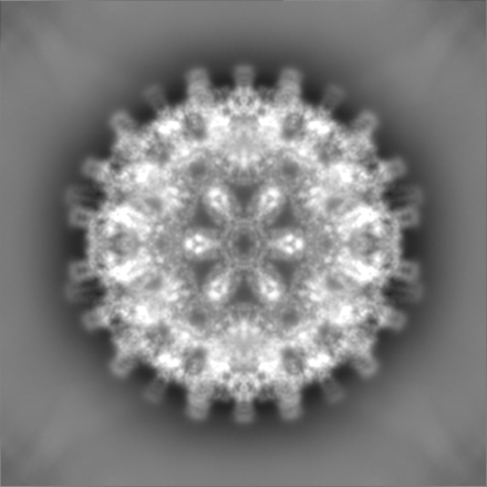
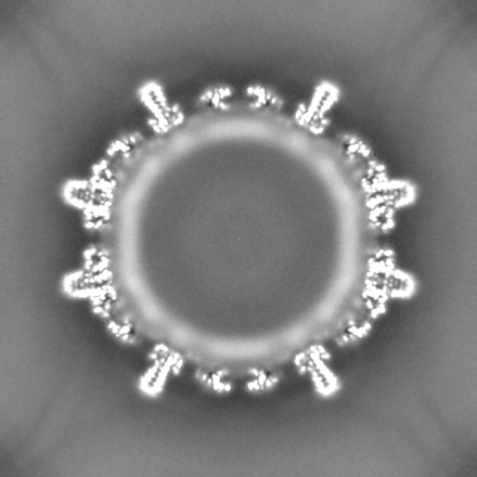
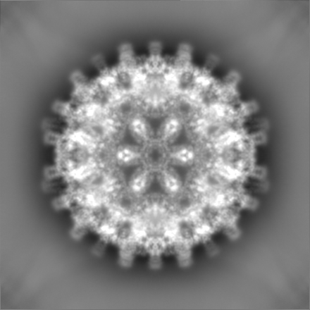
| Projections & Slices |
| ||||||||||||
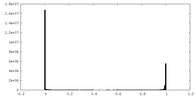
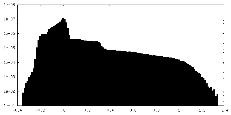
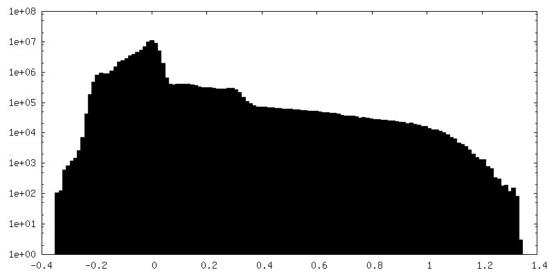
| Density Histograms |


-Half map: #1
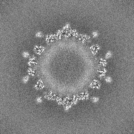
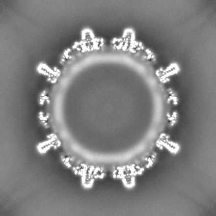
| File | emd_18001_half_map_1.map | ||||||||||||
|---|---|---|---|---|---|---|---|---|---|---|---|---|---|
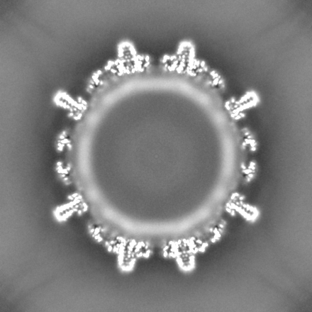
| Projections & Slices |
| ||||||||||||
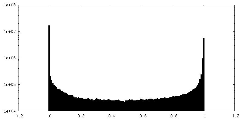
| Density Histograms |

-Half map: #2
| File | emd_18001_half_map_2.map | ||||||||||||
|---|---|---|---|---|---|---|---|---|---|---|---|---|---|
| Projections & Slices |
| ||||||||||||
| Density Histograms |
- Sample components
-Entire : Hepatitis B virus
| Entire | Name: Hepatitis B virus |
|---|---|
| Components |
|
-Supramolecule #1: Hepatitis B virus
| Supramolecule | Name: Hepatitis B virus / type: virus / ID: 1 / Parent: 0 / Macromolecule list: all / NCBI-ID: 10407 / Sci species name: Hepatitis B virus / Sci species strain: ayw/France/Tiollais/1979 / Virus type: VIRUS-LIKE PARTICLE / Virus isolate: OTHER / Virus enveloped: No / Virus empty: No |
|---|---|
| Host (natural) | Organism:  Homo sapiens (human) Homo sapiens (human) |
| Molecular weight | Theoretical: 5 MDa |
| Virus shell | Shell ID: 1 / Diameter: 360.0 Å / T number (triangulation number): 4 |
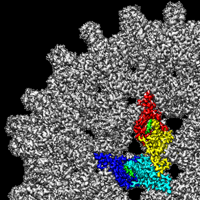
-Macromolecule #1: External core antigen
| Macromolecule | Name: External core antigen / type: protein_or_peptide / ID: 1 / Number of copies: 4 / Enantiomer: LEVO |
|---|---|
| Source (natural) | Organism: Hepatitis B virus |
| Molecular weight | Theoretical: 21.146217 KDa |
| Recombinant expression | Organism:  |
| Sequence | String: MDIDPYKEFG ATVELLSFLP SDFFPSVRDL LDTASALYRE ALESPEHCSP HHTALRQAIL CWGELMTLAT WVGVNLEDPA SRDLVVSYV NTNMGLKFRQ LLWFHISCLT FGRETVIEYL VSFGVWIRTP PAYRPPNAPI LSTLPETTVV RRRGRSPRRR T PSPRRRRS QSPRRRRSQS RESQC UniProtKB: External core antigen |
-Macromolecule #2: SLLGRM-dimer
| Macromolecule | Name: SLLGRM-dimer / type: protein_or_peptide / ID: 2 Details: This is a synthetic peptide with two moieties of SLLGRM that are linked by a PEG linker The density that is assigned to SLLGRM is modelled as Poly-Ala. Number of copies: 2 / Enantiomer: LEVO |
|---|---|
| Source (natural) | Organism: synthetic construct (others) |
| Molecular weight | Theoretical: 676.85 Da |
| Sequence | String: SLLGRM |
-Experimental details
-Structure determination
| Method | cryo EM |
|---|---|
 Processing Processing | single particle reconstruction |
| Aggregation state | particle |
-Sample preparation
| Concentration | 4 mg/mL |
|---|---|
| Buffer | pH: 7.5 |
| Grid | Model: Quantifoil R1.2/1.3 / Material: COPPER / Mesh: 300 / Support film - Material: CARBON / Support film - topology: HOLEY ARRAY |
| Vitrification | Cryogen name: ETHANE / Chamber humidity: 100 % / Chamber temperature: 277 K / Instrument: FEI VITROBOT MARK IV |
- Electron microscopy
| Microscope | FEI TITAN KRIOS |
|---|---|
| Image recording | Film or detector model: FEI FALCON III (4k x 4k) / Detector mode: INTEGRATING / Digitization - Dimensions - Width: 4096 pixel / Digitization - Dimensions - Height: 4096 pixel / Number grids imaged: 1 / Number real images: 4956 / Average exposure time: 2.6 sec. / Average electron dose: 40.0 e/Å2 |
| Electron beam | Acceleration voltage: 300 kV / Electron source:  FIELD EMISSION GUN FIELD EMISSION GUN |
| Electron optics | C2 aperture diameter: 70.0 µm / Illumination mode: FLOOD BEAM / Imaging mode: BRIGHT FIELD / Cs: 2.7 mm / Nominal defocus max: 1.4000000000000001 µm / Nominal defocus min: 0.6 µm / Nominal magnification: 75000 |
| Sample stage | Specimen holder model: FEI TITAN KRIOS AUTOGRID HOLDER / Cooling holder cryogen: NITROGEN |
| Experimental equipment |  Model: Titan Krios / Image courtesy: FEI Company |