 Movie
Movie Controller


+ Open data

- Basic information
| Entry | Database: PDB / ID: 8pwo | ||||||||||||||||||
|---|---|---|---|---|---|---|---|---|---|---|---|---|---|---|---|---|---|---|---|
| Title | Hepatitis B core protein with bound Geraniol | ||||||||||||||||||
 Components Components | Capsid protein | ||||||||||||||||||
 Keywords Keywords | VIRUS LIKE PARTICLE / geraniol Hepatitis B core protein hyfrophobic pocket pocket factor Hepatitis B virus | ||||||||||||||||||
| Function / homology |  Function and homology information Function and homology informationmicrotubule-dependent intracellular transport of viral material towards nucleus / T=4 icosahedral viral capsid / viral penetration into host nucleus / host cell / host cell cytoplasm / symbiont entry into host cell / structural molecule activity / DNA binding / RNA binding Similarity search - Function | ||||||||||||||||||
| Biological species |  Hepatitis B virus ayw/France/Tiollais/1979 Hepatitis B virus ayw/France/Tiollais/1979 | ||||||||||||||||||
| Method | ELECTRON MICROSCOPY / single particle reconstruction / cryo EM / Resolution: 2.8 Å | ||||||||||||||||||
 Authors Authors | Makbul, C. / Khayenko, V. / Maric, M.H. / Bottcher, B. | ||||||||||||||||||
| Funding support |  Germany, 5items Germany, 5items
| ||||||||||||||||||
 Citation Citation | Journal: Elife / Year: 2025 Title: Induction of hepatitis B core protein aggregation targeting an unconventional binding site Authors: Khayenko, V. / Makbul, C. / Schulte, C. / Hemmelmann, N. / Kachler, S. / Bottcher, B. / Maric, H.M. / Comas-Garcia, M. / Dotsch, V. | ||||||||||||||||||
| History |
|
- Structure visualization
| Structure viewer | Molecule: MolmilJmol/JSmol |
|---|
- Downloads & links
-Download
| PDBx/mmCIF format | 8pwo.cif.gz | 195.3 KB | Display | PDBx/mmCIF format |
|---|---|---|---|---|
| PDB format | pdb8pwo.ent.gz | 159.5 KB | Display | PDB format |
| PDBx/mmJSON format | 8pwo.json.gz | Tree view | PDBx/mmJSON format | |
| Others |  Other downloads Other downloads |
-Validation report
| Arichive directory | https://data.pdbj.org/pub/pdb/validation_reports/pw/8pwoftp://data.pdbj.org/pub/pdb/validation_reports/pw/8pwo | HTTPS FTP |
|---|
-Related structure data
| Related structure data | 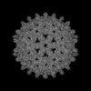 17996MC 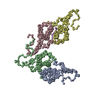 8px3C 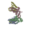 8px6C M: map data used to model this data C: citing same article ( |
|---|---|
| Similar structure data |

-Links
 PDBj
PDBj

- Assembly
| Deposited unit | 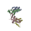
|
|---|---|
| 1 | x 60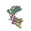
|
-Components
| #1: Protein | Mass: 21146.217 Da / Num. of mol.: 4 Source method: isolated from a genetically manipulated source Details: capsid has bound geraniol Source: (gene. exp.) Hepatitis B virus ayw/France/Tiollais/1979Production host:  #2: Chemical | ChemComp-64Z /   Mass: 154.249 Da / Num. of mol.: 4 / Source method: obtained synthetically / Formula: C10H18O / Feature type: SUBJECT OF INVESTIGATION Mass: 154.249 Da / Num. of mol.: 4 / Source method: obtained synthetically / Formula: C10H18O / Feature type: SUBJECT OF INVESTIGATIONHas ligand of interest | Y | Has protein modification | N | |
|---|
-Experimental details
-Experiment
| Experiment | Method: ELECTRON MICROSCOPY |
|---|---|
| EM experiment | Aggregation state: PARTICLE / 3D reconstruction method: single particle reconstruction |
- Sample preparation
| Component | Name: Hepatitis B virus / Type: VIRUS / Entity ID: #1 / Source: RECOMBINANT |
|---|---|
| Molecular weight | Value: 5 MDa / Experimental value: NO |
| Source (natural) | Organism:  Hepatitis B virus / Strain: ayw/France/Tiollais/1979 Hepatitis B virus / Strain: ayw/France/Tiollais/1979 |
| Source (recombinant) | Organism: |
| Details of virus | Empty: NO / Enveloped: NO / Isolate: OTHER / Type: VIRUS-LIKE PARTICLE |
| Natural host | Organism: Homo sapiens |
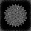
| Virus shell | Diameter: 360 nm / Triangulation number (T number): 4 |
| Buffer solution | pH: 7.5 |
| Specimen | Conc.: 4 mg/ml / Embedding applied: NO / Shadowing applied: NO / Staining applied: NO / Vitrification applied: YES |
| Specimen support | Details: plasma cleaner (model PDC-002. Harrick Plasma, Ithaca, NY, USA); at medium power of the instrument Grid material: COPPER / Grid mesh size: 300 divisions/in. / Grid type: Quantifoil R1.2/1.3 |
| Vitrification | Instrument: FEI VITROBOT MARK IV / Cryogen name: ETHANE / Humidity: 100 % / Chamber temperature: 277 K |
- Electron microscopy imaging
| Experimental equipment |  Model: Titan Krios / Image courtesy: FEI Company |
|---|---|
| Microscopy | Model: FEI TITAN KRIOS |
| Electron gun | Electron source:  FIELD EMISSION GUN / Accelerating voltage: 300 kV / Illumination mode: FLOOD BEAM FIELD EMISSION GUN / Accelerating voltage: 300 kV / Illumination mode: FLOOD BEAM |
| Electron lens | Mode: BRIGHT FIELD / Nominal magnification: 75000 X / Nominal defocus max: 1200 nm / Nominal defocus min: 600 nm / Cs: 2.7 mm / C2 aperture diameter: 70 µm / Alignment procedure: COMA FREE |
| Specimen holder | Cryogen: NITROGEN / Specimen holder model: FEI TITAN KRIOS AUTOGRID HOLDER |
| Image recording | Average exposure time: 3 sec. / Electron dose: 40 e/Å2 / Detector mode: INTEGRATING / Film or detector model: FEI FALCON III (4k x 4k) / Num. of grids imaged: 1 / Num. of real images: 4875 |
| Image scans | Width: 4096 / Height: 4096 |
- Processing
| EM software |
| |||||||||||||||||||||||||||||||||||||||||||||
|---|---|---|---|---|---|---|---|---|---|---|---|---|---|---|---|---|---|---|---|---|---|---|---|---|---|---|---|---|---|---|---|---|---|---|---|---|---|---|---|---|---|---|---|---|---|---|
| CTF correction | Type: PHASE FLIPPING AND AMPLITUDE CORRECTION | |||||||||||||||||||||||||||||||||||||||||||||
| Particle selection | Num. of particles selected: 331635 / Details: template picked | |||||||||||||||||||||||||||||||||||||||||||||
| Symmetry | Point symmetry: I (icosahedral) | |||||||||||||||||||||||||||||||||||||||||||||
| 3D reconstruction | Resolution: 2.8 Å / Resolution method: FSC 0.143 CUT-OFF / Num. of particles: 229646 / Algorithm: FOURIER SPACE / Symmetry type: POINT | |||||||||||||||||||||||||||||||||||||||||||||
| Atomic model building |
| |||||||||||||||||||||||||||||||||||||||||||||
| Refine LS restraints |
|
