 Movie
Movie Controller

[English] 日本語
 Yorodumi
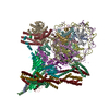
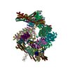
- EMDB-17368: Multibody map of the CENP-A nucleosome as part of the inner kinet... -
+ Open data

- Basic information
| Entry |  | |||||||||||||||
|---|---|---|---|---|---|---|---|---|---|---|---|---|---|---|---|---|
| Title | Multibody map of the CENP-A nucleosome as part of the inner kinetochore | |||||||||||||||
 Map data Map data | ||||||||||||||||
 Sample Sample |
| |||||||||||||||
 Keywords Keywords | kinetochore / point centromere / CENP-A nucleosome / topological entrapment / centromeric DNA / CELL CYCLE | |||||||||||||||
| Biological species |  | |||||||||||||||
| Method | single particle reconstruction / cryo EM / Resolution: 3.7 Å | |||||||||||||||
 Authors Authors | Dendooven TD / Zhang Z / Yang J / McLaughlin S / Schwabb J / Scheres S / Yatskevich S / Barford D | |||||||||||||||
| Funding support |  United Kingdom, United Kingdom,  Germany, 4 items Germany, 4 items
| |||||||||||||||
 Citation Citation | Journal: Sci Adv / Year: 2023 Title: Cryo-EM structure of the complete inner kinetochore of the budding yeast point centromere. Authors: Tom Dendooven / Ziguo Zhang / Jing Yang / Stephen H McLaughlin / Johannes Schwab / Sjors H W Scheres / Stanislau Yatskevich / David Barford / Abstract: The point centromere of budding yeast specifies assembly of the large kinetochore complex to mediate chromatid segregation. Kinetochores comprise the centromere-associated inner kinetochore (CCAN) ...The point centromere of budding yeast specifies assembly of the large kinetochore complex to mediate chromatid segregation. Kinetochores comprise the centromere-associated inner kinetochore (CCAN) complex and the microtubule-binding outer kinetochore KNL1-MIS12-NDC80 (KMN) network. The budding yeast inner kinetochore also contains the DNA binding centromere-binding factor 1 (CBF1) and CBF3 complexes. We determined the cryo-electron microscopy structure of the yeast inner kinetochore assembled onto the centromere-specific centromere protein A nucleosomes (CENP-A). This revealed a central CENP-A with extensively unwrapped DNA ends. These free DNA duplexes bind two CCAN protomers, one of which entraps DNA topologically, positioned on the centromere DNA element I (CDEI) motif by CBF1. The two CCAN protomers are linked through CBF3 forming an arch-like configuration. With a structural mechanism for how CENP-A can also be linked to KMN involving only CENP-QU, we present a model for inner kinetochore assembly onto a point centromere and how it organizes the outer kinetochore for chromosome attachment to the mitotic spindle. | |||||||||||||||
| History |
|
- Structure visualization
| Supplemental images |
|---|

- Downloads & links
-EMDB archive
| Map data | emd_17368.map.gz | 155.9 MB |  EMDB map data format EMDB map data format | |
|---|---|---|---|---|
| Header (meta data) | emd-17368-v30.xmlemd-17368.xml | 15.7 KB 15.7 KB | Display Display | EMDB header |
| Images | 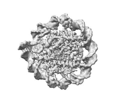 emd_17368.png emd_17368.png | 67.6 KB | ||
| Others | emd_17368_half_map_1.map.gzemd_17368_half_map_2.map.gz | 115.6 MB 115.7 MB | ||
| Archive directory |  http://ftp.pdbj.org/pub/emdb/structures/EMD-17368ftp://ftp.pdbj.org/pub/emdb/structures/EMD-17368 http://ftp.pdbj.org/pub/emdb/structures/EMD-17368ftp://ftp.pdbj.org/pub/emdb/structures/EMD-17368 | HTTPS FTP |
-Validation report
| Summary document | emd_17368_validation.pdf.gz | 967.6 KB | Display | EMDB validaton report |
|---|---|---|---|---|
| Full document | emd_17368_full_validation.pdf.gz | 967.1 KB | Display | |
| Data in XML | emd_17368_validation.xml.gz | 14.4 KB | Display | |
| Data in CIF | emd_17368_validation.cif.gz | 17 KB | Display | |
| Arichive directory | https://ftp.pdbj.org/pub/emdb/validation_reports/EMD-17368ftp://ftp.pdbj.org/pub/emdb/validation_reports/EMD-17368 | HTTPS FTP |
-Related structure data


-Links
| EMDB pages | EMDB (EBI/PDBe) / EMDataResource |
|---|
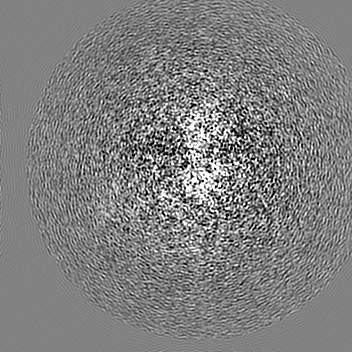
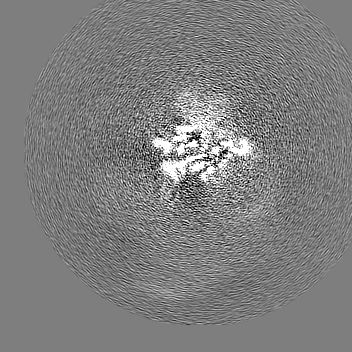
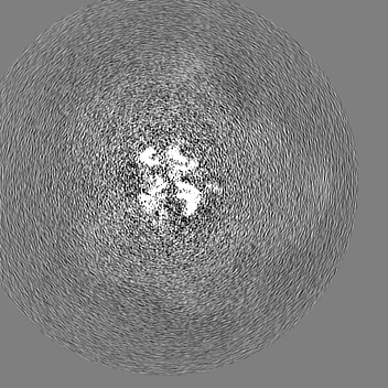
-Map
| File | Download / File: emd_17368.map.gz / Format: CCP4 / Size: 166.4 MB / Type: IMAGE STORED AS FLOATING POINT NUMBER (4 BYTES) | ||||||||||||||||||||||||||||||||||||
|---|---|---|---|---|---|---|---|---|---|---|---|---|---|---|---|---|---|---|---|---|---|---|---|---|---|---|---|---|---|---|---|---|---|---|---|---|---|
| Projections & slices | Image control
Images are generated by Spider. | ||||||||||||||||||||||||||||||||||||
| Voxel size | X=Y=Z: 1.08 Å | ||||||||||||||||||||||||||||||||||||
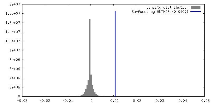
| Density |
| ||||||||||||||||||||||||||||||||||||
| Symmetry | Space group: 1 | ||||||||||||||||||||||||||||||||||||
| Details | EMDB XML:
|
 Z (Sec.)
 Y (Row.)
 X (Col.)


-Supplemental data
-Half map: #1
| File | emd_17368_half_map_1.map | ||||||||||||
|---|---|---|---|---|---|---|---|---|---|---|---|---|---|
| Projections & Slices |
| ||||||||||||
| Density Histograms |


-Half map: #2
| File | emd_17368_half_map_2.map | ||||||||||||
|---|---|---|---|---|---|---|---|---|---|---|---|---|---|
| Projections & Slices |
| ||||||||||||
| Density Histograms |


- Sample components
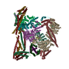
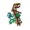
-Entire : A complex of CBF1-CCAN bound to centromeric C0N3 DNA
| Entire | Name: A complex of CBF1-CCAN bound to centromeric C0N3 DNA |
|---|---|
| Components |
|
-Supramolecule #1: A complex of CBF1-CCAN bound to centromeric C0N3 DNA
| Supramolecule | Name: A complex of CBF1-CCAN bound to centromeric C0N3 DNA / type: complex / ID: 1 / Parent: 0 Macromolecule list: #1-#5, #7, #9, #8, #10-#11, #6, #13-#15, #12, #16-#23 |
|---|---|
| Source (natural) | Organism: |
-Experimental details
-Structure determination
| Method | cryo EM |
|---|---|
 Processing Processing | single particle reconstruction |
| Aggregation state | particle |
-Sample preparation
| Buffer | pH: 8 |
|---|---|
| Vitrification | Cryogen name: ETHANE |
- Electron microscopy
| Microscope | FEI TITAN KRIOS |
|---|---|
| Image recording | Film or detector model: GATAN K3 BIOQUANTUM (6k x 4k) / Average electron dose: 40.0 e/Å2 |
| Electron beam | Acceleration voltage: 300 kV / Electron source:  FIELD EMISSION GUN FIELD EMISSION GUN |
| Electron optics | Illumination mode: SPOT SCAN / Imaging mode: BRIGHT FIELD / Nominal defocus max: 2.6 µm / Nominal defocus min: 1.0 µm |
| Experimental equipment |  Model: Titan Krios / Image courtesy: FEI Company |
-Image processing
| Startup model | Type of model: NONE |
|---|---|
| Final reconstruction | Resolution.type: BY AUTHOR / Resolution: 3.7 Å / Resolution method: FSC 0.143 CUT-OFF / Number images used: 108672 |
| Initial angle assignment | Type: MAXIMUM LIKELIHOOD |
| Final angle assignment | Type: MAXIMUM LIKELIHOOD |
