Movie
Movie Controller

[English] 日本語
 Yorodumi
- EMDB-15771: Type II amyloid-beta 42 filaments from high-spin supernatants of ... -
+ Open data

- Basic information
| Entry |  | ||||||||||||
|---|---|---|---|---|---|---|---|---|---|---|---|---|---|
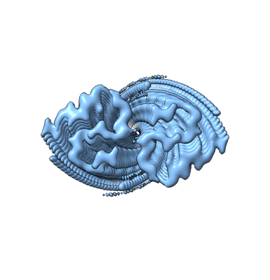
| Title | Type II amyloid-beta 42 filaments from high-spin supernatants of aqueous extracts from Alzheimer's disease brains | ABeta42 | ||||||||||||
 Map data Map data | |||||||||||||
 Sample Sample |
| ||||||||||||
 Keywords Keywords | amyloid / filaments / Abeta42 / amyloid-beta / cryo-EM / PROTEIN FIBRIL | ||||||||||||
| Function / homology |  Function and homology information Function and homology informationregulation of epidermal growth factor-activated receptor activity / cytosolic mRNA polyadenylation / collateral sprouting in absence of injury / microglia development / regulation of synapse structure or activity / regulation of Wnt signaling pathway / Formyl peptide receptors bind formyl peptides and many other ligands / axo-dendritic transport / synaptic assembly at neuromuscular junction / signaling receptor activator activity ...regulation of epidermal growth factor-activated receptor activity / cytosolic mRNA polyadenylation / collateral sprouting in absence of injury / microglia development / regulation of synapse structure or activity / regulation of Wnt signaling pathway / Formyl peptide receptors bind formyl peptides and many other ligands / axo-dendritic transport / synaptic assembly at neuromuscular junction / signaling receptor activator activity / smooth endoplasmic reticulum calcium ion homeostasis / axon midline choice point recognition / astrocyte activation involved in immune response / regulation of spontaneous synaptic transmission / mating behavior / NMDA selective glutamate receptor signaling pathway / ciliary rootlet / Lysosome Vesicle Biogenesis / PTB domain binding / Golgi-associated vesicle / positive regulation of amyloid fibril formation / neuron remodeling / : / Insertion of tail-anchored proteins into the endoplasmic reticulum membrane / Deregulated CDK5 triggers multiple neurodegenerative pathways in Alzheimer's disease models / suckling behavior / nuclear envelope lumen / dendrite development / COPII-coated ER to Golgi transport vesicle / presynaptic active zone / modulation of excitatory postsynaptic potential / TRAF6 mediated NF-kB activation / Advanced glycosylation endproduct receptor signaling / neuromuscular process controlling balance / The NLRP3 inflammasome / regulation of presynapse assembly / transition metal ion binding / negative regulation of long-term synaptic potentiation / regulation of multicellular organism growth / negative regulation of neuron differentiation / intracellular copper ion homeostasis / ECM proteoglycans / smooth endoplasmic reticulum / positive regulation of T cell migration / spindle midzone / Purinergic signaling in leishmaniasis infection / positive regulation of calcium-mediated signaling / protein serine/threonine kinase binding / positive regulation of chemokine production / clathrin-coated pit / regulation of peptidyl-tyrosine phosphorylation / forebrain development / Notch signaling pathway / Mitochondrial protein degradation / neuron projection maintenance / positive regulation of G2/M transition of mitotic cell cycle / positive regulation of protein metabolic process / ionotropic glutamate receptor signaling pathway / positive regulation of glycolytic process / cholesterol metabolic process / response to interleukin-1 / positive regulation of mitotic cell cycle / adult locomotory behavior / extracellular matrix organization / axonogenesis / platelet alpha granule lumen / trans-Golgi network membrane / positive regulation of peptidyl-threonine phosphorylation / dendritic shaft / learning / positive regulation of interleukin-1 beta production / locomotory behavior / positive regulation of long-term synaptic potentiation / central nervous system development / endosome lumen / astrocyte activation / positive regulation of JNK cascade / Post-translational protein phosphorylation / synapse organization / regulation of long-term neuronal synaptic plasticity / microglial cell activation / TAK1-dependent IKK and NF-kappa-B activation / visual learning / serine-type endopeptidase inhibitor activity / neuromuscular junction / recycling endosome / cognition / neuron cellular homeostasis / Golgi lumen / positive regulation of inflammatory response / positive regulation of non-canonical NF-kappaB signal transduction / endocytosis / cellular response to amyloid-beta / G2/M transition of mitotic cell cycle / Regulation of Insulin-like Growth Factor (IGF) transport and uptake by Insulin-like Growth Factor Binding Proteins (IGFBPs) / positive regulation of interleukin-6 production / positive regulation of tumor necrosis factor production / neuron projection development / cell-cell junction / synaptic vesicle Similarity search - Function | ||||||||||||
| Biological species |  Homo sapiens (human) / human (human) Homo sapiens (human) / human (human) | ||||||||||||
| Method | helical reconstruction / cryo EM / Resolution: 3.7 Å | ||||||||||||
 Authors Authors | Yang Y / Stern MA / Meunier LA / Liu W / Cai YQ / Ericsson M / Liu L / Selkoe JD / Goedert M / Scheres HWS | ||||||||||||
| Funding support |  United Kingdom, 3 items United Kingdom, 3 items
| ||||||||||||
 Citation Citation | Journal: Neuron / Year: 2023 Title: Abundant Aβ fibrils in ultracentrifugal supernatants of aqueous extracts from Alzheimer's disease brains. Authors: Andrew M Stern / Yang Yang / Shanxue Jin / Keitaro Yamashita / Angela L Meunier / Wen Liu / Yuqi Cai / Maria Ericsson / Lei Liu / Michel Goedert / Sjors H W Scheres / Dennis J Selkoe /  Abstract: Soluble oligomers of amyloid β-protein (Aβ) have been defined as aggregates in supernatants following ultracentrifugation of aqueous extracts from Alzheimer's disease (AD) brains and are believed ...Soluble oligomers of amyloid β-protein (Aβ) have been defined as aggregates in supernatants following ultracentrifugation of aqueous extracts from Alzheimer's disease (AD) brains and are believed to be upstream initiators of synaptic dysfunction, but little is known about their structures. We now report the unexpected presence of Aβ fibrils in synaptotoxic high-speed supernatants from AD brains extracted by soaking in an aqueous buffer. The fibrils did not appear to form during preparation, and their counts by EM correlated with Aβ ELISA quantification. Cryo-EM structures of aqueous Aβ fibrils were identical to those from sarkosyl-insoluble homogenates. The fibrils in aqueous extracts were labeled by lecanemab, an Aβ aggregate-directed antibody reported to improve AD cognitive outcomes. Lecanemab provided protection against aqueous fibril synaptotoxicity. We conclude that fibrils are abundant in aqueous extracts from AD brains and have the same structures as those from plaques. These findings have implications for AD pathogenesis and drug design. #1: Journal: Biorxiv / Year: 2022Title: Abundant A beta fibrils in ultracentrifugal supernatants of aqueous extracts from Alzheimer's disease brains Authors: Stern AM / Yang Y / Meunier AL / Liu W / Cai Y / Ericsson M / Liu L / Goedert M / Scheres SHW / Selkoe DJ | ||||||||||||
| History |
|
- Structure visualization
| Supplemental images |
|---|
- Downloads & links
-EMDB archive
| Map data | emd_15771.map.gz | 25.2 MB | EMDB map data format | |
|---|---|---|---|---|
| Header (meta data) | emd-15771-v30.xmlemd-15771.xml | 15.7 KB 15.7 KB | Display Display | EMDB header |
| FSC (resolution estimation) | emd_15771_fsc.xml | 9.2 KB | Display | FSC data file |
| Images |  emd_15771.png emd_15771.png | 76.6 KB | ||
| Masks | emd_15771_msk_1.map | 64 MB | Mask map | |
| Others | emd_15771_half_map_1.map.gzemd_15771_half_map_2.map.gz | 24.3 MB 24.3 MB | ||
| Archive directory |  http://ftp.pdbj.org/pub/emdb/structures/EMD-15771ftp://ftp.pdbj.org/pub/emdb/structures/EMD-15771 http://ftp.pdbj.org/pub/emdb/structures/EMD-15771ftp://ftp.pdbj.org/pub/emdb/structures/EMD-15771 | HTTPS FTP |
-Validation report
| Summary document | emd_15771_validation.pdf.gz | 851 KB | Display | EMDB validaton report |
|---|---|---|---|---|
| Full document | emd_15771_full_validation.pdf.gz | 850.5 KB | Display | |
| Data in XML | emd_15771_validation.xml.gz | 16.2 KB | Display | |
| Data in CIF | emd_15771_validation.cif.gz | 21.1 KB | Display | |
| Arichive directory | https://ftp.pdbj.org/pub/emdb/validation_reports/EMD-15771ftp://ftp.pdbj.org/pub/emdb/validation_reports/EMD-15771 | HTTPS FTP |
-Related structure data
| Related structure data |  8aztMC  8azsC  8azuC M: atomic model generated by this map C: citing same article ( |
|---|---|
| Similar structure data |
-Links
| EMDB pages | EMDB (EBI/PDBe) / EMDataResource |
|---|---|
| Related items in Molecule of the Month |


-Map
| File | Download / File: emd_15771.map.gz / Format: CCP4 / Size: 64 MB / Type: IMAGE STORED AS FLOATING POINT NUMBER (4 BYTES) | ||||||||||||||||||||
|---|---|---|---|---|---|---|---|---|---|---|---|---|---|---|---|---|---|---|---|---|---|
| Voxel size | X=Y=Z: 0.831 Å | ||||||||||||||||||||
| Density |
| ||||||||||||||||||||
| Symmetry | Space group: 1 | ||||||||||||||||||||
| Details | EMDB XML:
|
-Supplemental data
-Mask #1
| File | emd_15771_msk_1.map | ||||||||||||
|---|---|---|---|---|---|---|---|---|---|---|---|---|---|
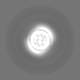
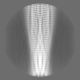
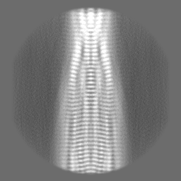
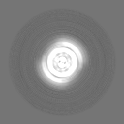
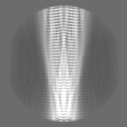
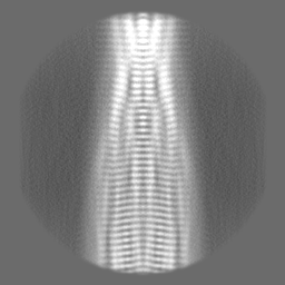
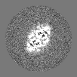
| Projections & Slices |
| ||||||||||||
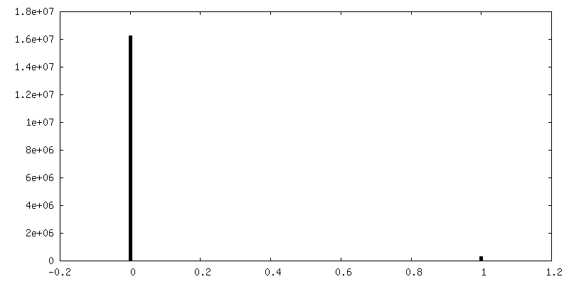
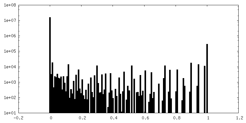
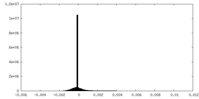
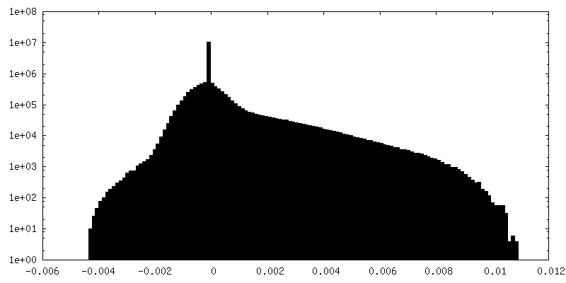
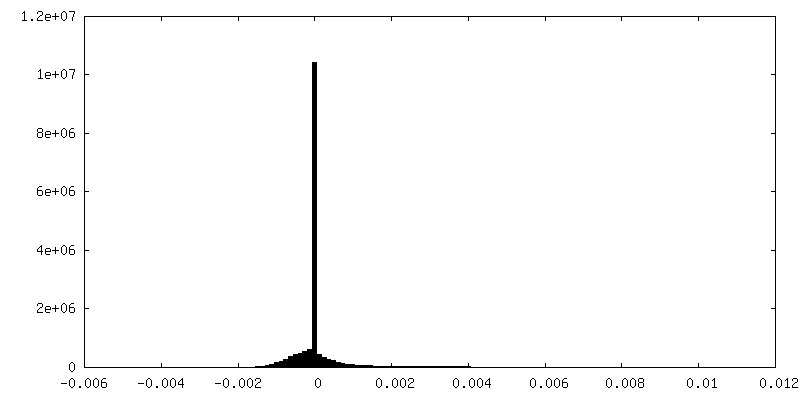
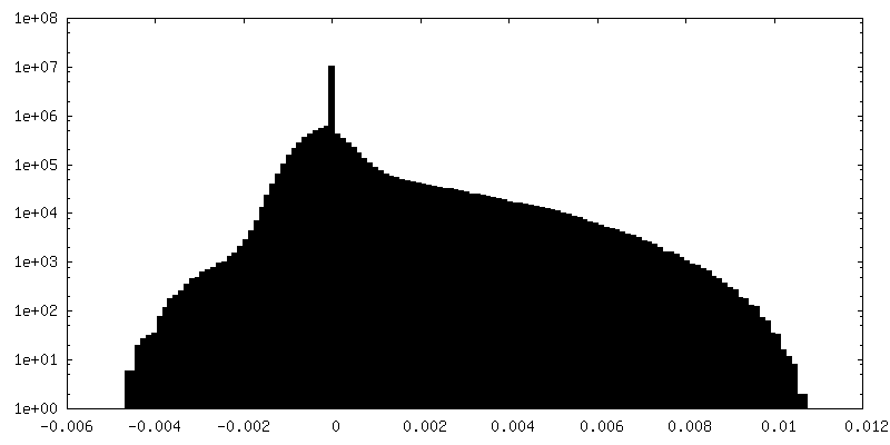
| Density Histograms |
 Z
Z Y
Y X
X


-Half map: #2
| File | emd_15771_half_map_1.map | ||||||||||||
|---|---|---|---|---|---|---|---|---|---|---|---|---|---|
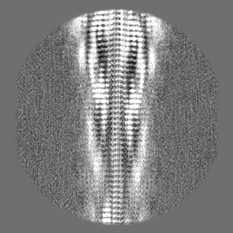
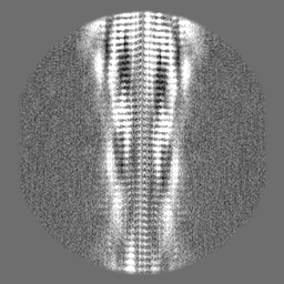
| Projections & Slices |
| ||||||||||||
| Density Histograms |

-Half map: #1
| File | emd_15771_half_map_2.map | ||||||||||||
|---|---|---|---|---|---|---|---|---|---|---|---|---|---|
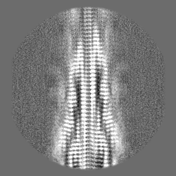
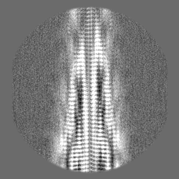
| Projections & Slices |
| ||||||||||||
| Density Histograms |
- Sample components
-Entire : Type II amyloid-beta 42 filaments in soluble high-molecular weigh...
| Entire | Name: Type II amyloid-beta 42 filaments in soluble high-molecular weight aggregate fractions extracted from Alzheimer's disease brain |
|---|---|
| Components |
|
-Supramolecule #1: Type II amyloid-beta 42 filaments in soluble high-molecular weigh...
| Supramolecule | Name: Type II amyloid-beta 42 filaments in soluble high-molecular weight aggregate fractions extracted from Alzheimer's disease brain type: tissue / ID: 1 / Parent: 0 / Macromolecule list: all |
|---|---|
| Source (natural) | Organism: Homo sapiens (human) |
-Macromolecule #1: Amyloid-beta precursor protein
| Macromolecule | Name: Amyloid-beta precursor protein / type: protein_or_peptide / ID: 1 / Number of copies: 1 / Enantiomer: LEVO |
|---|---|
| Source (natural) | Organism: human (human) |
| Molecular weight | Theoretical: 4.520087 KDa |
| Sequence | String: DAEFRHDSGY EVHHQKLVFF AEDVGSNKGA IIGLMVGGVV IA UniProtKB: Amyloid-beta precursor protein |
-Experimental details
-Structure determination
| Method | cryo EM |
|---|---|
 Processing Processing | helical reconstruction |
| Aggregation state | filament |
-Sample preparation
| Buffer | pH: 7.5 |
|---|---|
| Vitrification | Cryogen name: ETHANE |
- Electron microscopy
| Microscope | FEI TITAN KRIOS |
|---|---|
| Image recording | Film or detector model: GATAN K3 (6k x 4k) / Average electron dose: 40.0 e/Å2 |
| Electron beam | Acceleration voltage: 300 kV / Electron source:  FIELD EMISSION GUN FIELD EMISSION GUN |
| Electron optics | Illumination mode: FLOOD BEAM / Imaging mode: BRIGHT FIELD / Nominal defocus max: 2.4 µm / Nominal defocus min: 1.0 µm |
| Experimental equipment |  Model: Titan Krios / Image courtesy: FEI Company |
-Image processing
| Final reconstruction | Applied symmetry - Helical parameters - Δz: 4.8 Å Applied symmetry - Helical parameters - Δ&Phi: -2.8 ° Applied symmetry - Helical parameters - Axial symmetry: C2 (2 fold cyclic) Resolution.type: BY AUTHOR / Resolution: 3.7 Å / Resolution method: FSC 0.143 CUT-OFF / Software - Name: RELION / Number images used: 14740 |
|---|---|
| Startup model | Type of model: NONE |
| Final angle assignment | Type: NOT APPLICABLE |
| FSC plot (resolution estimation) | 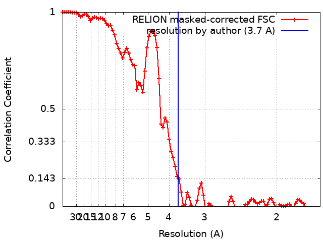 |