European Research Council (starting grant no. 716058)
European Union
Swiss National Science Foundation
NCCR transcure 185544
Switzerland
Swiss National Science Foundation
310030_188744
Switzerland
Swiss National Science Foundation
NCCR Molecular Systems Engineering
Switzerland
Swiss National Science Foundation
NCCR Chemical Biology
Switzerland
Citation
Journal: Nature / Year: 2023 Title: De novo design of protein interactions with learned surface fingerprints. Authors: Pablo Gainza / Sarah Wehrle / Alexandra Van Hall-Beauvais / Anthony Marchand / Andreas Scheck / Zander Harteveld / Stephen Buckley / Dongchun Ni / Shuguang Tan / Freyr Sverrisson / Casper ...Authors: Pablo Gainza / Sarah Wehrle / Alexandra Van Hall-Beauvais / Anthony Marchand / Andreas Scheck / Zander Harteveld / Stephen Buckley / Dongchun Ni / Shuguang Tan / Freyr Sverrisson / Casper Goverde / Priscilla Turelli / Charlène Raclot / Alexandra Teslenko / Martin Pacesa / Stéphane Rosset / Sandrine Georgeon / Jane Marsden / Aaron Petruzzella / Kefang Liu / Zepeng Xu / Yan Chai / Pu Han / George F Gao / Elisa Oricchio / Beat Fierz / Didier Trono / Henning Stahlberg / Michael Bronstein / Bruno E Correia / Abstract: Physical interactions between proteins are essential for most biological processes governing life. However, the molecular determinants of such interactions have been challenging to understand, even ...Physical interactions between proteins are essential for most biological processes governing life. However, the molecular determinants of such interactions have been challenging to understand, even as genomic, proteomic and structural data increase. This knowledge gap has been a major obstacle for the comprehensive understanding of cellular protein-protein interaction networks and for the de novo design of protein binders that are crucial for synthetic biology and translational applications. Here we use a geometric deep-learning framework operating on protein surfaces that generates fingerprints to describe geometric and chemical features that are critical to drive protein-protein interactions. We hypothesized that these fingerprints capture the key aspects of molecular recognition that represent a new paradigm in the computational design of novel protein interactions. As a proof of principle, we computationally designed several de novo protein binders to engage four protein targets: SARS-CoV-2 spike, PD-1, PD-L1 and CTLA-4. Several designs were experimentally optimized, whereas others were generated purely in silico, reaching nanomolar affinity with structural and mutational characterization showing highly accurate predictions. Overall, our surface-centric approach captures the physical and chemical determinants of molecular recognition, enabling an approach for the de novo design of protein interactions and, more broadly, of artificial proteins with function.
In the structure databanks used in Yorodumi, some data are registered as the other names, "COVID-19 virus" and "2019-nCoV". Here are the details of the virus and the list of structure data.
Jan 31, 2019. EMDB accession codes are about to change! (news from PDBe EMDB page)
EMDB accession codes are about to change! (news from PDBe EMDB page)
The allocation of 4 digits for EMDB accession codes will soon come to an end. Whilst these codes will remain in use, new EMDB accession codes will include an additional digit and will expand incrementally as the available range of codes is exhausted. The current 4-digit format prefixed with “EMD-” (i.e. EMD-XXXX) will advance to a 5-digit format (i.e. EMD-XXXXX), and so on. It is currently estimated that the 4-digit codes will be depleted around Spring 2019, at which point the 5-digit format will come into force.
The EM Navigator/Yorodumi systems omit the EMD- prefix.
Related info.:Q: What is EMD? / ID/Accession-code notation in Yorodumi/EM Navigator
Yorodumi is a browser for structure data from EMDB, PDB, SASBDB, etc.
This page is also the successor to EM Navigator detail page, and also detail information page/front-end page for Omokage search.
The word "yorodu" (or yorozu) is an old Japanese word meaning "ten thousand". "mi" (miru) is to see.
Related info.:EMDB / PDB / SASBDB / Comparison of 3 databanks / Yorodumi Search / Aug 31, 2016. New EM Navigator & Yorodumi / Yorodumi Papers / Jmol/JSmol / Function and homology information / Changes in new EM Navigator and Yorodumi
 Movie
Movie Controller

 Yorodumi
 Open data
 Basic information
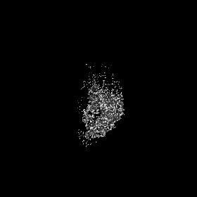
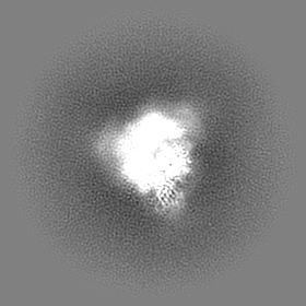
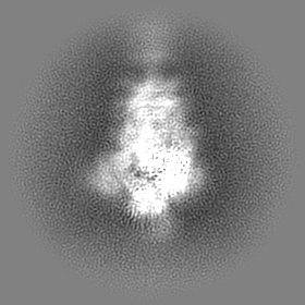
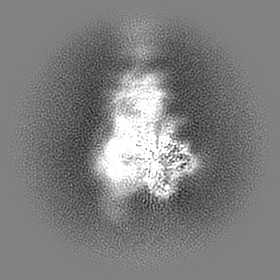
 Map data
 Sample
 Function and homology information
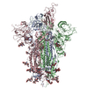
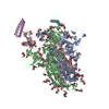
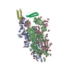
 Severe acute respiratory syndrome coronavirus 2 /
 Authors
 Switzerland, 5 items
  Citation

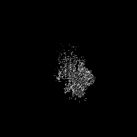
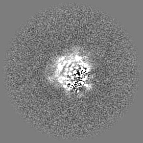
 Structure visualization
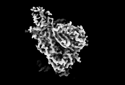
 Downloads & links
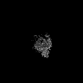
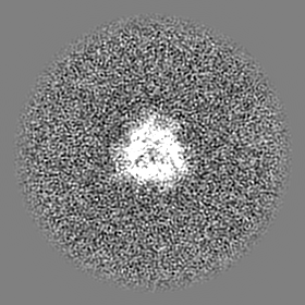
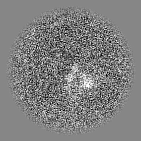
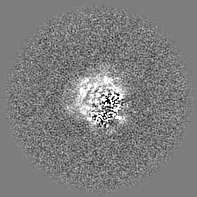
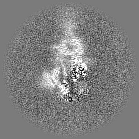
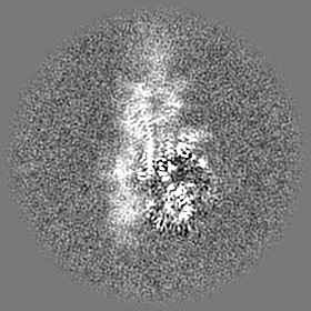
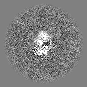
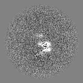
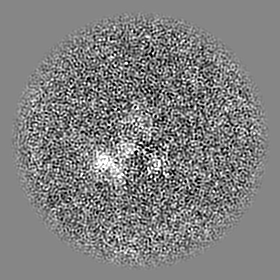
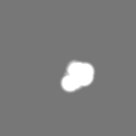
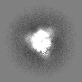
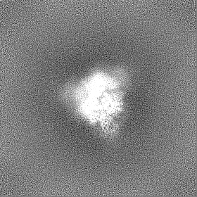
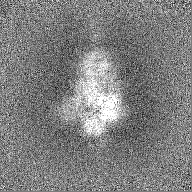
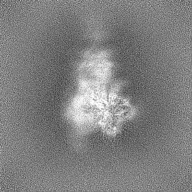
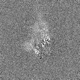
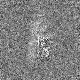
 emd_14930.png
 http://ftp.pdbj.org/pub/emdb/structures/EMD-14930
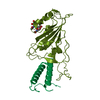


 Z (Sec.)
 Y (Row.)
 X (Col.)


 Sample components
 Homo sapiens (human)
 Processing
 Electron microscopy
 FIELD EMISSION GUN