Movie
Movie Controller


+ Open data

- Basic information
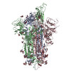
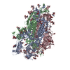
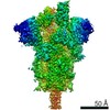
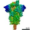
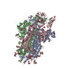
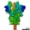
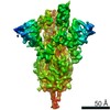
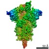
| Entry | Database: PDB / ID: 7qo7 | ||||||
|---|---|---|---|---|---|---|---|
| Title | SARS-CoV-2 S Omicron Spike B.1.1.529 | ||||||
 Components Components | Surface glycoprotein,Fibritin,SARS-CoV-2 S Omicron Spike B.1.1.529 | ||||||
 Keywords Keywords | VIRAL PROTEIN / SARS-CoV-2 S Omicron Spike B.1.1.529 | ||||||
| Function / homology | Fibritin C-terminal / Fibritin C-terminal region / Spike glycoprotein, N-terminal domain / Jelly Rolls / Sandwich / Mainly Beta / Fibritin / Surface glycoprotein Function and homology information Function and homology information | ||||||
| Biological species |   Severe acute respiratory syndrome coronavirus 2 Severe acute respiratory syndrome coronavirus 2 | ||||||
| Method | ELECTRON MICROSCOPY / single particle reconstruction / cryo EM / Resolution: 3.02 Å | ||||||
 Authors Authors | Ni, D. / Lau, K. / Turelli, P. / Beckert, B. / Nazarov, S. / Pojer, F. / Myasnikov, A. / Stahlberg, H. / Trono, D. | ||||||
| Funding support |  Switzerland, 1items Switzerland, 1items
| ||||||
 Citation Citation | Journal: Biorxiv / Year: 2021 Title: Structural analysis of the Spike of the Omicron SARS-COV-2 variant by cryo-EM and implications for immune evasion Authors: Ni, D. / Lau, K. / Turelli, P. / Raclot, C. / Beckert, B. / Nazarov, S. / Pojer, F. / Myasnikov, A. / Stahlberg, H. / Trono, D. | ||||||
| History |
|
- Structure visualization
| Movie |
Movie viewer |
|---|---|
| Structure viewer | Molecule: MolmilJmol/JSmol |
- Downloads & links
-Download
| PDBx/mmCIF format | 7qo7.cif.gz | 602.6 KB | Display | PDBx/mmCIF format |
|---|---|---|---|---|
| PDB format | pdb7qo7.ent.gz | 494.1 KB | Display | PDB format |
| PDBx/mmJSON format | 7qo7.json.gz | Tree view | PDBx/mmJSON format | |
| Others |  Other downloads Other downloads |
-Validation report
| Arichive directory | https://data.pdbj.org/pub/pdb/validation_reports/qo/7qo7ftp://data.pdbj.org/pub/pdb/validation_reports/qo/7qo7 | HTTPS FTP |
|---|
-Related structure data
| Related structure data | 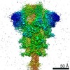 14086MC 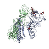 7qo9C M: map data used to model this data C: citing same article ( |
|---|---|
| Similar structure data |


-Links
 PDBj
PDBj

- Assembly
| Deposited unit | 
|
|---|---|
| 1 |
|
-Components
-Protein , 1 types, 3 molecules ABC
| #1: Protein | Mass: 142558.094 Da / Num. of mol.: 3 Source method: isolated from a genetically manipulated source Source: (gene. exp.) Severe acute respiratory syndrome coronavirus 2Gene: wac / Production host:   Cricetulus griseus (Chinese hamster) / References: UniProt: A0A8A4XEV3, UniProt: A0A2Z5WJZ7 Cricetulus griseus (Chinese hamster) / References: UniProt: A0A8A4XEV3, UniProt: A0A2Z5WJZ7 |
|---|
-Sugars , 5 types, 37 molecules 
| #2: Polysaccharide | alpha-D-mannopyranose-(1-3)-beta-D-mannopyranose-(1-4)-2-acetamido-2-deoxy-beta-D-glucopyranose-(1- ...alpha-D-mannopyranose-(1-3)-beta-D-mannopyranose-(1-4)-2-acetamido-2-deoxy-beta-D-glucopyranose-(1-4)-2-acetamido-2-deoxy-beta-D-glucopyranose Source method: isolated from a genetically manipulated source | ||||||
|---|---|---|---|---|---|---|---|
| #3: Polysaccharide | Source method: isolated from a genetically manipulated source #4: Polysaccharide | beta-D-mannopyranose-(1-4)-2-acetamido-2-deoxy-beta-D-glucopyranose-(1-4)-2-acetamido-2-deoxy-beta- ...beta-D-mannopyranose-(1-4)-2-acetamido-2-deoxy-beta-D-glucopyranose-(1-4)-2-acetamido-2-deoxy-beta-D-glucopyranose Source method: isolated from a genetically manipulated source #5: Polysaccharide | 2-acetamido-2-deoxy-beta-D-glucopyranose-(1-4)-[alpha-L-fucopyranose-(1-6)]2-acetamido-2-deoxy-beta- ...2-acetamido-2-deoxy-beta-D-glucopyranose-(1-4)-[alpha-L-fucopyranose-(1-6)]2-acetamido-2-deoxy-beta-D-glucopyranose | Source method: isolated from a genetically manipulated source #6: Sugar | ChemComp-NAG /  Type: D-saccharide, beta linking / Mass: 221.208 Da / Num. of mol.: 14 / Source method: obtained synthetically / Formula: C8H15NO6 Type: D-saccharide, beta linking / Mass: 221.208 Da / Num. of mol.: 14 / Source method: obtained synthetically / Formula: C8H15NO6 |
-Details
| Has ligand of interest | N |
|---|---|
| Has protein modification | Y |
-Experimental details
-Experiment
| Experiment | Method: ELECTRON MICROSCOPY |
|---|---|
| EM experiment | Aggregation state: PARTICLE / 3D reconstruction method: single particle reconstruction |
- Sample preparation
| Component | Name: Spike Trimer / Type: COMPLEX / Entity ID: #1 / Source: RECOMBINANT |
|---|---|
| Molecular weight | Value: 752 kDa/nm / Experimental value: YES |
| Source (natural) | Organism: Severe acute respiratory syndrome coronavirus 2 |
| Source (recombinant) | Organism: Cricetulus griseus (Chinese hamster) |
| Buffer solution | pH: 7.5 / Details: PBS |
| Specimen | Conc.: 0.35 mg/ml / Embedding applied: NO / Shadowing applied: NO / Staining applied: NO / Vitrification applied: YES |
| Specimen support | Grid material: COPPER / Grid mesh size: 400 divisions/in. / Grid type: Quantifoil R1.2/1.3 |
| Vitrification | Instrument: FEI VITROBOT MARK IV / Cryogen name: ETHANE / Humidity: 100 % |
- Electron microscopy imaging
| Experimental equipment |  Model: Titan Krios / Image courtesy: FEI Company |
|---|---|
| Microscopy | Model: TFS KRIOS |
| Electron gun | Electron source:  FIELD EMISSION GUN / Accelerating voltage: 300 kV / Illumination mode: OTHER FIELD EMISSION GUN / Accelerating voltage: 300 kV / Illumination mode: OTHER |
| Electron lens | Mode: BRIGHT FIELD / Nominal defocus max: 2500 nm / Nominal defocus min: 800 nm |
| Image recording | Electron dose: 60 e/Å2 / Film or detector model: FEI FALCON IV (4k x 4k) |
- Processing
| Software | Name: PHENIX / Version: 1.19.2_4158: / Classification: refinement | ||||||||||||||||||||||||
|---|---|---|---|---|---|---|---|---|---|---|---|---|---|---|---|---|---|---|---|---|---|---|---|---|---|
| EM software |
| ||||||||||||||||||||||||
| CTF correction | Type: NONE | ||||||||||||||||||||||||
| 3D reconstruction | Resolution: 3.02 Å / Resolution method: FSC 0.143 CUT-OFF / Num. of particles: 76450 / Symmetry type: POINT | ||||||||||||||||||||||||
| Atomic model building | B value: 120 / Protocol: AB INITIO MODEL / Space: REAL | ||||||||||||||||||||||||
| Atomic model building | PDB-ID: 6XM3 Accession code: 6XM3 / Source name: PDB / Type: experimental model | ||||||||||||||||||||||||
| Refine LS restraints |
|