 Movie
Movie Controller


+ Open data

- Basic information
| Entry | Database: EMDB / ID: EMD-13120 | |||||||||
|---|---|---|---|---|---|---|---|---|---|---|
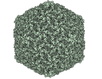
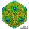
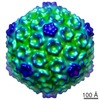
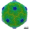
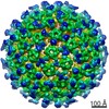
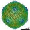
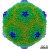
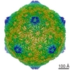
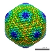
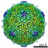
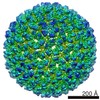
| Title | Mature capsid of bacteriophage phiRSA1 | |||||||||
 Map data Map data | Mature capsid of bacteriophage phiRSA1 | |||||||||
 Sample Sample |
| |||||||||
| Function / homology | Bacteriophage P2, capsid / Phage major capsid protein, P2 family / p2 family phage major capsid protein Function and homology information Function and homology information | |||||||||
| Biological species |  Ralstonia virus RSA1 / Ralstonia virus RSA1 /  Ralstonia solanacearum (bacteria) Ralstonia solanacearum (bacteria) | |||||||||
| Method | single particle reconstruction / cryo EM / Resolution: 3.9 Å | |||||||||
 Authors Authors | Effantin G / Fujiwara A / Kawsaki T / Yamada T / Schoehn G | |||||||||
 Citation Citation | Journal: Int J Mol Sci / Year: 2021 Title: High Resolution Structure of the Mature Capsid of Bacteriophage ϕRSA1 by Cryo-Electron Microscopy. Authors: Grégory Effantin / Akiko Fujiwara / Takeru Kawasaki / Takashi Yamada / Guy Schoehn /   Abstract: The ϕRSA1 bacteriophage has been isolated from , a gram negative bacteria having a significant economic impact on many important crops. We solved the three-dimensional structure of the ϕRSA1 mature ...The ϕRSA1 bacteriophage has been isolated from , a gram negative bacteria having a significant economic impact on many important crops. We solved the three-dimensional structure of the ϕRSA1 mature capsid to 3.9 Å resolution by cryo-electron microscopy. The capsid shell, that contains the 39 kbp of dsDNA genome, has an icosahedral symmetry characterized by an unusual triangulation number of T = 7, . The ϕRSA1 capsid is composed solely of the polymerization of the major capsid protein, gp8, which exhibits the typical "Johnson" fold first characterized in bacteriophage HK97. As opposed to the latter, the ϕRSA1 mature capsid is not stabilized by covalent crosslinking between its subunits, nor by the addition of a decoration protein. We further describe the molecular interactions occurring between the subunits of the ϕRSA1 capsid and their relationships with the other known bacteriophages. | |||||||||
| History |
|
- Structure visualization
| Movie |
Movie viewer |
|---|---|
| Structure viewer | EM map: SurfViewMolmilJmol/JSmol |
| Supplemental images |
- Downloads & links
-EMDB archive
| Map data | emd_13120.map.gz | 1012.6 MB | EMDB map data format | |
|---|---|---|---|---|
| Header (meta data) | emd-13120-v30.xmlemd-13120.xml | 9 KB 9 KB | Display Display | EMDB header |
| FSC (resolution estimation) | emd_13120_fsc.xml | 23.4 KB | Display | FSC data file |
| Images |  emd_13120.png emd_13120.png | 185.6 KB | ||
| Archive directory |  http://ftp.pdbj.org/pub/emdb/structures/EMD-13120ftp://ftp.pdbj.org/pub/emdb/structures/EMD-13120 http://ftp.pdbj.org/pub/emdb/structures/EMD-13120ftp://ftp.pdbj.org/pub/emdb/structures/EMD-13120 | HTTPS FTP |
-Validation report
| Summary document | emd_13120_validation.pdf.gz | 409.8 KB | Display | EMDB validaton report |
|---|---|---|---|---|
| Full document | emd_13120_full_validation.pdf.gz | 409.4 KB | Display | |
| Data in XML | emd_13120_validation.xml.gz | 17.8 KB | Display | |
| Data in CIF | emd_13120_validation.cif.gz | 25.2 KB | Display | |
| Arichive directory | https://ftp.pdbj.org/pub/emdb/validation_reports/EMD-13120ftp://ftp.pdbj.org/pub/emdb/validation_reports/EMD-13120 | HTTPS FTP |
-Related structure data
| Related structure data |  7oz4MC M: atomic model generated by this map C: citing same article ( |
|---|---|
| Similar structure data |
-Links
| EMDB pages | EMDB (EBI/PDBe) / EMDataResource |
|---|
-Map
| File | Download / File: emd_13120.map.gz / Format: CCP4 / Size: 1.1 GB / Type: IMAGE STORED AS FLOATING POINT NUMBER (4 BYTES) | ||||||||||||||||||||||||||||||||||||||||||||||||||||||||||||
|---|---|---|---|---|---|---|---|---|---|---|---|---|---|---|---|---|---|---|---|---|---|---|---|---|---|---|---|---|---|---|---|---|---|---|---|---|---|---|---|---|---|---|---|---|---|---|---|---|---|---|---|---|---|---|---|---|---|---|---|---|---|
| Annotation | Mature capsid of bacteriophage phiRSA1 | ||||||||||||||||||||||||||||||||||||||||||||||||||||||||||||
| Voxel size | X=Y=Z: 1.21 Å | ||||||||||||||||||||||||||||||||||||||||||||||||||||||||||||
| Density |
| ||||||||||||||||||||||||||||||||||||||||||||||||||||||||||||
| Symmetry | Space group: 1 | ||||||||||||||||||||||||||||||||||||||||||||||||||||||||||||
| Details | EMDB XML:
CCP4 map header:
| ||||||||||||||||||||||||||||||||||||||||||||||||||||||||||||
-Supplemental data
- Sample components
-Entire : Ralstonia solanacearum
| Entire | Name: Ralstonia solanacearum (bacteria) |
|---|---|
| Components |
|
-Supramolecule #1: Ralstonia solanacearum
| Supramolecule | Name: Ralstonia solanacearum / type: virus / ID: 1 / Parent: 0 / Macromolecule list: all / NCBI-ID: 305 / Sci species name: Ralstonia solanacearum / Virus type: VIRION / Virus isolate: STRAIN / Virus enveloped: No / Virus empty: No |
|---|---|
| Virus shell | Shell ID: 1 / Name: gp8 / Diameter: 640.0 Å / T number (triangulation number): 7 |
-Macromolecule #1: p2 family phage major capsid protein
| Macromolecule | Name: p2 family phage major capsid protein / type: protein_or_peptide / ID: 1 / Number of copies: 7 / Enantiomer: LEVO |
|---|---|
| Source (natural) | Organism: Ralstonia virus RSA1 |
| Molecular weight | Theoretical: 37.831531 KDa |
| Sequence | String: MRNDTRRLFA AYKAAIAKLN GVERVDEKFS VAPSVQQKLE TKVQESSDFL KSINFYGVPE QEGEKIGLGV SGPVASTTDT TQQDRETSD ISTMDGRRYR CEQTNSDTHI TYQKLDAWAK FADFQTRIRD AIIKRQALDR IMIGFNGVSR AATSDRVANP M LQDVNKGW ...String: MRNDTRRLFA AYKAAIAKLN GVERVDEKFS VAPSVQQKLE TKVQESSDFL KSINFYGVPE QEGEKIGLGV SGPVASTTDT TQQDRETSD ISTMDGRRYR CEQTNSDTHI TYQKLDAWAK FADFQTRIRD AIIKRQALDR IMIGFNGVSR AATSDRVANP M LQDVNKGW LQNLREQAPQ RVMKEGKAAA GKITVGGAGA DYGNLDALVY DITNHLVEPW YAEDPDLVVV CGRNLLSDKY FP LVNRDRD PVQQIAADLI ISQKRIGNLP AIRVPYFPAN GLLVTRLDNL SIYYQEGGRR RTILDNAKRD RIENYESSND AYV IEDLAC AAMAENIALA AAA |
-Experimental details
-Structure determination
| Method | cryo EM |
|---|---|
 Processing Processing | single particle reconstruction |
| Aggregation state | particle |
-Sample preparation
| Buffer | pH: 7.5 |
|---|---|
| Vitrification | Cryogen name: ETHANE / Instrument: FEI VITROBOT MARK IV |
- Electron microscopy
| Microscope | FEI TECNAI F30 |
|---|---|
| Image recording | Film or detector model: GATAN K2 SUMMIT (4k x 4k) / Detector mode: COUNTING / Average electron dose: 1.0 e/Å2 |
| Electron beam | Acceleration voltage: 300 kV / Electron source:  FIELD EMISSION GUN FIELD EMISSION GUN |
| Electron optics | Illumination mode: FLOOD BEAM / Imaging mode: BRIGHT FIELD |
| Experimental equipment |  Model: Tecnai F30 / Image courtesy: FEI Company |
-Image processing
| Final reconstruction | Resolution.type: BY AUTHOR / Resolution: 3.9 Å / Resolution method: FSC 0.143 CUT-OFF / Number images used: 3399 |
|---|---|
| Initial angle assignment | Type: PROJECTION MATCHING |
| Final angle assignment | Type: PROJECTION MATCHING |
| FSC plot (resolution estimation) | 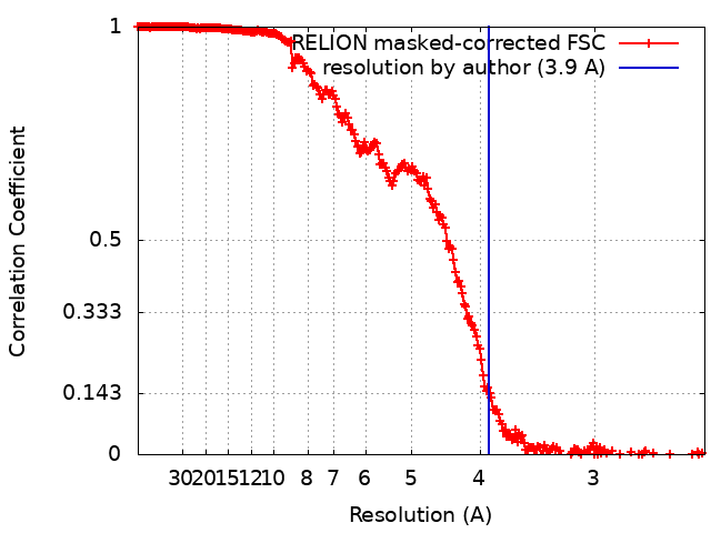 |
