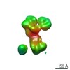
Journal: Int J Mol Sci / Year: 2021 Title: High Resolution Structure of the Mature Capsid of Bacteriophage ϕRSA1 by Cryo-Electron Microscopy. Authors: Grégory Effantin / Akiko Fujiwara / Takeru Kawasaki / Takashi Yamada / Guy Schoehn / Abstract: The ϕRSA1 bacteriophage has been isolated from , a gram negative bacteria having a significant economic impact on many important crops. We solved the three-dimensional structure of the ϕRSA1 mature ...The ϕRSA1 bacteriophage has been isolated from , a gram negative bacteria having a significant economic impact on many important crops. We solved the three-dimensional structure of the ϕRSA1 mature capsid to 3.9 Å resolution by cryo-electron microscopy. The capsid shell, that contains the 39 kbp of dsDNA genome, has an icosahedral symmetry characterized by an unusual triangulation number of T = 7, . The ϕRSA1 capsid is composed solely of the polymerization of the major capsid protein, gp8, which exhibits the typical "Johnson" fold first characterized in bacteriophage HK97. As opposed to the latter, the ϕRSA1 mature capsid is not stabilized by covalent crosslinking between its subunits, nor by the addition of a decoration protein. We further describe the molecular interactions occurring between the subunits of the ϕRSA1 capsid and their relationships with the other known bacteriophages.
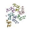
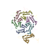
A: p2 family phage major capsid protein G: p2 family phage major capsid protein F: p2 family phage major capsid protein B: p2 family phage major capsid protein E: p2 family phage major capsid protein C: p2 family phage major capsid protein D: p2 family phage major capsid protein
In the structure databanks used in Yorodumi, some data are registered as the other names, "COVID-19 virus" and "2019-nCoV". Here are the details of the virus and the list of structure data.
Jan 31, 2019. EMDB accession codes are about to change! (news from PDBe EMDB page)
EMDB accession codes are about to change! (news from PDBe EMDB page)
The allocation of 4 digits for EMDB accession codes will soon come to an end. Whilst these codes will remain in use, new EMDB accession codes will include an additional digit and will expand incrementally as the available range of codes is exhausted. The current 4-digit format prefixed with “EMD-” (i.e. EMD-XXXX) will advance to a 5-digit format (i.e. EMD-XXXXX), and so on. It is currently estimated that the 4-digit codes will be depleted around Spring 2019, at which point the 5-digit format will come into force.
The EM Navigator/Yorodumi systems omit the EMD- prefix.
Related info.:Q: What is EMD? / ID/Accession-code notation in Yorodumi/EM Navigator
Yorodumi is a browser for structure data from EMDB, PDB, SASBDB, etc.
This page is also the successor to EM Navigator detail page, and also detail information page/front-end page for Omokage search.
The word "yorodu" (or yorozu) is an old Japanese word meaning "ten thousand". "mi" (miru) is to see.
Related info.:EMDB / PDB / SASBDB / Comparison of 3 databanks / Yorodumi Search / Aug 31, 2016. New EM Navigator & Yorodumi / Yorodumi Papers / Jmol/JSmol / Function and homology information / Changes in new EM Navigator and Yorodumi
 Movie
Movie Controller


 Open data
 Basic information
 Components
 Keywords
 Function and homology information
 Ralstonia virus RSA1
 Authors
 Citation

 Structure visualization
 Downloads & links
 Other downloads
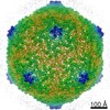


 PDBj
PDBj Assembly
 Sample preparation
 Ralstonia solanacearum (bacteria)
 Electron microscopy imaging
 FIELD EMISSION GUN / Accelerating voltage: 300 kV / Illumination mode: FLOOD BEAM
 Processing