 Movie
Movie Controller

[English] 日本語
 Yorodumi
- EMDB-12579: Cryo electron tomogram of Shigella flexneri drfaC mutant entrappe... -
+ Open data

- Basic information
| Entry | Database: EMDB / ID: EMD-12579 | ||||||||||||||||||
|---|---|---|---|---|---|---|---|---|---|---|---|---|---|---|---|---|---|---|---|
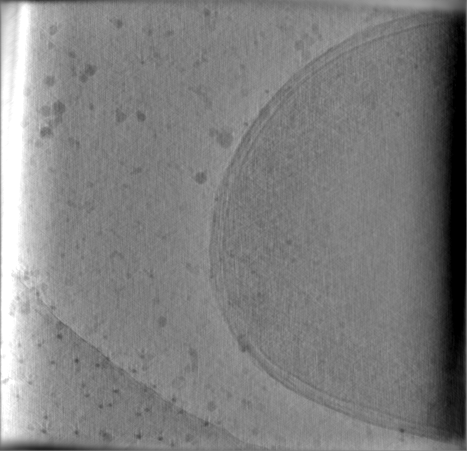
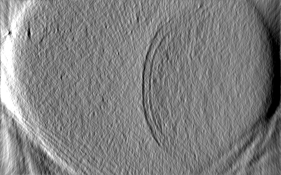
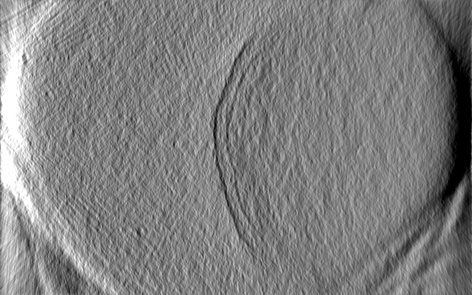
| Title | Cryo electron tomogram of Shigella flexneri drfaC mutant entrapped in septin cages. | ||||||||||||||||||
 Map data Map data | |||||||||||||||||||
 Sample Sample |
| ||||||||||||||||||
| Biological species |  Shigella flexneri (bacteria) Shigella flexneri (bacteria) | ||||||||||||||||||
| Method | electron tomography / cryo EM | ||||||||||||||||||
 Authors Authors | Lobato-Marquez D / Xu J / Ojiakor A / Pilhofer M / Mostowy S | ||||||||||||||||||
| Funding support | European Union,  Switzerland, 5 items Switzerland, 5 items
| ||||||||||||||||||
 Citation Citation | Journal: Nat Commun / Year: 2021 Title: Mechanistic insight into bacterial entrapment by septin cage reconstitution. Authors: Damián Lobato-Márquez / Jingwei Xu / Gizem Özbaykal Güler / Adaobi Ojiakor / Martin Pilhofer / Serge Mostowy /  Abstract: Septins are cytoskeletal proteins that assemble into hetero-oligomeric complexes and sense micron-scale membrane curvature. During infection with Shigella flexneri, an invasive enteropathogen, ...Septins are cytoskeletal proteins that assemble into hetero-oligomeric complexes and sense micron-scale membrane curvature. During infection with Shigella flexneri, an invasive enteropathogen, septins restrict actin tail formation by entrapping bacteria in cage-like structures. Here, we reconstitute septin cages in vitro using purified recombinant septin complexes (SEPT2-SEPT6-SEPT7), and study how these recognize bacterial cells and assemble on their surface. We show that septin complexes recognize the pole of growing Shigella cells. An amphipathic helix domain in human SEPT6 enables septins to sense positively curved membranes and entrap bacterial cells. Shigella strains lacking lipopolysaccharide components are more efficiently entrapped in septin cages. Finally, cryo-electron tomography of in vitro cages reveals how septins assemble as filaments on the bacterial cell surface. | ||||||||||||||||||
| History |
|
- Structure visualization
| Movie |
 Movie viewer Movie viewer |
|---|---|
| Supplemental images |

- Downloads & links
-EMDB archive
| Map data | emd_12579.map.gz | 1.9 GB | EMDB map data format | |
|---|---|---|---|---|
| Header (meta data) | emd-12579-v30.xmlemd-12579.xml | 9.7 KB 9.7 KB | Display Display | EMDB header |
| Images | 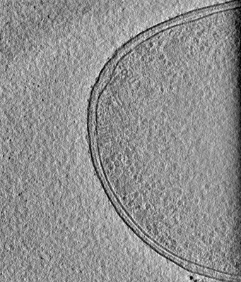 emd_12579.png emd_12579.png | 118.4 KB | ||
| Archive directory |  http://ftp.pdbj.org/pub/emdb/structures/EMD-12579ftp://ftp.pdbj.org/pub/emdb/structures/EMD-12579 http://ftp.pdbj.org/pub/emdb/structures/EMD-12579ftp://ftp.pdbj.org/pub/emdb/structures/EMD-12579 | HTTPS FTP |
-Validation report
| Summary document | emd_12579_validation.pdf.gz | 284.6 KB | Display | EMDB validaton report |
|---|---|---|---|---|
| Full document | emd_12579_full_validation.pdf.gz | 284.2 KB | Display | |
| Data in XML | emd_12579_validation.xml.gz | 4.9 KB | Display | |
| Data in CIF | emd_12579_validation.cif.gz | 5.5 KB | Display | |
| Arichive directory | https://ftp.pdbj.org/pub/emdb/validation_reports/EMD-12579ftp://ftp.pdbj.org/pub/emdb/validation_reports/EMD-12579 | HTTPS FTP |
-Related structure data
-Links
| EMDB pages | EMDB (EBI/PDBe) / EMDataResource |
|---|
-Map
| File | Download / File: emd_12579.map.gz / Format: CCP4 / Size: 2 GB / Type: IMAGE STORED AS FLOATING POINT NUMBER (4 BYTES) | ||||||||||||||||||||||||||||||||||||||||||||||||||||||||||||
|---|---|---|---|---|---|---|---|---|---|---|---|---|---|---|---|---|---|---|---|---|---|---|---|---|---|---|---|---|---|---|---|---|---|---|---|---|---|---|---|---|---|---|---|---|---|---|---|---|---|---|---|---|---|---|---|---|---|---|---|---|---|
| Projections & slices | Image control
Images are generated by Spider. generated in cubic-lattice coordinate | ||||||||||||||||||||||||||||||||||||||||||||||||||||||||||||
| Voxel size | X=Y=Z: 11 Å | ||||||||||||||||||||||||||||||||||||||||||||||||||||||||||||
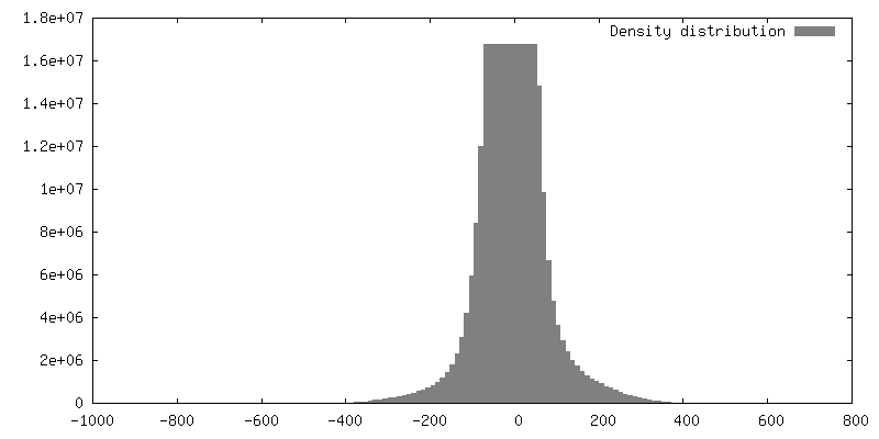
| Density |
| ||||||||||||||||||||||||||||||||||||||||||||||||||||||||||||
| Symmetry | Space group: 1 | ||||||||||||||||||||||||||||||||||||||||||||||||||||||||||||
| Details | EMDB XML:
CCP4 map header:
| ||||||||||||||||||||||||||||||||||||||||||||||||||||||||||||
 Z (Sec.)
 Y (Row.)
 X (Col.)


-Supplemental data
- Sample components
-Entire : Shigella flexneri drfaC mutant entrapped in septin cages.
| Entire | Name: Shigella flexneri drfaC mutant entrapped in septin cages. |
|---|---|
| Components |
|
-Supramolecule #1: Shigella flexneri drfaC mutant entrapped in septin cages.
| Supramolecule | Name: Shigella flexneri drfaC mutant entrapped in septin cages. type: cell / ID: 1 / Parent: 0 |
|---|---|
| Source (natural) | Organism: Shigella flexneri (bacteria) |
-Experimental details
-Structure determination
| Method | cryo EM |
|---|---|
 Processing Processing | electron tomography |
| Aggregation state | cell |
-Sample preparation
| Buffer | pH: 8 |
|---|---|
| Vitrification | Cryogen name: ETHANE-PROPANE |
| Sectioning | Other: NO SECTIONING |
| Fiducial marker | Manufacturer: cytodiagnostics / Diameter: 10 nm |
- Electron microscopy
| Microscope | FEI TITAN KRIOS |
|---|---|
| Image recording | Film or detector model: GATAN K2 SUMMIT (4k x 4k) / Detector mode: COUNTING / Average electron dose: 2.1 e/Å2 |
| Electron beam | Acceleration voltage: 300 kV / Electron source:  FIELD EMISSION GUN FIELD EMISSION GUN |
| Electron optics | Illumination mode: FLOOD BEAM / Imaging mode: BRIGHT FIELD |
| Experimental equipment |  Model: Titan Krios / Image courtesy: FEI Company |
-Image processing
| Final reconstruction | Algorithm: BACK PROJECTION / Software - Name: IMOD / Number images used: 61 |
|---|
