H2020 Marie Curie Actions of the European Commission
H2020-MSCA-IF-2016-752022
European Union
European Research Council (ERC)
772853-ENTRAPMENT
European Union
Wellcome Trust
206444/Z/17/Z
European Union
European Research Council (ERC)
679209
European Union
Swiss National Science Foundation
31003A_179255
Switzerland
Citation
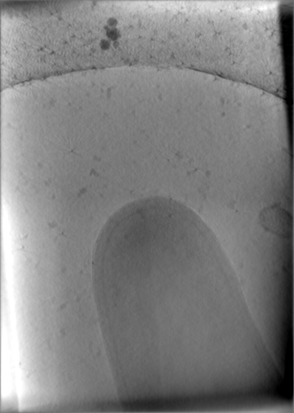
Journal: Nat Commun / Year: 2021 Title: Mechanistic insight into bacterial entrapment by septin cage reconstitution. Authors: Damián Lobato-Márquez / Jingwei Xu / Gizem Özbaykal Güler / Adaobi Ojiakor / Martin Pilhofer / Serge Mostowy / Abstract: Septins are cytoskeletal proteins that assemble into hetero-oligomeric complexes and sense micron-scale membrane curvature. During infection with Shigella flexneri, an invasive enteropathogen, ...Septins are cytoskeletal proteins that assemble into hetero-oligomeric complexes and sense micron-scale membrane curvature. During infection with Shigella flexneri, an invasive enteropathogen, septins restrict actin tail formation by entrapping bacteria in cage-like structures. Here, we reconstitute septin cages in vitro using purified recombinant septin complexes (SEPT2-SEPT6-SEPT7), and study how these recognize bacterial cells and assemble on their surface. We show that septin complexes recognize the pole of growing Shigella cells. An amphipathic helix domain in human SEPT6 enables septins to sense positively curved membranes and entrap bacterial cells. Shigella strains lacking lipopolysaccharide components are more efficiently entrapped in septin cages. Finally, cryo-electron tomography of in vitro cages reveals how septins assemble as filaments on the bacterial cell surface.
In the structure databanks used in Yorodumi, some data are registered as the other names, "COVID-19 virus" and "2019-nCoV". Here are the details of the virus and the list of structure data.
Jan 31, 2019. EMDB accession codes are about to change! (news from PDBe EMDB page)
EMDB accession codes are about to change! (news from PDBe EMDB page)
The allocation of 4 digits for EMDB accession codes will soon come to an end. Whilst these codes will remain in use, new EMDB accession codes will include an additional digit and will expand incrementally as the available range of codes is exhausted. The current 4-digit format prefixed with “EMD-” (i.e. EMD-XXXX) will advance to a 5-digit format (i.e. EMD-XXXXX), and so on. It is currently estimated that the 4-digit codes will be depleted around Spring 2019, at which point the 5-digit format will come into force.
The EM Navigator/Yorodumi systems omit the EMD- prefix.
Related info.:Q: What is EMD? / ID/Accession-code notation in Yorodumi/EM Navigator
Yorodumi is a browser for structure data from EMDB, PDB, SASBDB, etc.
This page is also the successor to EM Navigator detail page, and also detail information page/front-end page for Omokage search.
The word "yorodu" (or yorozu) is an old Japanese word meaning "ten thousand". "mi" (miru) is to see.
Related info.:EMDB / PDB / SASBDB / Comparison of 3 databanks / Yorodumi Search / Aug 31, 2016. New EM Navigator & Yorodumi / Yorodumi Papers / Jmol/JSmol / Function and homology information / Changes in new EM Navigator and Yorodumi
 Movie
Movie Controller

 Yorodumi
 Open data
 Basic information
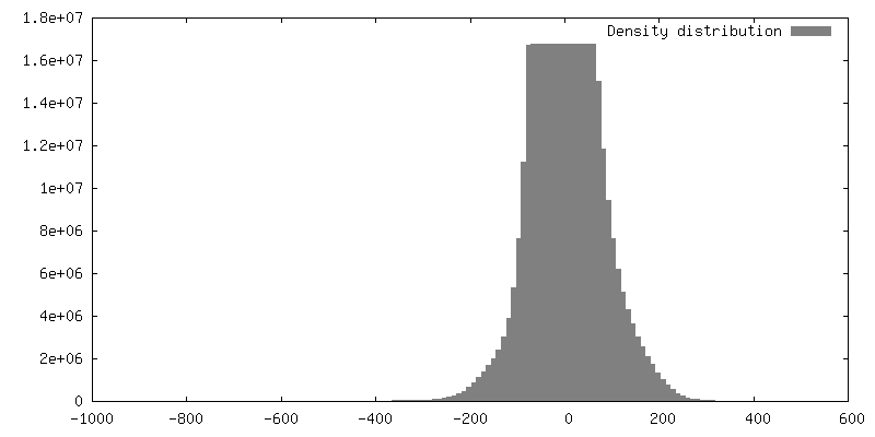
 Map data
 Sample
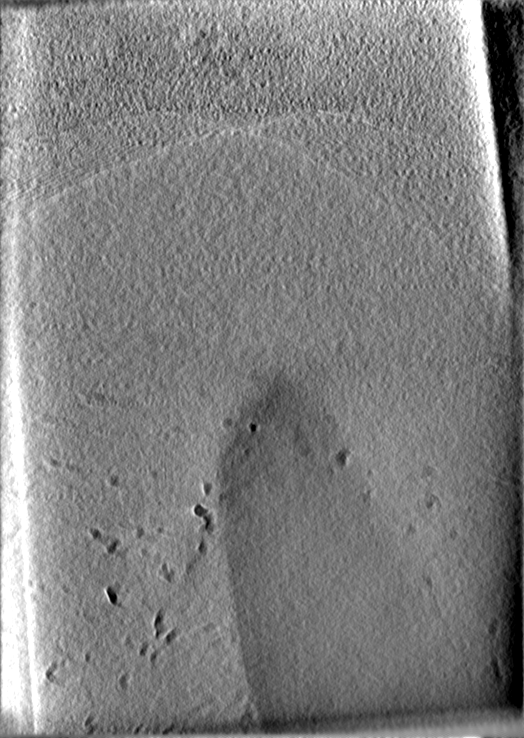
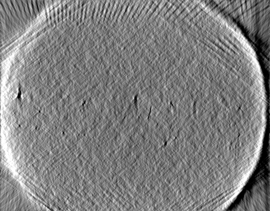
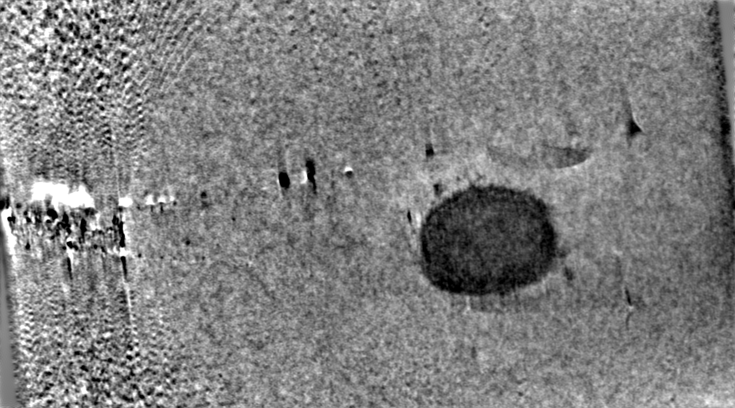
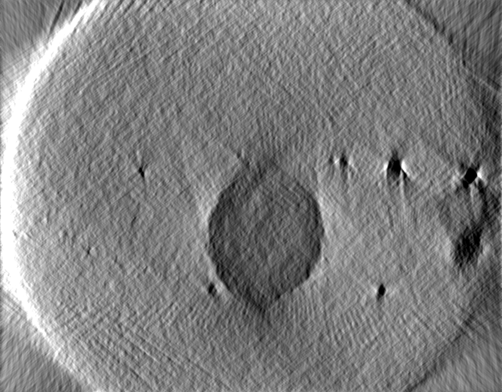
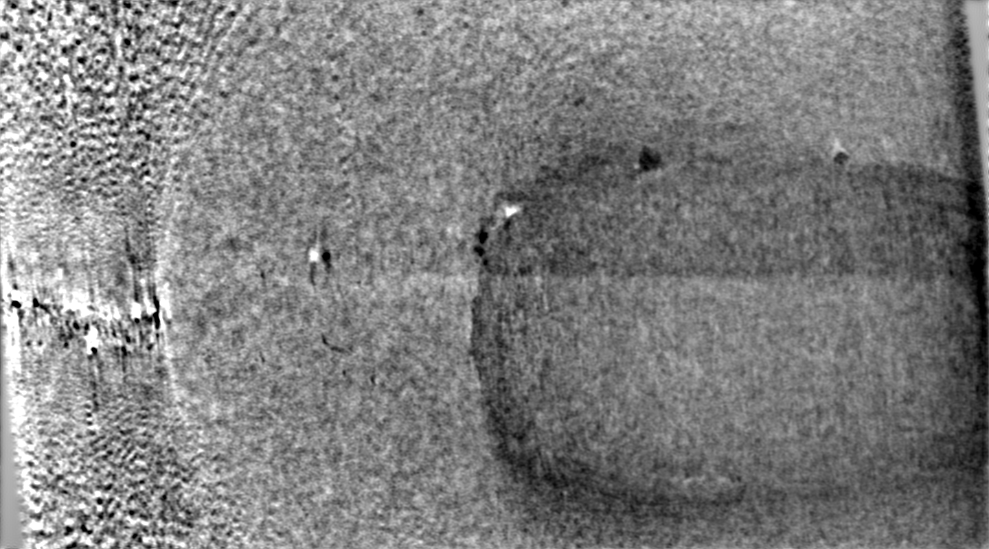
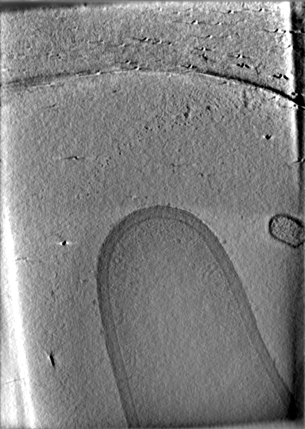
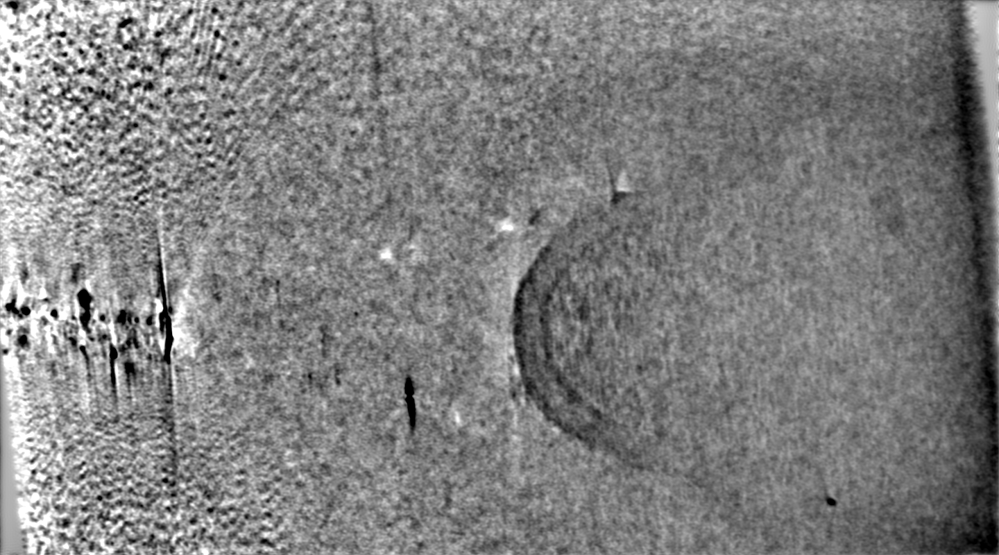
 Mycolicibacterium smegmatis (bacteria)
 Authors
 Switzerland, 5 items
  Citation
 Structure visualization
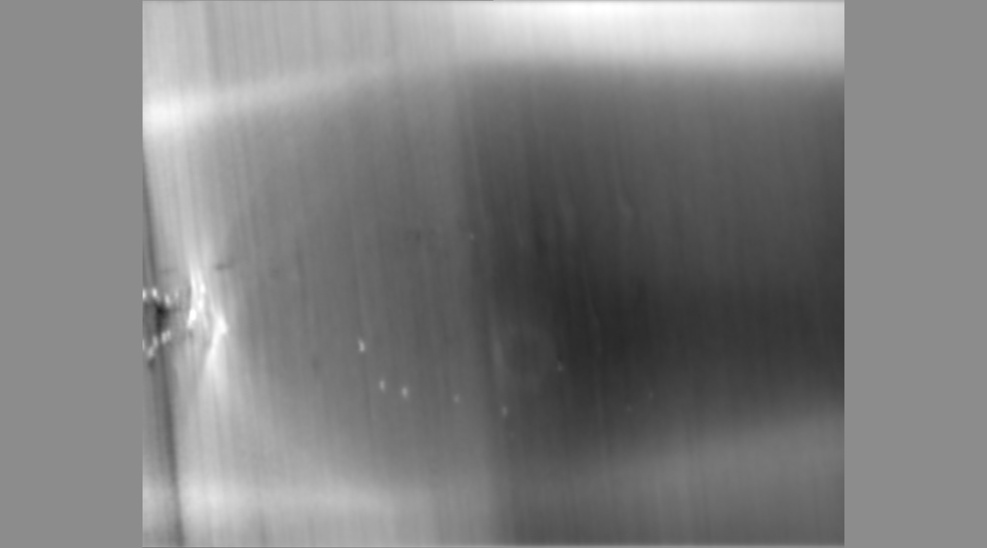
 Movie viewer
 Downloads & links
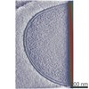
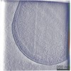
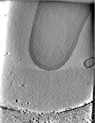 emd_12562.png
 http://ftp.pdbj.org/pub/emdb/structures/EMD-12562
 Z (Sec.)
 Y (Row.)
 X (Col.)


 Sample components
 Processing
 Electron microscopy
 FIELD EMISSION GUN