Movie
Movie Controller

[English] 日本語
 Yorodumi
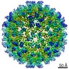
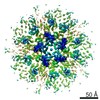
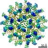
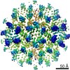
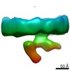
- EMDB-12487: Structure of the mature RSV CA lattice: hexamer derived from tube... -
+ Open data

- Basic information
| Entry | Database: EMDB / ID: EMD-12487 | ||||||||||||||||||
|---|---|---|---|---|---|---|---|---|---|---|---|---|---|---|---|---|---|---|---|
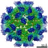
| Title | Structure of the mature RSV CA lattice: hexamer derived from tubes (C2-symmetric) | ||||||||||||||||||
 Map data Map data | RSV CA lattice: hexamer derived from tubes | ||||||||||||||||||
 Sample Sample |
| ||||||||||||||||||
| Function / homology |  Function and homology information Function and homology informationhost cell nucleoplasm / viral procapsid maturation / host cell nucleolus /  Hydrolases; Acting on peptide bonds (peptidases); Aspartic endopeptidases / viral capsid / structural constituent of virion / nucleic acid binding / aspartic-type endopeptidase activity / host cell plasma membrane / proteolysis ...host cell nucleoplasm / viral procapsid maturation / host cell nucleolus / Hydrolases; Acting on peptide bonds (peptidases); Aspartic endopeptidases / viral capsid / structural constituent of virion / nucleic acid binding / aspartic-type endopeptidase activity / host cell plasma membrane / proteolysis / zinc ion binding / membrane Hydrolases; Acting on peptide bonds (peptidases); Aspartic endopeptidases / viral capsid / structural constituent of virion / nucleic acid binding / aspartic-type endopeptidase activity / host cell plasma membrane / proteolysis ...host cell nucleoplasm / viral procapsid maturation / host cell nucleolus / Hydrolases; Acting on peptide bonds (peptidases); Aspartic endopeptidases / viral capsid / structural constituent of virion / nucleic acid binding / aspartic-type endopeptidase activity / host cell plasma membrane / proteolysis / zinc ion binding / membraneSimilarity search - Function | ||||||||||||||||||
| Biological species |  Rous sarcoma virus (strain Prague C) / Rous sarcoma virus - Prague C Rous sarcoma virus (strain Prague C) / Rous sarcoma virus - Prague C | ||||||||||||||||||
| Method | subtomogram averaging / cryo EM / Resolution: 4.3 Å | ||||||||||||||||||
 Authors Authors | Obr M / Ricana CL / Nikulin N / Feathers J-PR / Klanschnig M / Thader A / Johnson MC / Vogt VM / Schur FKM / Dick RA | ||||||||||||||||||
| Funding support |  Austria, Austria,  United States, European Union, 5 items United States, European Union, 5 items
| ||||||||||||||||||
 Citation Citation | Journal: Nat Commun / Year: 2021 Title: Structure of the mature Rous sarcoma virus lattice reveals a role for IP6 in the formation of the capsid hexamer. Authors: Martin Obr / Clifton L Ricana / Nadia Nikulin / Jon-Philip R Feathers / Marco Klanschnig / Andreas Thader / Marc C Johnson / Volker M Vogt / Florian K M Schur / Robert A Dick / Abstract: Inositol hexakisphosphate (IP6) is an assembly cofactor for HIV-1. We report here that IP6 is also used for assembly of Rous sarcoma virus (RSV), a retrovirus from a different genus. IP6 is ~100-fold ...Inositol hexakisphosphate (IP6) is an assembly cofactor for HIV-1. We report here that IP6 is also used for assembly of Rous sarcoma virus (RSV), a retrovirus from a different genus. IP6 is ~100-fold more potent at promoting RSV mature capsid protein (CA) assembly than observed for HIV-1 and removal of IP6 in cells reduces infectivity by 100-fold. Here, visualized by cryo-electron tomography and subtomogram averaging, mature capsid-like particles show an IP6-like density in the CA hexamer, coordinated by rings of six lysines and six arginines. Phosphate and IP6 have opposing effects on CA in vitro assembly, inducing formation of T = 1 icosahedrons and tubes, respectively, implying that phosphate promotes pentamer and IP6 hexamer formation. Subtomogram averaging and classification optimized for analysis of pleomorphic retrovirus particles reveal that the heterogeneity of mature RSV CA polyhedrons results from an unexpected, intrinsic CA hexamer flexibility. In contrast, the CA pentamer forms rigid units organizing the local architecture. These different features of hexamers and pentamers determine the structural mechanism to form CA polyhedrons of variable shape in mature RSV particles. | ||||||||||||||||||
| History |
|
- Structure visualization
| Movie |
Movie viewer |
|---|---|
| Structure viewer | EM map: SurfViewMolmilJmol/JSmol |
| Supplemental images |

- Downloads & links
-EMDB archive
| Map data | emd_12487.map.gz | 40 MB | EMDB map data format | |
|---|---|---|---|---|
| Header (meta data) | emd-12487-v30.xmlemd-12487.xml | 18.9 KB 18.9 KB | Display Display | EMDB header |
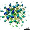
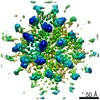
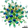
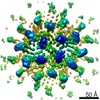
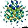
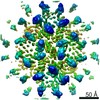
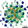
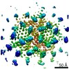
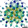
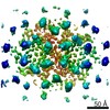
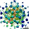
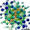
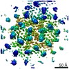
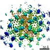
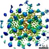
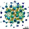
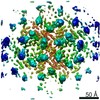
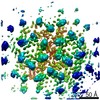
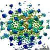
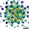
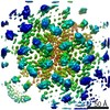
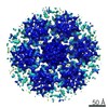
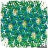
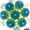
| Images | 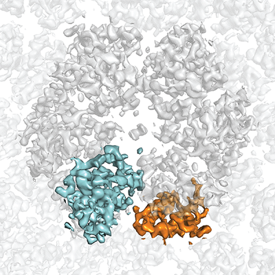 emd_12487.png emd_12487.png | 281.9 KB | ||
| Archive directory |  http://ftp.pdbj.org/pub/emdb/structures/EMD-12487ftp://ftp.pdbj.org/pub/emdb/structures/EMD-12487 http://ftp.pdbj.org/pub/emdb/structures/EMD-12487ftp://ftp.pdbj.org/pub/emdb/structures/EMD-12487 | HTTPS FTP |
-Related structure data
| Related structure data |  7no2MC 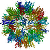 7no0C 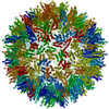 7no1C 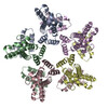 7no3C 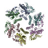 7no4C 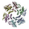 7no5C  7no6C  7no7C  7no8C  7no9C  7noaC 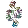 7nobC 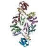 7nocC 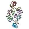 7nodC 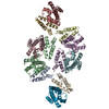 7noeC  7nofC 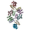 7nogC 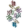 7nohC 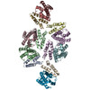 7noiC 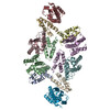 7nojC 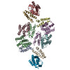 7nokC 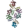 7nolC 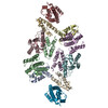 7nomC 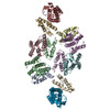 7nonC 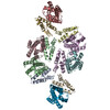 7nooC 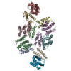 7nopC 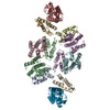 7noqC M: atomic model generated by this map C: citing same article ( |
|---|---|
| Similar structure data |
-Links
| EMDB pages | EMDB (EBI/PDBe) / EMDataResource |
|---|
-Map
| File | Download / File: emd_12487.map.gz / Format: CCP4 / Size: 42.9 MB / Type: IMAGE STORED AS FLOATING POINT NUMBER (4 BYTES) | ||||||||||||||||||||||||||||||||||||||||||||||||||||||||||||
|---|---|---|---|---|---|---|---|---|---|---|---|---|---|---|---|---|---|---|---|---|---|---|---|---|---|---|---|---|---|---|---|---|---|---|---|---|---|---|---|---|---|---|---|---|---|---|---|---|---|---|---|---|---|---|---|---|---|---|---|---|---|
| Annotation | RSV CA lattice: hexamer derived from tubes | ||||||||||||||||||||||||||||||||||||||||||||||||||||||||||||
| Voxel size | X=Y=Z: 1.3278 Å | ||||||||||||||||||||||||||||||||||||||||||||||||||||||||||||
| Density |
| ||||||||||||||||||||||||||||||||||||||||||||||||||||||||||||
| Symmetry | Space group: 1 | ||||||||||||||||||||||||||||||||||||||||||||||||||||||||||||
| Details | EMDB XML:
CCP4 map header:
| ||||||||||||||||||||||||||||||||||||||||||||||||||||||||||||
-Supplemental data
- Sample components
-Entire : Rous sarcoma virus - Prague C
| Entire | Name: Rous sarcoma virus - Prague C |
|---|---|
| Components |
|
-Supramolecule #1: Rous sarcoma virus - Prague C
| Supramolecule | Name: Rous sarcoma virus - Prague C / type: virus / ID: 1 / Parent: 0 / Macromolecule list: all / NCBI-ID: 11888 / Sci species name: Rous sarcoma virus - Prague C / Virus type: VIRUS-LIKE PARTICLE / Virus isolate: OTHER / Virus enveloped: No / Virus empty: Yes |
|---|---|
| Host (natural) | Organism: unidentified (others) |
| Host system | Organism:  Escherichia coli BL21(DE3) (bacteria) Escherichia coli BL21(DE3) (bacteria) |
| Virus shell | Shell ID: 1 / Name: CANC tubes |
-Macromolecule #1: Capsid protein p27, alternate cleaved 1
| Macromolecule | Name: Capsid protein p27, alternate cleaved 1 / type: protein_or_peptide / ID: 1 / Number of copies: 3 / Enantiomer: LEVO |
|---|---|
| Source (natural) | Organism: Rous sarcoma virus (strain Prague C) / Strain: Prague C |
| Molecular weight | Theoretical: 24.773594 KDa |
| Recombinant expression | Organism: Escherichia coli BL21(DE3) (bacteria) |
| Sequence | String: PVVIKTEGPA WTPLEPKLIT RLADTVRTKG LRSPITMAEV EALMSSPLLP HDVTNLMRVI LGPAPYALWM DAWGVQLQTV IAAATRDPR HPANGQGRGE RTNLNRLKGL ADGMVGNPQG QAALLRPGEL VAITASALQA FREVARLAEP AGPWADIMQG P SESFVDFA ...String: PVVIKTEGPA WTPLEPKLIT RLADTVRTKG LRSPITMAEV EALMSSPLLP HDVTNLMRVI LGPAPYALWM DAWGVQLQTV IAAATRDPR HPANGQGRGE RTNLNRLKGL ADGMVGNPQG QAALLRPGEL VAITASALQA FREVARLAEP AGPWADIMQG P SESFVDFA NRLIKAVEGS DLPPSARAPV IIDCFRQKSQ PDIQQLIRTA PSTLTTPGEI IKYVLDRQKT A |
-Experimental details
-Structure determination
| Method | cryo EM |
|---|---|
 Processing Processing | subtomogram averaging |
| Aggregation state | particle |
-Sample preparation
| Buffer | pH: 6.2 Component:
| |||||||||||||||
|---|---|---|---|---|---|---|---|---|---|---|---|---|---|---|---|---|
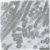
| Grid | Model: C-flat-2/2 / Material: COPPER / Mesh: 200 / Support film - Material: CARBON / Support film - topology: HOLEY / Pretreatment - Type: GLOW DISCHARGE / Pretreatment - Atmosphere: AIR | |||||||||||||||
| Vitrification | Cryogen name: ETHANE / Chamber humidity: 100 % / Chamber temperature: 277 K / Instrument: FEI VITROBOT MARK IV / Details: 2.5 seconds blotting time. |
- Electron microscopy
| Microscope | FEI TITAN KRIOS |
|---|---|
| Electron beam | Acceleration voltage: 300 kV / Electron source: FIELD EMISSION GUN |
| Electron optics | C2 aperture diameter: 100.0 µm / Illumination mode: FLOOD BEAM / Imaging mode: BRIGHT FIELDBright-field microscopy / Cs: 2.7 mm / Nominal defocus max: 4.0 µm / Nominal defocus min: 1.5 µm / Nominal magnification: 105000 |
| Specialist optics | Energy filter - Name: GIF Quantum LS / Energy filter - Slit width: 20 eV |
| Sample stage | Specimen holder model: FEI TITAN KRIOS AUTOGRID HOLDER / Cooling holder cryogen: NITROGEN |
| Details | Areas of interest for high-resolution data collection were identified in low magnification montages. Prior to tomogram acquisition, gain references were acquired and the filter was fully tuned. Microscope tuning was performed using the FEI AutoCTF software. The ilumination mode used during acquisition was nanoprobe. |
| Image recording | Film or detector model: GATAN K2 QUANTUM (4k x 4k) / Detector mode: COUNTING / Digitization - Dimensions - Width: 3708 pixel / Digitization - Dimensions - Height: 3838 pixel / Digitization - Frames/image: 1-10 / Number grids imaged: 1 / Average exposure time: 1.4 sec. / Average electron dose: 3.5 e/Å2 |
| Experimental equipment |  Model: Titan Krios / Image courtesy: FEI Company |
-Image processing
| Extraction | Number tomograms: 44 / Number images used: 45088 Software: (Name: MATLAB (ver. R2018b), IMOD (ver. 4.9), Dynamo (ver. 1.1.333)) | ||||||
|---|---|---|---|---|---|---|---|
| CTF correction | Software: (Name: CTFFIND (ver. 4.1.10), IMOD (ver. 4.9), NOVACTF) Details: CTF-correction was initially performed using ctfphaseflip in IMOD and NovaCTF in the final steps | ||||||
| Final 3D classification | Software - Name: MATLAB (ver. R2018b) | ||||||
| Final angle assignment | Type: OTHER / Software - Name: Dynamo (ver. 1.1.333) Details: Subtomogram alignment using Dynamo alignment project. | ||||||
| Final reconstruction | Applied symmetry - Point group: C2 (2 fold cyclic) / Algorithm: BACK PROJECTION / Resolution.type: BY AUTHOR / Resolution: 4.3 Å / Resolution method: FSC 0.143 CUT-OFF Software:
Number subtomograms used: 40962 |
-Atomic model buiding 1
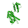
| Initial model | PDB ID: Chain - Chain ID: A / Chain - Residue range: 10-226 |
|---|---|
| Details | Three copies of CA monomer were rigid body-fitted into the EM density to accommodate the 3 symmetry independent CA copies. The fit was further refined in Coot. The symmetry independent copies were expanded according to the C2 symmetry, and an additional ring of CTDs adjacent to the CA hexamer was added to account for the continuous lattice during the refinement. The model was refined by iterating between automatic real space refinement in Phenix and manual model inspection/editing in Coot. |
| Refinement | Space: REAL / Protocol: AB INITIO MODEL / Target criteria: Correlation coefficient |
| Output model | PDB-7no2: |