[English] 日本語
Yorodumi
- EMDB-11768: Bacterial 30S ribosomal subunit assembly complex state D (head domain) -

+
Open data


ID or keywords:

Loading...

-
Basic information

Entry
Database: EMDB / ID: EMD-11768
TitleBacterial 30S ribosomal subunit assembly complex state D (head domain)
Map dataMultibody refinement map for 30S (head domain)
Sample
  • Complex: Bacterial 30S ribosomal subunit assembly complex state D (head domain)
    • RNA: x 1 types
    • Protein or peptide: x 8 types
  • Ligand: x 2 types
KeywordsCryo-EM / 30S biogenesis / ribosome assembly / RbfA / RsgA / YjeQ / RimP / KsgA / RsmA / RIBOSOME
Function / homology
Function and homology information


ribosomal small subunit assembly / small ribosomal subunit / cytosolic small ribosomal subunit / tRNA binding / rRNA binding / structural constituent of ribosome / translation / mRNA binding / RNA binding
Similarity search - Function
Ribosomal protein S14, bacterial/plastid / Ribosomal protein S3, bacterial-type / Ribosomal protein S19, bacterial-type / Ribosomal protein S13, bacterial-type / Ribosomal protein S7, bacterial/organellar-type / Ribosomal protein S9, bacterial/plastid / Ribosomal protein S2, bacteria/mitochondria/plastid / Ribosomal protein S2 signature 2. / K Homology domain / K homology RNA-binding domain ...Ribosomal protein S14, bacterial/plastid / Ribosomal protein S3, bacterial-type / Ribosomal protein S19, bacterial-type / Ribosomal protein S13, bacterial-type / Ribosomal protein S7, bacterial/organellar-type / Ribosomal protein S9, bacterial/plastid / Ribosomal protein S2, bacteria/mitochondria/plastid / Ribosomal protein S2 signature 2. / K Homology domain / K homology RNA-binding domain / Ribosomal protein S10, conserved site / Ribosomal protein S10 signature. / Ribosomal protein S3, conserved site / Ribosomal protein S3 signature. / Ribosomal protein S14, conserved site / Ribosomal protein S14 signature. / Ribosomal protein S2 signature 1. / KH domain / Type-2 KH domain profile. / K Homology domain, type 2 / Ribosomal protein S3, C-terminal / Ribosomal protein S3, C-terminal domain / Ribosomal protein S3, C-terminal domain superfamily / Ribosomal protein S15/S19, conserved site / Ribosomal protein S19 signature. / Ribosomal protein S10 / Ribosomal protein S19/S15 / Ribosomal protein S19/S15, superfamily / Ribosomal protein S19 / Ribosomal protein S2, conserved site / Ribosomal protein S7, conserved site / Ribosomal protein S7 signature. / Ribosomal protein S2 / Ribosomal protein S2, flavodoxin-like domain superfamily / Ribosomal protein S2 / K homology domain superfamily, prokaryotic type / Ribosomal protein S13, conserved site / Ribosomal protein S13 signature. / Ribosomal protein S13 / 30s ribosomal protein S13, C-terminal / Ribosomal protein S13/S18 / Ribosomal protein S13 family profile. / Ribosomal protein S14 / Ribosomal protein S14p/S29e / K homology domain-like, alpha/beta / Ribosomal protein S10p/S20e / Ribosomal protein S10 domain / Ribosomal protein S10 domain superfamily / Ribosomal protein S10p/S20e / Ribosomal protein S9, conserved site / Ribosomal protein S9 signature. / Ribosomal protein S13-like, H2TH / Ribosomal protein S5/S7 / Ribosomal protein S7 domain / Ribosomal protein S7 domain superfamily / Ribosomal protein S7p/S5e / Ribosomal protein S9 / Ribosomal protein S9/S16 / Ribosomal protein S5 domain 2-type fold, subgroup / Ribosomal protein S5 domain 2-type fold
Similarity search - Domain/homology
30S ribosomal protein S7 / Small ribosomal subunit protein uS10 / Small ribosomal subunit protein uS19 / Small ribosomal subunit protein uS3 / Small ribosomal subunit protein uS14 / Small ribosomal subunit protein uS13 / Small ribosomal subunit protein uS9 / Small ribosomal subunit protein uS2
Similarity search - Component
Biological speciesEscherichia coli (E. coli)
Methodsingle particle reconstruction / cryo EM / Resolution: 4.9 Å
AuthorsSchedlbauer A / Iturrioz I
Funding support Spain, 2 items
OrganizationGrant numberCountry
Ministry of Economy and Competitiveness (MINECO)CTQ2017-82222-R Spain
European CommissionPCIG14-GA-2013-632072 Spain
CitationJournal: Sci Adv / Year: 2021
Title: A conserved rRNA switch is central to decoding site maturation on the small ribosomal subunit.
Authors: Andreas Schedlbauer / Idoia Iturrioz / Borja Ochoa-Lizarralde / Tammo Diercks / Jorge Pedro López-Alonso / José Luis Lavin / Tatsuya Kaminishi / Retina Çapuni / Neha Dhimole / Elisa de ...Authors: Andreas Schedlbauer / Idoia Iturrioz / Borja Ochoa-Lizarralde / Tammo Diercks / Jorge Pedro López-Alonso / José Luis Lavin / Tatsuya Kaminishi / Retina Çapuni / Neha Dhimole / Elisa de Astigarraga / David Gil-Carton / Paola Fucini / Sean R Connell /
Abstract: While a structural description of the molecular mechanisms guiding ribosome assembly in eukaryotic systems is emerging, bacteria use an unrelated core set of assembly factors for which high- ...While a structural description of the molecular mechanisms guiding ribosome assembly in eukaryotic systems is emerging, bacteria use an unrelated core set of assembly factors for which high-resolution structural information is still missing. To address this, we used single-particle cryo-electron microscopy to visualize the effects of bacterial ribosome assembly factors RimP, RbfA, RsmA, and RsgA on the conformational landscape of the 30 ribosomal subunit and obtained eight snapshots representing late steps in the folding of the decoding center. Analysis of these structures identifies a conserved secondary structure switch in the 16 ribosomal RNA central to decoding site maturation and suggests both a sequential order of action and molecular mechanisms for the assembly factors in coordinating and controlling this switch. Structural and mechanistic parallels between bacterial and eukaryotic systems indicate common folding features inherent to all ribosomes.
History
DepositionSep 19, 2020-
Header (metadata) releaseJul 7, 2021-
Map releaseJul 7, 2021-
UpdateApr 24, 2024-
Current statusApr 24, 2024Processing site: PDBe / Status: Released

-
Structure visualization

Movie
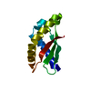
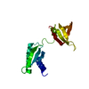
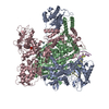
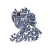
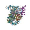
  • Surface view with section colored by density value
  • Surface level: 0.018
  • Imaged by UCSF Chimera
  • Download
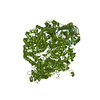
  • Surface view colored by height
  • Surface level: 0.018
  • Imaged by UCSF Chimera
  • Download
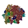
  • Surface view with fitted model
  • Atomic models: PDB-7afk
  • Surface level: 0.018
  • Imaged by UCSF Chimera
  • Download
Movie viewer
Structure viewerEM map:
SurfViewMolmilJmol/JSmol
Supplemental images

Downloads & links

-
Map

FileDownload / File: emd_11768.map.gz / Format: CCP4 / Size: 216 MB / Type: IMAGE STORED AS FLOATING POINT NUMBER (4 BYTES)
AnnotationMultibody refinement map for 30S (head domain)
Projections & slices

Image control

Size
Brightness
Contrast
Others
AxesZ (Sec.)Y (Row.)X (Col.)
1.09 Å/pix.
x 384 pix.
= 416.64 Å
1.09 Å/pix.
x 384 pix.
= 416.64 Å
1.09 Å/pix.
x 384 pix.
= 416.64 Å

Surface

Projections

Slices (1/3)

Slices (1/2)

Slices (2/3)

Images are generated by Spider.

Voxel sizeX=Y=Z: 1.085 Å
Density
Contour LevelBy AUTHOR: 0.018 / Movie #1: 0.018
Minimum - Maximum-0.026022926 - 0.08595441
Average (Standard dev.)-0.000142686 (±0.003322889)
SymmetrySpace group: 1
Details

EMDB XML:

Map geometry
Axis orderXYZ
Origin000
Dimensions384384384
Spacing384384384
CellA=B=C: 416.64 Å
α=β=γ: 90.0 °

CCP4 map header:

modeImage stored as Reals
Å/pix. X/Y/Z1.0851.0851.085
M x/y/z384384384
origin x/y/z0.0000.0000.000
length x/y/z416.640416.640416.640
α/β/γ90.00090.00090.000
MAP C/R/S123
start NC/NR/NS000
NC/NR/NS384384384
D min/max/mean-0.0260.086-0.000

-
Supplemental data

-
Mask #1

Fileemd_11768_msk_1.map
Projections & Slices
AxesZYX

Projections

Slices (1/2)
Density Histograms

-
Half map: Multibody refinement half map 1 for 30S ribosome (head domain)

Fileemd_11768_half_map_1.map
AnnotationMultibody refinement half map 1 for 30S ribosome (head domain)
Projections & Slices
AxesZYX

Projections

Slices (1/2)
Density Histograms

-
Half map: Multibody refinement half map 2 for 30S ribosome (head domain)

Fileemd_11768_half_map_2.map
AnnotationMultibody refinement half map 2 for 30S ribosome (head domain)
Projections & Slices
AxesZYX

Projections

Slices (1/2)
Density Histograms

-
Sample components

+
Entire : Bacterial 30S ribosomal subunit assembly complex state D (head domain)

EntireName: Bacterial 30S ribosomal subunit assembly complex state D (head domain)
Components
  • Complex: Bacterial 30S ribosomal subunit assembly complex state D (head domain)
    • RNA: 16SrRNA (head domain of the 30S ribosome)
    • Protein or peptide: 30S ribosomal protein S2
    • Protein or peptide: 30S ribosomal protein S3
    • Protein or peptide: 30S ribosomal protein S7
    • Protein or peptide: 30S ribosomal protein S9
    • Protein or peptide: 30S ribosomal protein S10
    • Protein or peptide: 30S ribosomal protein S13
    • Protein or peptide: 30S ribosomal protein S14
    • Protein or peptide: 30S ribosomal protein S19
  • Ligand: MAGNESIUM ION
  • Ligand: ZINC ION

+
Supramolecule #1: Bacterial 30S ribosomal subunit assembly complex state D (head domain)

SupramoleculeName: Bacterial 30S ribosomal subunit assembly complex state D (head domain)
type: complex / ID: 1 / Parent: 0 / Macromolecule list: #1-#9 / Details: 30S head from multibody refinement
Source (natural)Organism: Escherichia coli (E. coli)

+
Macromolecule #1: 16SrRNA (head domain of the 30S ribosome)

MacromoleculeName: 16SrRNA (head domain of the 30S ribosome) / type: rna / ID: 1 / Number of copies: 1
Source (natural)Organism: Escherichia coli (E. coli)
Molecular weightTheoretical: 499.52825 KDa
SequenceString: AAAUUGAAGA GUUUGAUCAU GGCUCAGAUU GAACGCUGGC GGCAGGCCUA ACACAUGCAA GUCGAACGGU AACAGGAAGA AGCUUGCUU CUUUGCUGAC GAGUGGCGGA CGGGUGAGUA AUGUCUGGGA AACUGCCUGA UGGAGGGGGA UAACUACUGG A AACGGUAG ...String:
AAAUUGAAGA GUUUGAUCAU GGCUCAGAUU GAACGCUGGC GGCAGGCCUA ACACAUGCAA GUCGAACGGU AACAGGAAGA AGCUUGCUU CUUUGCUGAC GAGUGGCGGA CGGGUGAGUA AUGUCUGGGA AACUGCCUGA UGGAGGGGGA UAACUACUGG A AACGGUAG CUAAUACCGC AUAACGUCGC AAGACCAAAG AGGGGGACCU UCGGGCCUCU UGCCAUCGGA UGUGCCCAGA UG GGAUUAG CUAGUAGGUG GGGUAACGGC UCACCUAGGC GACGAUCCCU AGCUGGUCUG AGAGGAUGAC CAGCCACACU GGA ACUGAG ACACGGUCCA GACUCCUACG GGAGGCAGCA GUGGGGAAUA UUGCACAAUG GGCGCAAGCC UGAUGCAGCC AUGC CGCGU GUAUGAAGAA GGCCUUCGGG UUGUAAAGUA CUUUCAGCGG GGAGGAAGGG AGUAAAGUUA AUACCUUUGC UCAUU GACG UUACCCGCAG AAGAAGCACC GGCUAACUCC G(PSU)GCCAGCAG CC(G7M)CGGUAAU ACGGAGGGUG CAAGCGUU A AUCGGAAUUA CUGGGCGUAA AGCGCACGCA GGCGGUUUGU UAAGUCAGAU GUGAAAUCCC CGGGCUCAAC CUGGGAACU GCAUCUGAUA CUGGCAAGCU UGAGUCUCGU AGAGGGGGGU AGAAUUCCAG GUGUAGCGGU GAAAUGCGUA GAGAUCUGGA GGAAUACCG GUGGCGAAGG CGGCCCCCUG GACGAAGACU GACGCUCAGG UGCGAAAGCG UGGGGAGCAA ACAGGAUUAG A UACCCUGG UAGUCCACGC CGUAAACGAU GUCGACUUGG AGGUUGUGCC CUUGAGGCGU GGCUUCCGGA GCUAACGCGU UA AGUCGAC CGCCUGGGGA GUACGGCCGC AAGGUUAAAA CUCAAAUGAA UUGACGGGGG CCCGCACAAG CGGUGGAGCA UGU GGUUUA AUUCGAU(2MG)(5MC)A ACGCGAAGAA CCUUACCUGG UCUUGACAUC CACGGAAGUU UUCAGAGAUG AGAAUG UGC CUUCGGGAAC CGUGAGACAG GUGCUGCAUG GCUGUCGUCA GCUCGUGUUG UGAAAUGUUG GGUUAAGUCC CGCAACG AG CGCAACCCUU AUCCUUUGUU GCCAGCGGUC CGGCCGGGAA CUCAAAGGAG ACUGCCAGUG AUAAACUGGA GGAAGGUG G GGAUGACGUC AAGUCAUCAU G(2MG)CCCUUACG ACCAGGGCUA CACACGUGCU ACAAUGGCGC AUACAAAGAG AAGCG ACCU CGCGAGAGCA AGCGGACCUC AUAAAGUGCG UCGUAGUCCG GAUUGGAGUC UGCAACUCGA CUCCAUGAAG UCGGAA UCG CUAGUAAUCG UGGAUCAGAA UGCCACGGUG AAUACGUUCC CGGCCUUGUA CACACCG(4OC)CC GU(5MC)ACACCA UGGGAGUGGG UUGCAAAAGA AGUAGGUAGC UUAACCUUCG GGAGGGCGCU UACCACUUUG UGAUUCAUGA CUGGGGUGAA GUCG(UR3)AACA AGGUAACCGU AGG(2MG)G(MA6)(MA6)CCU GCGGUUGGAU CACCUCCUUA

+
Macromolecule #2: 30S ribosomal protein S2

MacromoleculeName: 30S ribosomal protein S2 / type: protein_or_peptide / ID: 2 / Number of copies: 1 / Enantiomer: LEVO
Source (natural)Organism: Escherichia coli (E. coli)
Molecular weightTheoretical: 26.78167 KDa
SequenceString: MATVSMRDML KAGVHFGHQT RYWNPKMKPF IFGARNKVHI INLEKTVPMF NEALAELNKI ASRKGKILFV GTKRAASEAV KDAALSCDQ FFVNHRWLGG MLTNWKTVRQ SIKRLKDLET QSQDGTFDKL TKKEALMRTR ELEKLENSLG GIKDMGGLPD A LFVIDADH ...String:
MATVSMRDML KAGVHFGHQT RYWNPKMKPF IFGARNKVHI INLEKTVPMF NEALAELNKI ASRKGKILFV GTKRAASEAV KDAALSCDQ FFVNHRWLGG MLTNWKTVRQ SIKRLKDLET QSQDGTFDKL TKKEALMRTR ELEKLENSLG GIKDMGGLPD A LFVIDADH EHIAIKEANN LGIPVFAIVD TNSDPDGVDF VIPGNDDAIR AVTLYLGAVA ATVREGRSQD LASQAEESFV EA E

UniProtKB: Small ribosomal subunit protein uS2

+
Macromolecule #3: 30S ribosomal protein S3

MacromoleculeName: 30S ribosomal protein S3 / type: protein_or_peptide / ID: 3 / Number of copies: 1 / Enantiomer: LEVO
Source (natural)Organism: Escherichia coli (E. coli)
Molecular weightTheoretical: 26.031316 KDa
SequenceString: MGQKVHPNGI RLGIVKPWNS TWFANTKEFA DNLDSDFKVR QYLTKELAKA SVSRIVIERP AKSIRVTIHT ARPGIVIGKK GEDVEKLRK VVADIAGVPA QINIAEVRKP ELDAKLVADS ITSQLERRVM FRRAMKRAVQ NAMRLGAKGI KVEVSGRLGG A EIARTEWY ...String:
MGQKVHPNGI RLGIVKPWNS TWFANTKEFA DNLDSDFKVR QYLTKELAKA SVSRIVIERP AKSIRVTIHT ARPGIVIGKK GEDVEKLRK VVADIAGVPA QINIAEVRKP ELDAKLVADS ITSQLERRVM FRRAMKRAVQ NAMRLGAKGI KVEVSGRLGG A EIARTEWY REGRVPLHTL RADIDYNTSE AHTTYGVIGV KVWIFKGEIL GGMAAVEQPE KPAAQPKKQQ RKGRK

UniProtKB: Small ribosomal subunit protein uS3

+
Macromolecule #4: 30S ribosomal protein S7

MacromoleculeName: 30S ribosomal protein S7 / type: protein_or_peptide / ID: 4 / Number of copies: 1 / Enantiomer: LEVO
Source (natural)Organism: Escherichia coli (E. coli)
Molecular weightTheoretical: 20.055156 KDa
SequenceString:
MPRRRVIGQR KILPDPKFGS ELLAKFVNIL MVDGKKSTAE SIVYSALETL AQRSGKSELE AFEVALENVR PTVEVKSRRV GGSTYQVPV EVRPVRRNAL AMRWIVEAAR KRGDKSMALR LANELSDAAE NKGTAVKKRE DVHRMAEANK AFAHYRWLSL R SFSHQAGA SSKQPALGYL N

UniProtKB: 30S ribosomal protein S7

+
Macromolecule #5: 30S ribosomal protein S9

MacromoleculeName: 30S ribosomal protein S9 / type: protein_or_peptide / ID: 5 / Number of copies: 1 / Enantiomer: LEVO
Source (natural)Organism: Escherichia coli (E. coli)
Molecular weightTheoretical: 14.88627 KDa
SequenceString:
MAENQYYGTG RRKSSAARVF IKPGNGKIVI NQRSLEQYFG RETARMVVRQ PLELVDMVEK LDLYITVKGG GISGQAGAIR HGITRALME YDESLRSELR KAGFVTRDAR QVERKKVGLR KARRRPQFSK R

UniProtKB: Small ribosomal subunit protein uS9

+
Macromolecule #6: 30S ribosomal protein S10

MacromoleculeName: 30S ribosomal protein S10 / type: protein_or_peptide / ID: 6 / Number of copies: 1 / Enantiomer: LEVO
Source (natural)Organism: Escherichia coli (E. coli)
Molecular weightTheoretical: 11.755597 KDa
SequenceString:
MQNQRIRIRL KAFDHRLIDQ ATAEIVETAK RTGAQVRGPI PLPTRKERFT VLISPHVNKD ARDQYEIRTH LRLVDIVEPT EKTVDALMR LDLAAGVDVQ ISLG

UniProtKB: Small ribosomal subunit protein uS10

+
Macromolecule #7: 30S ribosomal protein S13

MacromoleculeName: 30S ribosomal protein S13 / type: protein_or_peptide / ID: 7 / Number of copies: 1 / Enantiomer: LEVO
Source (natural)Organism: Escherichia coli (E. coli)
Molecular weightTheoretical: 13.128467 KDa
SequenceString:
MARIAGINIP DHKHAVIALT SIYGVGKTRS KAILAAAGIA EDVKISELSE GQIDTLRDEV AKFVVEGDLR REISMSIKRL MDLGCYRGL RHRRGLPVRG QRTKTNARTR KGPRKPIKK

UniProtKB: Small ribosomal subunit protein uS13

+
Macromolecule #8: 30S ribosomal protein S14

MacromoleculeName: 30S ribosomal protein S14 / type: protein_or_peptide / ID: 8 / Number of copies: 1 / Enantiomer: LEVO
Source (natural)Organism: Escherichia coli (E. coli)
Molecular weightTheoretical: 11.60656 KDa
SequenceString:
MAKQSMKARE VKRVALADKY FAKRAELKAI ISDVNASDED RWNAVLKLQT LPRDSSPSRQ RNRCRQTGRP HGFLRKFGLS RIKVREAAM RGEIPGLKKA SW

UniProtKB: Small ribosomal subunit protein uS14

+
Macromolecule #9: 30S ribosomal protein S19

MacromoleculeName: 30S ribosomal protein S19 / type: protein_or_peptide / ID: 9 / Number of copies: 1 / Enantiomer: LEVO
Source (natural)Organism: Escherichia coli (E. coli)
Molecular weightTheoretical: 10.455355 KDa
SequenceString:
MPRSLKKGPF IDLHLLKKVE KAVESGDKKP LRTWSRRSTI FPNMIGLTIA VHNGRQHVPV FVTDEMVGHK LGEFAPTRTY RGHAADKKA KKK

UniProtKB: Small ribosomal subunit protein uS19

+
Macromolecule #10: MAGNESIUM ION

MacromoleculeName: MAGNESIUM ION / type: ligand / ID: 10 / Number of copies: 12 / Formula: MG
Molecular weightTheoretical: 24.305 Da

+
Macromolecule #11: ZINC ION

MacromoleculeName: ZINC ION / type: ligand / ID: 11 / Number of copies: 1 / Formula: ZN
Molecular weightTheoretical: 65.409 Da

-
Experimental details

-
Structure determination

Methodcryo EM
Processingsingle particle reconstruction
Aggregation stateparticle

-
Sample preparation

BufferpH: 7.8
GridModel: Quantifoil R2/2 / Support film - Material: CARBON / Support film - topology: CONTINUOUS
VitrificationCryogen name: ETHANE / Chamber humidity: 100 % / Chamber temperature: 277 K / Instrument: FEI VITROBOT MARK III

-
Electron microscopy

MicroscopeFEI TITAN KRIOS
Image recordingFilm or detector model: GATAN K2 SUMMIT (4k x 4k) / Detector mode: COUNTING / Average electron dose: 46.1 e/Å2
Electron beamAcceleration voltage: 300 kV / Electron source: FIELD EMISSION GUN
Electron opticsIllumination mode: SPOT SCAN / Imaging mode: BRIGHT FIELD
Sample stageSpecimen holder model: FEI TITAN KRIOS AUTOGRID HOLDER / Cooling holder cryogen: NITROGEN
Experimental equipment
Model: Titan Krios / Image courtesy: FEI Company

+
Image processing

Particle selectionNumber selected: 200953
Startup modelType of model: OTHER / Details: Relion initial model
Final reconstructionApplied symmetry - Point group: C1 (asymmetric) / Resolution.type: BY AUTHOR / Resolution: 4.9 Å / Resolution method: FSC 0.143 CUT-OFF / Software - Name: RELION (ver. 3.0) / Number images used: 4573
Initial angle assignmentType: MAXIMUM LIKELIHOOD / Software - Name: RELION (ver. 3.0)
Final angle assignmentType: MAXIMUM LIKELIHOOD / Software - Name: RELION (ver. 3.0)
Final 3D classificationSoftware - Name: RELION (ver. 3.0)
FSC plot (resolution estimation)

-
Atomic model buiding 1

Initial modelPDB ID:

Chain - Source name: PDB / Chain - Initial model type: experimental model
RefinementSpace: REAL / Protocol: BACKBONE TRACE
Output model

PDB-7afk:
Bacterial 30S ribosomal subunit assembly complex state D (head domain)

+
About Yorodumi

-
News

-
Feb 9, 2022. New format data for meta-information of EMDB entries

New format data for meta-information of EMDB entries

  • Version 3 of the EMDB header file is now the official format.
  • The previous official version 1.9 will be removed from the archive.

Related info.:EMDB header

External links:wwPDB to switch to version 3 of the EMDB data model

-
Aug 12, 2020. Covid-19 info

Covid-19 info

URL: https://pdbj.org/emnavi/covid19.php

New page: Covid-19 featured information page in EM Navigator.

Related info.:Covid-19 info / Mar 5, 2020. Novel coronavirus structure data

+
Mar 5, 2020. Novel coronavirus structure data

Novel coronavirus structure data

Related info.:Yorodumi Speices / Aug 12, 2020. Covid-19 info

External links:COVID-19 featured content - PDBj / Molecule of the Month (242):Coronavirus Proteases

+
Jan 31, 2019. EMDB accession codes are about to change! (news from PDBe EMDB page)

EMDB accession codes are about to change! (news from PDBe EMDB page)

  • The allocation of 4 digits for EMDB accession codes will soon come to an end. Whilst these codes will remain in use, new EMDB accession codes will include an additional digit and will expand incrementally as the available range of codes is exhausted. The current 4-digit format prefixed with “EMD-” (i.e. EMD-XXXX) will advance to a 5-digit format (i.e. EMD-XXXXX), and so on. It is currently estimated that the 4-digit codes will be depleted around Spring 2019, at which point the 5-digit format will come into force.
  • The EM Navigator/Yorodumi systems omit the EMD- prefix.

Related info.:Q: What is EMD? / ID/Accession-code notation in Yorodumi/EM Navigator

External links:EMDB Accession Codes are Changing Soon! / Contact to PDBj

+
Jul 12, 2017. Major update of PDB

Major update of PDB

  • wwPDB released updated PDB data conforming to the new PDBx/mmCIF dictionary.
  • This is a major update changing the version number from 4 to 5, and with Remediation, in which all the entries are updated.
  • In this update, many items about electron microscopy experimental information are reorganized (e.g. em_software).
  • Now, EM Navigator and Yorodumi are based on the updated data.

External links:wwPDB Remediation / Enriched Model Files Conforming to OneDep Data Standards Now Available in the PDB FTP Archive

-
Yorodumi

Thousand views of thousand structures

  • Yorodumi is a browser for structure data from EMDB, PDB, SASBDB, etc.
  • This page is also the successor to EM Navigator detail page, and also detail information page/front-end page for Omokage search.
  • The word "yorodu" (or yorozu) is an old Japanese word meaning "ten thousand". "mi" (miru) is to see.

Related info.:EMDB / PDB / SASBDB / Comparison of 3 databanks / Yorodumi Search / Aug 31, 2016. New EM Navigator & Yorodumi / Yorodumi Papers / Jmol/JSmol / Function and homology information / Changes in new EM Navigator and Yorodumi

Read more