ムービー
ムービー コントローラー


+ データを開く

- 基本情報
基本情報
| 登録情報 | データベース: EMDB / ID: EMD-1126 | |||||||||
|---|---|---|---|---|---|---|---|---|---|---|
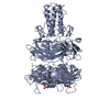
| タイトル | The tail structure of bacteriophage T4 and its mechanism of contraction. | |||||||||
 マップデータ マップデータ | EM density map for the extended tail of bacteriophage T4 | |||||||||
 試料 試料 |
| |||||||||
| 機能・相同性 |  機能・相同性情報 機能・相同性情報virus tail, sheath / symbiont genome ejection through host cell envelope, contractile tail mechanism / virus tail, baseplate / viral tail assembly / viral release from host cell / virion component 類似検索 - 分子機能 | |||||||||
| 生物種 |  Enterobacteria phage T4 (ファージ) Enterobacteria phage T4 (ファージ) | |||||||||
| 手法 | 単粒子再構成法 / クライオ電子顕微鏡法 / ネガティブ染色法 / 解像度: 15.0 Å | |||||||||
 データ登録者 データ登録者 | Kostyuchenko VA / Chipman PR / Leiman PG / Arisaka F / Mesyanzhinov VV / Rossmann MG | |||||||||
 引用 引用 | ジャーナル: Nat Struct Mol Biol / 年: 2005 タイトル: The tail structure of bacteriophage T4 and its mechanism of contraction. 著者: Victor A Kostyuchenko / Paul R Chipman / Petr G Leiman / Fumio Arisaka / Vadim V Mesyanzhinov / Michael G Rossmann /  要旨: Bacteriophage T4 and related viruses have a contractile tail that serves as an efficient mechanical device for infecting bacteria. A three-dimensional cryo-EM reconstruction of the mature T4 tail ...Bacteriophage T4 and related viruses have a contractile tail that serves as an efficient mechanical device for infecting bacteria. A three-dimensional cryo-EM reconstruction of the mature T4 tail assembly at 15-A resolution shows the hexagonal dome-shaped baseplate, the extended contractile sheath, the long tail fibers attached to the baseplate and the collar formed by six whiskers that interact with the long tail fibers. Comparison with the structure of the contracted tail shows that tail contraction is associated with a substantial rearrangement of the domains within the sheath protein and results in shortening of the sheath to about one-third of its original length. During contraction, the tail tube extends beneath the baseplate by about one-half of its total length and rotates by 345 degrees , allowing it to cross the host's periplasmic space. | |||||||||
| 履歴 |
|
- 構造の表示
構造の表示
| ムービー |
ムービービューア |
|---|---|
| 構造ビューア | EMマップ: SurfViewMolmilJmol/JSmol |
| 添付画像 |

- ダウンロードとリンク
-EMDBアーカイブ
| マップデータ | emd_1126.map.gz | 18.4 MB | EMDBマップデータ形式 | |
|---|---|---|---|---|
| ヘッダ (付随情報) | emd-1126-v30.xmlemd-1126.xml | 9.2 KB 9.2 KB | 表示 表示 | EMDBヘッダ |
| 画像 | 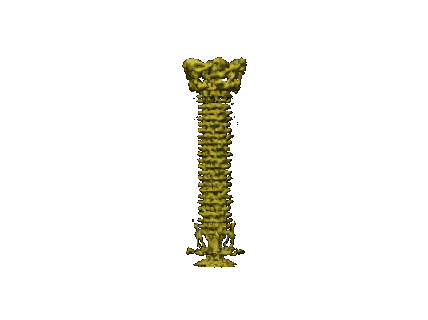 1126.gif 1126.gif | 11.6 KB | ||
| アーカイブディレクトリ |  http://ftp.pdbj.org/pub/emdb/structures/EMD-1126ftp://ftp.pdbj.org/pub/emdb/structures/EMD-1126 http://ftp.pdbj.org/pub/emdb/structures/EMD-1126ftp://ftp.pdbj.org/pub/emdb/structures/EMD-1126 | HTTPS FTP |
-関連構造データ
-リンク
| EMDBのページ | EMDB (EBI/PDBe) / EMDataResource |
|---|---|
| 「今月の分子」の関連する項目 |


-マップ
| ファイル | ダウンロード / ファイル: emd_1126.map.gz / 形式: CCP4 / 大きさ: 45.9 MB / タイプ: IMAGE STORED AS FLOATING POINT NUMBER (4 BYTES) | ||||||||||||||||||||||||||||||||||||||||||||||||||||||||||||||||||||
|---|---|---|---|---|---|---|---|---|---|---|---|---|---|---|---|---|---|---|---|---|---|---|---|---|---|---|---|---|---|---|---|---|---|---|---|---|---|---|---|---|---|---|---|---|---|---|---|---|---|---|---|---|---|---|---|---|---|---|---|---|---|---|---|---|---|---|---|---|---|
| 注釈 | EM density map for the extended tail of bacteriophage T4 | ||||||||||||||||||||||||||||||||||||||||||||||||||||||||||||||||||||
| 投影像・断面図 | 画像のコントロール
画像は Spider により作成 これらの図は立方格子座標系で作成されたものです | ||||||||||||||||||||||||||||||||||||||||||||||||||||||||||||||||||||
| ボクセルのサイズ | X=Y=Z: 3.97 Å | ||||||||||||||||||||||||||||||||||||||||||||||||||||||||||||||||||||
| 密度 |
| ||||||||||||||||||||||||||||||||||||||||||||||||||||||||||||||||||||
| 対称性 | 空間群: 1 | ||||||||||||||||||||||||||||||||||||||||||||||||||||||||||||||||||||
| 詳細 | EMDB XML:
CCP4マップ ヘッダ情報:
| ||||||||||||||||||||||||||||||||||||||||||||||||||||||||||||||||||||
 Z (Sec.)
 X (Row.)
 Y (Col.)


-添付データ
- 試料の構成要素
-全体 : native T4 phage
| 全体 | 名称: native T4 phage |
|---|---|
| 要素 |
|
-超分子 #1000: native T4 phage
| 超分子 | 名称: native T4 phage / タイプ: sample / ID: 1000 / 集合状態: hexameric tails bound to 5-2 capsids / Number unique components: 1 |
|---|
-超分子 #1: Enterobacteria phage T4
| 超分子 | 名称: Enterobacteria phage T4 / タイプ: virus / ID: 1 / 詳細: native phage / NCBI-ID: 10665 / 生物種: Enterobacteria phage T4 / ウイルスタイプ: VIRION / ウイルス・単離状態: STRAIN / ウイルス・エンベロープ: No / ウイルス・中空状態: No |
|---|---|
| 宿主 | 生物種:  |
-実験情報
-構造解析
| 手法 | ネガティブ染色法, クライオ電子顕微鏡法 |
|---|---|
 解析 解析 | 単粒子再構成法 |
| 試料の集合状態 | particle |
-試料調製
| 緩衝液 | pH: 7 / 詳細: H20 |
|---|---|
| 染色 | タイプ: NEGATIVE / 詳細: cryo |
| グリッド | 詳細: 200 mesh copper grids |
| 凍結 | 凍結剤: ETHANE |
- 電子顕微鏡法
| 顕微鏡 | FEI/PHILIPS CM300FEG/ST |
|---|---|
| 日付 | 2003年1月10日 |
| 撮影 | デジタル化 - スキャナー: ZEISS SCAI / デジタル化 - サンプリング間隔: 7 µm / 実像数: 89 / ビット/ピクセル: 12 |
| 電子線 | 加速電圧: 300 kV / 電子線源:  FIELD EMISSION GUN FIELD EMISSION GUN |
| 電子光学系 | 倍率(補正後): 47000 / 照射モード: SPOT SCAN / 撮影モード: BRIGHT FIELD / Cs: 2 mm / 最大 デフォーカス(公称値): 4.0 µm / 最小 デフォーカス(公称値): 1.0 µm / 倍率(公称値): 45000 |
| 試料ステージ | 試料ホルダーモデル: GATAN LIQUID NITROGEN |
-画像解析
| CTF補正 | 詳細: each particle image |
|---|---|
| 最終 再構成 | 想定した対称性 - 点群: C6 (6回回転対称) / アルゴリズム: OTHER / 解像度のタイプ: BY AUTHOR / 解像度: 15.0 Å / 解像度の算出法: FSC 0.5 CUT-OFF / ソフトウェア - 名称: SPIDER-like 詳細: reconstruction done with in-house implementation of BP RP algorithm from SPIDER to support non-cubicreconstruction volumes 使用した粒子像数: 3029 |
-原子モデル構築 1
| 初期モデル | PDB ID: |
|---|---|
| 精密化 | 空間: REAL |
| 得られたモデル |  PDB-1zku: 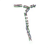 PDB-2bsg:  PDB-3foh:  PDB-3j2m: |

-原子モデル構築 2
| 初期モデル | PDB ID: |
|---|---|
| 精密化 | 空間: REAL |
| 得られたモデル | PDB-1zku: PDB-2bsg: PDB-3foh: PDB-3j2m: |