Movie
Movie Controller

[English] 日本語
 Yorodumi
- EMDB-10863: Structure of the SARS-CoV-2 spike S1 protein in complex with CR30... -
+ Open data

- Basic information
| Entry | Database: EMDB / ID: EMD-10863 | |||||||||
|---|---|---|---|---|---|---|---|---|---|---|
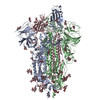
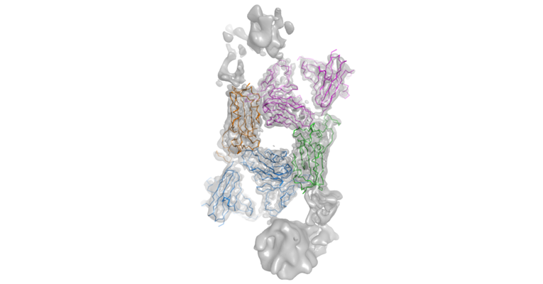
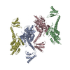
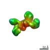
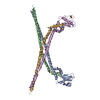
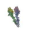
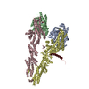
| Title | Structure of the SARS-CoV-2 spike S1 protein in complex with CR3022 Fab | |||||||||
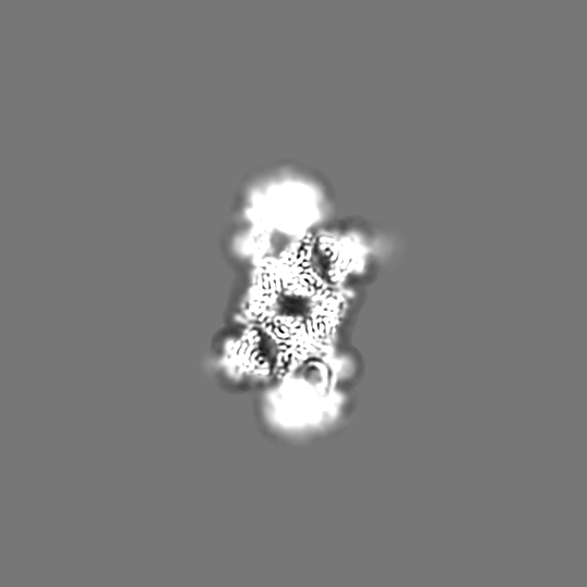
 Map data Map data | The map has strongly anisotropic resolution. RBD and CR3022 Fab are fitted. The S1 N-terminal domains are not well defined and can be viewed at lower contour level, 0.345 | |||||||||
 Sample Sample |
| |||||||||
 Keywords Keywords | SARS-CoV-2 Spike protein / RBD / CR3022 / complex / VIRAL PROTEIN | |||||||||
| Function / homology |  Function and homology information Function and homology informationsymbiont-mediated disruption of host tissue / Maturation of spike protein / Translation of Structural Proteins / Virion Assembly and Release / host cell surface / host extracellular region / symbiont-mediated-mediated suppression of host tetherin activity / Induction of Cell-Cell Fusion / structural constituent of virion / positive regulation of viral entry into host cell ...symbiont-mediated disruption of host tissue / Maturation of spike protein / Translation of Structural Proteins / Virion Assembly and Release / host cell surface / host extracellular region / symbiont-mediated-mediated suppression of host tetherin activity / Induction of Cell-Cell Fusion / structural constituent of virion / positive regulation of viral entry into host cell / membrane fusion / host cell endoplasmic reticulum-Golgi intermediate compartment membrane / Attachment and Entry / entry receptor-mediated virion attachment to host cell / receptor-mediated virion attachment to host cell / host cell surface receptor binding / symbiont-mediated suppression of host innate immune response / endocytosis involved in viral entry into host cell / receptor ligand activity / fusion of virus membrane with host plasma membrane / fusion of virus membrane with host endosome membrane / viral envelope / symbiont entry into host cell / virion attachment to host cell / host cell plasma membrane / SARS-CoV-2 activates/modulates innate and adaptive immune responses / virion membrane / membrane / identical protein binding / plasma membrane Similarity search - Function | |||||||||
| Biological species |   Severe acute respiratory syndrome coronavirus 2 / Severe acute respiratory syndrome coronavirus 2 /  Homo sapiens (human) Homo sapiens (human) | |||||||||
| Method | single particle reconstruction / cryo EM / Resolution: 3.3 Å | |||||||||
 Authors Authors | Huo J / Zhao Y | |||||||||
| Funding support |  United Kingdom, 2 items United Kingdom, 2 items
| |||||||||
 Citation Citation | Journal: Cell Host Microbe / Year: 2020 Title: Neutralization of SARS-CoV-2 by Destruction of the Prefusion Spike. Authors: Jiandong Huo / Yuguang Zhao / Jingshan Ren / Daming Zhou / Helen M E Duyvesteyn / Helen M Ginn / Loic Carrique / Tomas Malinauskas / Reinis R Ruza / Pranav N M Shah / Tiong Kit Tan / Pramila ...Authors: Jiandong Huo / Yuguang Zhao / Jingshan Ren / Daming Zhou / Helen M E Duyvesteyn / Helen M Ginn / Loic Carrique / Tomas Malinauskas / Reinis R Ruza / Pranav N M Shah / Tiong Kit Tan / Pramila Rijal / Naomi Coombes / Kevin R Bewley / Julia A Tree / Julika Radecke / Neil G Paterson / Piyada Supasa / Juthathip Mongkolsapaya / Gavin R Screaton / Miles Carroll / Alain Townsend / Elizabeth E Fry / Raymond J Owens / David I Stuart /  Abstract: There are as yet no licensed therapeutics for the COVID-19 pandemic. The causal coronavirus (SARS-CoV-2) binds host cells via a trimeric spike whose receptor binding domain (RBD) recognizes ...There are as yet no licensed therapeutics for the COVID-19 pandemic. The causal coronavirus (SARS-CoV-2) binds host cells via a trimeric spike whose receptor binding domain (RBD) recognizes angiotensin-converting enzyme 2, initiating conformational changes that drive membrane fusion. We find that the monoclonal antibody CR3022 binds the RBD tightly, neutralizing SARS-CoV-2, and report the crystal structure at 2.4 Å of the Fab/RBD complex. Some crystals are suitable for screening for entry-blocking inhibitors. The highly conserved, structure-stabilizing CR3022 epitope is inaccessible in the prefusion spike, suggesting that CR3022 binding facilitates conversion to the fusion-incompetent post-fusion state. Cryogenic electron microscopy (cryo-EM) analysis confirms that incubation of spike with CR3022 Fab leads to destruction of the prefusion trimer. Presentation of this cryptic epitope in an RBD-based vaccine might advantageously focus immune responses. Binders at this epitope could be useful therapeutically, possibly in synergy with an antibody that blocks receptor attachment. | |||||||||
| History |
|
- Structure visualization
| Movie |
Movie viewer |
|---|---|
| Structure viewer | EM map: SurfViewMolmilJmol/JSmol |
| Supplemental images |
- Downloads & links
-EMDB archive
| Map data | emd_10863.map.gz | 9 MB | EMDB map data format | |
|---|---|---|---|---|
| Header (meta data) | emd-10863-v30.xmlemd-10863.xml | 24.6 KB 24.6 KB | Display Display | EMDB header |
| Images |  emd_10863.png emd_10863.png | 145.7 KB | ||
| Filedesc metadata | emd-10863.cif.gz | 7.2 KB | ||
| Archive directory |  http://ftp.pdbj.org/pub/emdb/structures/EMD-10863ftp://ftp.pdbj.org/pub/emdb/structures/EMD-10863 http://ftp.pdbj.org/pub/emdb/structures/EMD-10863ftp://ftp.pdbj.org/pub/emdb/structures/EMD-10863 | HTTPS FTP |
-Related structure data
| Related structure data |  6yorMC  6ylaC  6ym0C  6z97C M: atomic model generated by this map C: citing same article ( |
|---|---|
| Similar structure data |


-Links
| EMDB pages | EMDB (EBI/PDBe) / EMDataResource |
|---|---|
| Related items in Molecule of the Month |


-Map
| File | Download / File: emd_10863.map.gz / Format: CCP4 / Size: 600.7 MB / Type: IMAGE STORED AS FLOATING POINT NUMBER (4 BYTES) | ||||||||||||||||||||||||||||||||||||||||||||||||||||||||||||
|---|---|---|---|---|---|---|---|---|---|---|---|---|---|---|---|---|---|---|---|---|---|---|---|---|---|---|---|---|---|---|---|---|---|---|---|---|---|---|---|---|---|---|---|---|---|---|---|---|---|---|---|---|---|---|---|---|---|---|---|---|---|
| Annotation | The map has strongly anisotropic resolution. RBD and CR3022 Fab are fitted. The S1 N-terminal domains are not well defined and can be viewed at lower contour level, 0.345 | ||||||||||||||||||||||||||||||||||||||||||||||||||||||||||||
| Projections & slices | Image control
Images are generated by Spider. | ||||||||||||||||||||||||||||||||||||||||||||||||||||||||||||
| Voxel size | X=Y=Z: 0.83 Å | ||||||||||||||||||||||||||||||||||||||||||||||||||||||||||||
| Density |
| ||||||||||||||||||||||||||||||||||||||||||||||||||||||||||||
| Symmetry | Space group: 1 | ||||||||||||||||||||||||||||||||||||||||||||||||||||||||||||
| Details | EMDB XML:
CCP4 map header:
| ||||||||||||||||||||||||||||||||||||||||||||||||||||||||||||
 Z (Sec.)
 Y (Row.)
 X (Col.)


-Supplemental data
- Sample components
-Entire : Binary complex of SARS-CoV-2 spike S1 with CR3022 Fab
| Entire | Name: Binary complex of SARS-CoV-2 spike S1 with CR3022 Fab |
|---|---|
| Components |
|
-Supramolecule #1: Binary complex of SARS-CoV-2 spike S1 with CR3022 Fab
| Supramolecule | Name: Binary complex of SARS-CoV-2 spike S1 with CR3022 Fab / type: complex / ID: 1 / Parent: 0 / Macromolecule list: all |
|---|
-Supramolecule #2: RBD
| Supramolecule | Name: RBD / type: complex / ID: 2 / Parent: 1 / Macromolecule list: #1 |
|---|---|
| Source (natural) | Organism: Severe acute respiratory syndrome coronavirus 2 |
-Supramolecule #3: Immunoblobulin heavy chain
| Supramolecule | Name: Immunoblobulin heavy chain / type: complex / ID: 3 / Parent: 1 / Macromolecule list: #2 |
|---|---|
| Source (natural) | Organism: Homo sapiens (human) |
-Supramolecule #4: Immunoblobulin light chain
| Supramolecule | Name: Immunoblobulin light chain / type: complex / ID: 4 / Parent: 1 / Macromolecule list: #3 |
|---|---|
| Source (natural) | Organism: Homo sapiens (human) |
-Macromolecule #1: Spike glycoprotein
| Macromolecule | Name: Spike glycoprotein / type: protein_or_peptide / ID: 1 / Number of copies: 2 / Enantiomer: LEVO |
|---|---|
| Source (natural) | Organism: Severe acute respiratory syndrome coronavirus 2 |
| Molecular weight | Theoretical: 22.758518 KDa |
| Recombinant expression | Organism: Homo sapiens (human) |
| Sequence | String: PNITNLCPFG EVFNATRFAS VYAWNRKRIS NCVADYSVLY NSASFSTFKC YGVSPTKLND LCFTNVYADS FVIRGDEVRQ IAPGQTGKI ADYNYKLPDD FTGCVIAWNS NNLDSKVGGN YNYLYRLFRK SNLKPFERDI STEIYQAGST PCNGVEGFNC Y FPLQSYGF ...String: PNITNLCPFG EVFNATRFAS VYAWNRKRIS NCVADYSVLY NSASFSTFKC YGVSPTKLND LCFTNVYADS FVIRGDEVRQ IAPGQTGKI ADYNYKLPDD FTGCVIAWNS NNLDSKVGGN YNYLYRLFRK SNLKPFERDI STEIYQAGST PCNGVEGFNC Y FPLQSYGF QPTNGVGYQP YRVVVLSFEL LHAPATVCGP KKSTN UniProtKB: Spike glycoprotein |
-Macromolecule #2: IgG H chain
| Macromolecule | Name: IgG H chain / type: protein_or_peptide / ID: 2 / Number of copies: 2 / Enantiomer: LEVO |
|---|---|
| Source (natural) | Organism: Homo sapiens (human) |
| Molecular weight | Theoretical: 24.45552 KDa |
| Recombinant expression | Organism: Homo sapiens (human) |
| Sequence | String: TQMQLVQSGT EVKKPGESLK ISCKGSGYGF ITYWIGWVRQ MPGKGLEWMG IIYPGDSETR YSPSFQGQVT ISADKSINTA YLQWSSLKA SDTAIYYCAG GSGISTPMDV WGQGTTVTVA STKGPSVFPL APSSKSTSGG TAALGCLVKD YFPEPVTVSW N SGALTSGV ...String: TQMQLVQSGT EVKKPGESLK ISCKGSGYGF ITYWIGWVRQ MPGKGLEWMG IIYPGDSETR YSPSFQGQVT ISADKSINTA YLQWSSLKA SDTAIYYCAG GSGISTPMDV WGQGTTVTVA STKGPSVFPL APSSKSTSGG TAALGCLVKD YFPEPVTVSW N SGALTSGV HTFPAVLQSS GLYSLSSVVT VPSSSLGTQT YICNVNHKPS NTKVDKKVEP KSCDKHHHHH H |
-Macromolecule #3: IgG L chain
| Macromolecule | Name: IgG L chain / type: protein_or_peptide / ID: 3 / Number of copies: 2 / Enantiomer: LEVO |
|---|---|
| Source (natural) | Organism: Homo sapiens (human) |
| Molecular weight | Theoretical: 24.289885 KDa |
| Recombinant expression | Organism: Homo sapiens (human) |
| Sequence | String: DIQLTQSPDS LAVSLGERAT INCKSSQSVL YSSINKNYLA WYQQKPGQPP KLLIYWASTR ESGVPDRFSG SGSGTDFTLT ISSLQAEDV AVYYCQQYYS TPYTFGQGTK VEIKRTVAAP SVFIFPPSDE QLKSGTASVV CLLNNFYPRE AKVQWKVDNA L QSGNSQES ...String: DIQLTQSPDS LAVSLGERAT INCKSSQSVL YSSINKNYLA WYQQKPGQPP KLLIYWASTR ESGVPDRFSG SGSGTDFTLT ISSLQAEDV AVYYCQQYYS TPYTFGQGTK VEIKRTVAAP SVFIFPPSDE QLKSGTASVV CLLNNFYPRE AKVQWKVDNA L QSGNSQES VTEQDSKDST YSLSSTLTLS KADYEKHKVY ACEVTHQGLS SPVTKSFNRG EC |
-Experimental details
-Structure determination
| Method | cryo EM |
|---|---|
 Processing Processing | single particle reconstruction |
| Aggregation state | particle |
-Sample preparation
| Buffer | pH: 8 / Details: 2 mM Tris pH 8.0, 200 mM NaCl, 0.02 % NaN3 |
|---|---|
| Vitrification | Cryogen name: ETHANE |
- Electron microscopy
| Microscope | FEI TITAN KRIOS |
|---|---|
| Image recording | Film or detector model: GATAN K3 (6k x 4k) / Number grids imaged: 1 / Number real images: 13307 / Average exposure time: 1.4 sec. / Average electron dose: 42.0 e/Å2 |
| Electron beam | Acceleration voltage: 300 kV / Electron source:  FIELD EMISSION GUN FIELD EMISSION GUN |
| Electron optics | Illumination mode: FLOOD BEAM / Imaging mode: BRIGHT FIELD |
| Experimental equipment |  Model: Titan Krios / Image courtesy: FEI Company |