 Movie
Movie Controller

[English] 日本語
 Yorodumi
- EMDB-1057: The structure of echovirus type 12 bound to a two-domain fragment... -
+ Open data

- Basic information
| Entry | Database: EMDB / ID: EMD-1057 | |||||||||
|---|---|---|---|---|---|---|---|---|---|---|
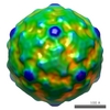
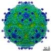
| Title | The structure of echovirus type 12 bound to a two-domain fragment of its cellular attachment protein decay-accelerating factor (CD 55). | |||||||||
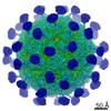
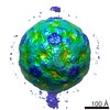
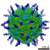
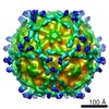
 Map data Map data | Three dimensional reconstruction of echovirus type 12 bound to domains three and four of its cellular receptor decay-accelerating factor. Calculated from cryo-negative stain images | |||||||||
 Sample Sample |
| |||||||||
| Function / homology |  Function and homology information Function and homology informationregulation of lipopolysaccharide-mediated signaling pathway / negative regulation of complement activation / regulation of complement-dependent cytotoxicity / regulation of complement activation / respiratory burst / positive regulation of CD4-positive, alpha-beta T cell activation / positive regulation of CD4-positive, alpha-beta T cell proliferation / Class B/2 (Secretin family receptors) / symbiont-mediated suppression of host cytoplasmic pattern recognition receptor signaling pathway via inhibition of RIG-I activity / ficolin-1-rich granule membrane ...regulation of lipopolysaccharide-mediated signaling pathway / negative regulation of complement activation / regulation of complement-dependent cytotoxicity / regulation of complement activation / respiratory burst / positive regulation of CD4-positive, alpha-beta T cell activation / positive regulation of CD4-positive, alpha-beta T cell proliferation / Class B/2 (Secretin family receptors) / symbiont-mediated suppression of host cytoplasmic pattern recognition receptor signaling pathway via inhibition of RIG-I activity / ficolin-1-rich granule membrane / complement activation, classical pathway / transport vesicle / side of membrane / COPI-mediated anterograde transport / endoplasmic reticulum-Golgi intermediate compartment membrane / Regulation of Complement cascade / secretory granule membrane / picornain 2A / symbiont-mediated suppression of host mRNA export from nucleus / symbiont genome entry into host cell via pore formation in plasma membrane / picornain 3C / T=pseudo3 icosahedral viral capsid / positive regulation of T cell cytokine production / host cell cytoplasmic vesicle membrane / ribonucleoside triphosphate phosphatase activity / nucleoside-triphosphate phosphatase / virus receptor activity / positive regulation of cytosolic calcium ion concentration / channel activity / monoatomic ion transmembrane transport / DNA replication / RNA helicase activity / membrane raft / endocytosis involved in viral entry into host cell / Golgi membrane / symbiont-mediated activation of host autophagy / innate immune response / RNA-directed RNA polymerase / cysteine-type endopeptidase activity / viral RNA genome replication / RNA-directed RNA polymerase activity / Neutrophil degranulation / DNA-templated transcription / lipid binding / virion attachment to host cell / host cell nucleus / structural molecule activity / cell surface / proteolysis / RNA binding / extracellular exosome / extracellular region / zinc ion binding / ATP binding / plasma membrane Similarity search - Function | |||||||||
| Biological species |  Homo sapiens (human) / Homo sapiens (human) /  Human echovirus 12 Human echovirus 12 | |||||||||
| Method | single particle reconstruction / cryo EM / negative staining / Resolution: 16.0 Å | |||||||||
 Authors Authors | Bhella D / Goodfellow IG / Roversi P / Pettigrew D / Chaudhry Y / Evans DJ / Lea SM | |||||||||
 Citation Citation | Journal: J Biol Chem / Year: 2004 Title: The structure of echovirus type 12 bound to a two-domain fragment of its cellular attachment protein decay-accelerating factor (CD 55). Authors: David Bhella / Ian G Goodfellow / Pietro Roversi / David Pettigrew / Yasmin Chaudhry / David J Evans / Susan M Lea /  Abstract: Echovirus type 12 (EV12), an Enterovirus of the Picornaviridae family, uses the complement regulator decay-accelerating factor (DAF, CD55) as a cellular receptor. We have calculated a three- ...Echovirus type 12 (EV12), an Enterovirus of the Picornaviridae family, uses the complement regulator decay-accelerating factor (DAF, CD55) as a cellular receptor. We have calculated a three-dimensional reconstruction of EV12 bound to a fragment of DAF consisting of short consensus repeat domains 3 and 4 from cryo-negative stain electron microscopy data (EMD code 1057). This shows that, as for an earlier reconstruction of the related echovirus type 7 bound to DAF, attachment is not within the viral canyon but occurs close to the 2-fold symmetry axes. Despite this general similarity our reconstruction reveals a receptor interaction that is quite different from that observed for EV7. Fitting of the crystallographic co-ordinates for DAF(34) and EV11 into the reconstruction shows a close agreement between the crystal structure of the receptor fragment and the density for the virus-bound receptor, allowing unambiguous positioning of the receptor with respect to the virion (PDB code 1UPN). Our finding that the mode of virus-receptor interaction in EV12 is distinct from that seen for EV7 raises interesting questions regarding the evolution and biological significance of the DAF binding phenotype in these viruses. | |||||||||
| History |
|
- Structure visualization
| Movie |
Movie viewer |
|---|---|
| Structure viewer | EM map: SurfViewMolmilJmol/JSmol |
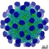
| Supplemental images |
 UCSF Chimera
- Downloads & links
-EMDB archive
| Map data | emd_1057.map.gz | 1.3 MB | EMDB map data format | |
|---|---|---|---|---|
| Header (meta data) | emd-1057-v30.xmlemd-1057.xml | 10.6 KB 10.6 KB | Display Display | EMDB header |
| Images | 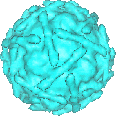 1057.gif 1057.gif | 74.1 KB | ||
| Archive directory |  http://ftp.pdbj.org/pub/emdb/structures/EMD-1057ftp://ftp.pdbj.org/pub/emdb/structures/EMD-1057 http://ftp.pdbj.org/pub/emdb/structures/EMD-1057ftp://ftp.pdbj.org/pub/emdb/structures/EMD-1057 | HTTPS FTP |
-Related structure data
| Related structure data | 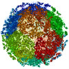 1upnMC 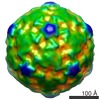 1058C M: atomic model generated by this map C: citing same article ( |
|---|---|
| Similar structure data |
-Links
| EMDB pages | EMDB (EBI/PDBe) / EMDataResource |
|---|---|
| Related items in Molecule of the Month |


-Map
| File | Download / File: emd_1057.map.gz / Format: CCP4 / Size: 20 MB / Type: IMAGE STORED AS FLOATING POINT NUMBER (4 BYTES) | ||||||||||||||||||||||||||||||||||||||||||||||||||||||||||||||||||||
|---|---|---|---|---|---|---|---|---|---|---|---|---|---|---|---|---|---|---|---|---|---|---|---|---|---|---|---|---|---|---|---|---|---|---|---|---|---|---|---|---|---|---|---|---|---|---|---|---|---|---|---|---|---|---|---|---|---|---|---|---|---|---|---|---|---|---|---|---|---|
| Annotation | Three dimensional reconstruction of echovirus type 12 bound to domains three and four of its cellular receptor decay-accelerating factor. Calculated from cryo-negative stain images | ||||||||||||||||||||||||||||||||||||||||||||||||||||||||||||||||||||
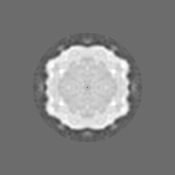
| Projections & slices | Image control
Images are generated by Spider. | ||||||||||||||||||||||||||||||||||||||||||||||||||||||||||||||||||||
| Voxel size | X=Y=Z: 3.42 Å | ||||||||||||||||||||||||||||||||||||||||||||||||||||||||||||||||||||
| Density |
| ||||||||||||||||||||||||||||||||||||||||||||||||||||||||||||||||||||
| Symmetry | Space group: 1 | ||||||||||||||||||||||||||||||||||||||||||||||||||||||||||||||||||||
| Details | EMDB XML:
CCP4 map header:
| ||||||||||||||||||||||||||||||||||||||||||||||||||||||||||||||||||||
 Z (Sec.)
 Y (Row.)
 X (Col.)


-Supplemental data
- Sample components
-Entire : Echovirus type 12 bound to decay accelerating factor domains 3 and 4
| Entire | Name: Echovirus type 12 bound to decay accelerating factor domains 3 and 4 |
|---|---|
| Components |
|
-Supramolecule #1000: Echovirus type 12 bound to decay accelerating factor domains 3 and 4
| Supramolecule | Name: Echovirus type 12 bound to decay accelerating factor domains 3 and 4 type: sample / ID: 1000 / Number unique components: 2 |
|---|
-Supramolecule #1: Human echovirus 12
| Supramolecule | Name: Human echovirus 12 / type: virus / ID: 1 / Name.synonym: EV12 / NCBI-ID: 35293 / Sci species name: Human echovirus 12 / Virus type: VIRION / Virus isolate: SEROTYPE / Virus enveloped: No / Virus empty: No / Syn species name: EV12 |
|---|---|
| Host (natural) | Organism: Homo sapiens (human) / synonym: VERTEBRATES |
-Supramolecule #2: Decay accelerating factor domains 3 and 4
| Supramolecule | Name: Decay accelerating factor domains 3 and 4 / type: organelle_or_cellular_component / ID: 2 / Name.synonym: DAF34 / Number of copies: 60 / Oligomeric state: Monomer / Recombinant expression: Yes |
|---|---|
| Source (natural) | Organism: Homo sapiens (human) / synonym: Human |
| Recombinant expression | Organism:  Komagataella pastoris (fungus) Komagataella pastoris (fungus) |
-Experimental details
-Structure determination
| Method | negative staining, cryo EM |
|---|---|
 Processing Processing | single particle reconstruction |
| Aggregation state | particle |
-Sample preparation
| Concentration | 0.2 mg/mL |
|---|---|
| Buffer | pH: 7.4 / Details: PBS A |
| Staining | Type: NEGATIVE Details: Protein absorbed to grid, floated onto 20% ammonium molybdate (pH 7.4), blotted and plunged into liquid ethane |
| Grid | Details: 400 mesh Quantifoils |
| Vitrification | Cryogen name: ETHANE / Method: blot for 2 seconds, wait for 2 seconds plunge |
- Electron microscopy
| Microscope | JEOL 1200EXII |
|---|---|
| Alignment procedure | Legacy - Astigmatism: objective astigmatism corrected at 200k x |
| Details | MICROSCOPE JEOL 1200 EX with OXFORD INSTRUMENTS CRYO-TRANSFER STAGE |
| Image recording | Category: FILM / Film or detector model: KODAK SO-163 FILM / Digitization - Scanner: OTHER / Digitization - Sampling interval: 3.42 µm / Number real images: 16 / Details: Images scanned on a Dunvegan HiScan / Bits/pixel: 16 |
| Electron beam | Acceleration voltage: 120 kV / Electron source: LAB6 |
| Electron optics | Calibrated magnification: 29200 / Illumination mode: FLOOD BEAM / Imaging mode: BRIGHT FIELD / Cs: 3.4 mm / Nominal defocus max: 2.0 µm / Nominal defocus min: 0.3 µm / Nominal magnification: 30000 |
| Sample stage | Specimen holder: side entry / Specimen holder model: OTHER |
-Image processing
| CTF correction | Details: Merged individual particles from focal pairs |
|---|---|
| Final reconstruction | Applied symmetry - Point group: I (icosahedral) / Algorithm: OTHER / Resolution.type: BY AUTHOR / Resolution: 16.0 Å / Resolution method: FSC 0.5 CUT-OFF / Software - Name: EM3DR2, PFT2, CTFMIX Details: Particles were aligned using a model based strategy starting with a model derived from the crystallographic co-ordinates of EV-1, filtered to 16 Angstroms resolution. The program is called ...Details: Particles were aligned using a model based strategy starting with a model derived from the crystallographic co-ordinates of EV-1, filtered to 16 Angstroms resolution. The program is called PFT (Polar Fourier Transform). The reconstructions were calculated using the EM3DR2 program which is based on the standard method of calculating icosahedral reconstructions as described by Crowther,'Fourier-Bessel'. Number images used: 903 |
-Atomic model buiding 1
| Details | 3D crystal structure fitting details lodged with PDB 1UPN |
|---|---|
| Output model | PDB-1upn: |
