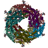
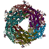
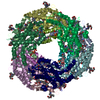
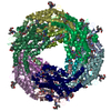
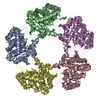
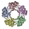
ジャーナル: Structure / 年: 1998 タイトル: Three-dimensional reconstructions from cryoelectron microscopy images reveal an intimate complex between helicase DnaB and its loading partner DnaC. 著者: C San Martin / M Radermacher / B Wolpensinger / A Engel / C S Miles / N E Dixon / J M Carazo / 要旨: BACKGROUND: DNA helicases play a fundamental role in all aspects of nucleic acid metabolism and defects in these enzymes have been implicated in a number of inherited human disorders. DnaB is the ...BACKGROUND: DNA helicases play a fundamental role in all aspects of nucleic acid metabolism and defects in these enzymes have been implicated in a number of inherited human disorders. DnaB is the major replicative DNA helicase in Escherichia coli and has been used as a model system for studying the structure and function of hexameric helicases. The native protein is a hexamer of identical subunits, which in solution forms a complex with six molecules of the loading protein DnaC. DnaB is delivered from this complex onto the DNA template, with the subsequent release of DnaC. We report here the structures of the DnaB helicase hexamer and its complex with DnaC under a defined set of experimental conditions, as determined by three-dimensional cryoelectron microscopy. It was hoped that the structures would provide insight into the mechanisms of helicase activity. RESULTS: The DnaB structure reveals that six DnaB monomers assemble as three asymmetric dimers to form a polar, ring-like hexamer. The hexamer has two faces, one displaying threefold and the other ...RESULTS: The DnaB structure reveals that six DnaB monomers assemble as three asymmetric dimers to form a polar, ring-like hexamer. The hexamer has two faces, one displaying threefold and the other sixfold symmetry. The six DnaC protomers bind tightly to the sixfold face of the DnaB hexamer. This is the first report of a three-dimensional structure of a helicase obtained using cryoelectron microscopy, and the first report of the structure of a helicase in complex with a loading protein. CONCLUSIONS: The structures of the DnaB helicase and its complex with DnaC reveal some interesting structural features relevant to helicase function and to the assembly of the two-protein complex. ...CONCLUSIONS: The structures of the DnaB helicase and its complex with DnaC reveal some interesting structural features relevant to helicase function and to the assembly of the two-protein complex. The results presented here provide a basis for a more complete understanding of the structure and function of these important proteins.
GO: DNA helicase activity / InterPro: INTERPRO: IPR001198
-
実験情報
-
構造解析
手法
クライオ電子顕微鏡法
解析
単粒子再構成法
試料の集合状態
particle
-
試料調製
濃度
.04 mg/mL
緩衝液
pH: 7.6 詳細: 50 mM Tris.HCl pH 7.6 2 mM DTT 5 mM MgCl2 200 mM NaCl 0.25 mM ADP
凍結
凍結剤: ETHANE / 装置: HOMEMADE PLUNGER 詳細: Vitrification instrument: double side blotting device 手法: samples were adsorbed onto carbon-coated molybdenum holey grids after 30s glow discharge, and vitrified by quick plunging in liquid ethane in a double-side blotting device
 ムービー
ムービー コントローラー


 データを開く
 基本情報
基本情報 マップデータ
 試料
試料 機能・相同性情報
 データ登録者
 引用
引用
 構造の表示
構造の表示 UCSF Chimera
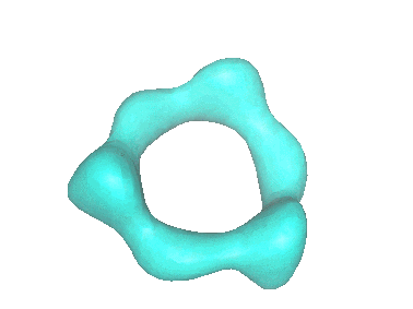
 ダウンロードとリンク
 1022.gif
 http://ftp.pdbj.org/pub/emdb/structures/EMD-1022
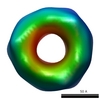


 Z (Sec.)
 Y (Row.)
 X (Col.)


 試料の構成要素
 解析
解析 電子顕微鏡法