 ムービー
ムービー コントローラー


+ データを開く

- 基本情報
基本情報
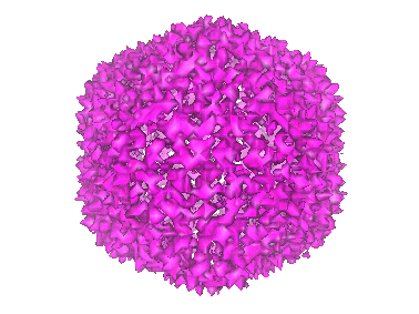
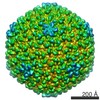
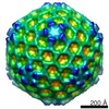
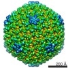
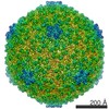
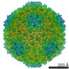
| 登録情報 | データベース: EMDB / ID: EMD-1012 | |||||||||
|---|---|---|---|---|---|---|---|---|---|---|
| タイトル | Minor proteins, mobile arms and membrane-capsid interactions in the bacteriophage PRD1 capsid. | |||||||||
 マップデータ マップデータ | This is a map of the sus607 mutant | |||||||||
 試料 試料 |
| |||||||||
| 機能・相同性 | Bacteriophage PRD1, P3 / Bacteriophage PRD1, P3, N-terminal / : / P3 major capsid protein N-terminal / P3 major capsid protein C-terminal / Group II dsDNA virus coat/capsid protein / Viral coat protein subunit / viral capsid / Major capsid protein P3 機能・相同性情報 機能・相同性情報 | |||||||||
| 生物種 |   Enterobacteria phage PRD1 (ファージ) Enterobacteria phage PRD1 (ファージ) | |||||||||
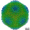
| 手法 | 単粒子再構成法 / クライオ電子顕微鏡法 / 解像度: 13.4 Å | |||||||||
 データ登録者 データ登録者 | San Martin C / Huiskonen JT / Bamford JK / Butcher SJ / Fuller SD / Bamford DH / Burnett RM | |||||||||
 引用 引用 | ジャーナル: Nat Struct Biol / 年: 2002 タイトル: Minor proteins, mobile arms and membrane-capsid interactions in the bacteriophage PRD1 capsid. 著者: Carmen San Martín / Juha T Huiskonen / Jaana K H Bamford / Sarah J Butcher / Stephen D Fuller / Dennis H Bamford / Roger M Burnett /  要旨: Bacteriophage PRD1 shares many structural and functional similarities with adenovirus. A major difference is the PRD1 internal membrane, which acts in concert with vertex proteins to translocate the ...Bacteriophage PRD1 shares many structural and functional similarities with adenovirus. A major difference is the PRD1 internal membrane, which acts in concert with vertex proteins to translocate the phage genome into the host. Multiresolution models of the PRD1 capsid, together with genetic analyses, provide fine details of the molecular interactions associated with particle stability and membrane dynamics. The N- and C-termini of the major coat protein (P3), which are required for capsid assembly, act as conformational switches bridging capsid to membrane and linking P3 trimers. Electrostatic P3-membrane interactions increase virion stability upon DNA packaging. Newly revealed proteins suggest how the metastable vertex works and how the capsid edges are stabilized. | |||||||||
| 履歴 |
|
- 構造の表示
構造の表示
| ムービー |
ムービービューア |
|---|---|
| 構造ビューア | EMマップ: SurfViewMolmilJmol/JSmol |
| 添付画像 |
- ダウンロードとリンク
-EMDBアーカイブ
| マップデータ | emd_1012.map.gz | 59.1 MB | EMDBマップデータ形式 | |
|---|---|---|---|---|
| ヘッダ (付随情報) | emd-1012-v30.xmlemd-1012.xml | 19.8 KB 19.8 KB | 表示 表示 | EMDBヘッダ |
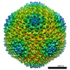
| 画像 |  1012.gif 1012.gif | 48.2 KB | ||
| その他 | emd_1012_additional_1.map.gzemd_1012_additional_2.map.gzemd_1012_additional_3.map.gz | 238.5 KB 238.8 KB 238.4 KB | ||
| アーカイブディレクトリ |  http://ftp.pdbj.org/pub/emdb/structures/EMD-1012ftp://ftp.pdbj.org/pub/emdb/structures/EMD-1012 http://ftp.pdbj.org/pub/emdb/structures/EMD-1012ftp://ftp.pdbj.org/pub/emdb/structures/EMD-1012 | HTTPS FTP |
-関連構造データ
-リンク
| EMDBのページ | EMDB (EBI/PDBe) / EMDataResource |
|---|---|
| 「今月の分子」の関連する項目 |


-マップ
| ファイル | ダウンロード / ファイル: emd_1012.map.gz / 形式: CCP4 / 大きさ: 62.5 MB / タイプ: IMAGE STORED AS FLOATING POINT NUMBER (4 BYTES) | ||||||||||||||||||||||||||||||||||||||||||||||||||||||||||||
|---|---|---|---|---|---|---|---|---|---|---|---|---|---|---|---|---|---|---|---|---|---|---|---|---|---|---|---|---|---|---|---|---|---|---|---|---|---|---|---|---|---|---|---|---|---|---|---|---|---|---|---|---|---|---|---|---|---|---|---|---|---|
| 注釈 | This is a map of the sus607 mutant | ||||||||||||||||||||||||||||||||||||||||||||||||||||||||||||
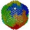
| 投影像・断面図 | 画像のコントロール
画像は Spider により作成 | ||||||||||||||||||||||||||||||||||||||||||||||||||||||||||||
| ボクセルのサイズ | X=Y=Z: 3.42 Å | ||||||||||||||||||||||||||||||||||||||||||||||||||||||||||||
| 密度 |
| ||||||||||||||||||||||||||||||||||||||||||||||||||||||||||||
| 対称性 | 空間群: 1 | ||||||||||||||||||||||||||||||||||||||||||||||||||||||||||||
| 詳細 | EMDB XML:
CCP4マップ ヘッダ情報:
| ||||||||||||||||||||||||||||||||||||||||||||||||||||||||||||
 Z (Sec.)
 Y (Row.)
 X (Col.)


-添付データ
-添付マップデータ: emd 1012 additional 1.map
| ファイル | emd_1012_additional_1.map | ||||||||||||
|---|---|---|---|---|---|---|---|---|---|---|---|---|---|
| 投影像・断面図 |
| ||||||||||||
| 密度ヒストグラム |


-添付マップデータ: emd 1012 additional 2.map
| ファイル | emd_1012_additional_2.map | ||||||||||||
|---|---|---|---|---|---|---|---|---|---|---|---|---|---|
| 投影像・断面図 |
| ||||||||||||
| 密度ヒストグラム |


-添付マップデータ: emd 1012 additional 3.map
| ファイル | emd_1012_additional_3.map | ||||||||||||
|---|---|---|---|---|---|---|---|---|---|---|---|---|---|
| 投影像・断面図 |
| ||||||||||||
| 密度ヒストグラム |


- 試料の構成要素
-全体 : Bacteriophage PRD1 sus607 mutant
| 全体 | 名称: Bacteriophage PRD1 sus607 mutant |
|---|---|
| 要素 |
|
-超分子 #1000: Bacteriophage PRD1 sus607 mutant
| 超分子 | 名称: Bacteriophage PRD1 sus607 mutant / タイプ: sample / ID: 1000 詳細: The sample is a virion containing at least 19 structural proteins and a double stranded DNA genome. This map is from the sus607 mutant and lacks protein P11 集合状態: A pseudo T=25 assembly / Number unique components: 1 |
|---|---|
| 分子量 | 理論値: 70 MDa |
-超分子 #1: Enterobacteria phage PRD1
| 超分子 | 名称: Enterobacteria phage PRD1 / タイプ: virus / ID: 1 / Name.synonym: bacteriophage PRD1 / 詳細: a T=25 virion / NCBI-ID: 10658 / 生物種: Enterobacteria phage PRD1 / ウイルスタイプ: VIRION / ウイルス・単離状態: STRAIN / ウイルス・エンベロープ: No / ウイルス・中空状態: No / Syn species name: bacteriophage PRD1 |
|---|---|
| 宿主 | 生物種:  Salmonella enterica (サルモネラ菌) / 別称: BACTERIA(EUBACTERIA) Salmonella enterica (サルモネラ菌) / 別称: BACTERIA(EUBACTERIA) |
| ウイルス殻 | Shell ID: 1 / 名称: capsid (contains proteins P3 and P30) / 直径: 70 Å / T番号(三角分割数): 25 |
-実験情報
-構造解析
| 手法 | クライオ電子顕微鏡法 |
|---|---|
 解析 解析 | 単粒子再構成法 |
| 試料の集合状態 | particle |
-試料調製
| 濃度 | 1 mg/mL |
|---|---|
| 緩衝液 | pH: 7.2 / 詳細: 20 mM Tris HCl |
| 凍結 | 凍結剤: ETHANE / チャンバー内湿度: 60 % / チャンバー内温度: 23 K / 装置: HOMEMADE PLUNGER 詳細: Vitrification instrument: EMBL plunger. vitrification carried out at 23 degrees at ambient humidity 手法: Blot for 1 s before plunging into ethane slush |
- 電子顕微鏡法
| 顕微鏡 | FEI/PHILIPS CM200FEG/ST |
|---|---|
| 温度 | 平均: 105 K |
| 日付 | 2001年11月1日 |
| 撮影 | カテゴリ: FILM / フィルム・検出器のモデル: KODAK SO-163 FILM / デジタル化 - スキャナー: ZEISS SCAI / デジタル化 - サンプリング間隔: 7.0 µm / 実像数: 15 / 平均電子線量: 6 e/Å2 / カメラ長: 44 詳細: images were scanned at 7 micron steps size and then averaged to give a final size of 14 microns Od range: 1 / ビット/ピクセル: 8 |
| 電子線 | 加速電圧: 200 kV / 電子線源:  FIELD EMISSION GUN FIELD EMISSION GUN |
| 電子光学系 | 照射モード: FLOOD BEAM / 撮影モード: BRIGHT FIELD / Cs: 2 mm / 最大 デフォーカス(公称値): 4.13 µm / 最小 デフォーカス(公称値): 0.77 µm / 倍率(公称値): 50000 |
| 試料ステージ | 試料ホルダー: eucentric / 試料ホルダーモデル: GATAN LIQUID NITROGEN |
-画像解析
| 詳細 | The particles were purified by rate-zonal centrifugation and ion-exchange chromatography Walin,Tuma,Thomas and Bamford Virology (1994) 201:1-7 |
|---|---|
| CTF補正 | 詳細: normalized sum of ctf multiplied images |
| 最終 再構成 | 想定した対称性 - 点群: I (正20面体型対称) / アルゴリズム: OTHER / 解像度のタイプ: BY AUTHOR / 解像度: 13.4 Å / 解像度の算出法: OTHER / ソフトウェア - 名称: EMBL-ICOS, MRC 詳細: final maps were calculated by making a normalized sum of seperate ctf multiplied maps Baker, T. S., Olson, N. H., and Fuller, S. D. (1999). Adding the third dimension to virus life cycles: ...詳細: final maps were calculated by making a normalized sum of seperate ctf multiplied maps Baker, T. S., Olson, N. H., and Fuller, S. D. (1999). Adding the third dimension to virus life cycles: Three-Dimensional Reconstruction of Icosahedral Viruses from Cryo-Electron Micrographs. Microbiology and Molecular Biology Reviews 63, 862-922. Butcher, S. J., Bamford, D. H., and Fuller, S. D. (1995). DNA packaging orders the membrane of bacteriophage PRD1. Embo J 14, 6078-6086. Ferlenghi, I., Gowen, B., de Haas, F., Mancini, E. J., Garoff, H., Sjoberg, M., and Fuller, S. D. (1998). The first step: activation of the Semliki Forest virus spike protein precursor causes a localized conformational change in the trimeric spike. J Mol Biol 283, 71-81. Fuller, S. D., Berriman, J. A., Butcher, S. J., and Gowen, B. E. (1995). Low pH induces swiveling of the glycoprotein heterodimers in the Semliki Forest virus spike complex. Cell 81, 715-725. Fuller, S. D., Butcher, S. J., Cheng, R. H., and Baker, T. S. (1996). Three-dimensional reconstruction of icosahedral particles--the uncommon line. J Struct Biol 116, 48-55. Mancini, E. J., Clarke, M., Gowen, B., Rutten, T., and Fuller, S. D. (2000). Cryo-electron microscopy reveals the functional organization of an enveloped virus, Semliki Forest virus. Molecular Cell 5, 255-266. Mancini, E. J., de Haas, F., and Fuller, S. D. (1997). High-resolution icosahedral reconstruction: fulfilling the promise of cryo-electron microscopy. Structure 5, 741-750. San Martin, C., Burnett, R. M., de Haas, F., Heinkel, R., Rutten, T., Fuller, S. D., Butcher, S. J., and Bamford, D. H. (2001). Combined EM/X-ray imaging yields a quasi-atomic model of the adenovirus-related bacteriophage PRD1 and shows key capsid and membrane interactions. Structure (Camb) 9, 917-930. San Martin, C., Huiskonen, J. T., Bamford, J. K., Butcher, S. J., Fuller, S. D., Bamford, D. H., and Burnett, R. M. (2002). Minor proteins, mobile arms and membrane-capsid interactions in the bacteriophage PRD1 capsid. Nat Struct Biol 9, 756-763. Sheehan, B., Fuller, S. D., Pique, M. E., and Yeager, M. (1996). AVS software for visualization in molecular microscopy. J Struct Biol 116, 99-106. 使用した粒子像数: 1729 |
| 最終 角度割当 | 詳細: range giving min eigenvalues for inversion of less than .01 |
-原子モデル構築 1
| 初期モデル | PDB ID: Chain - #0 - Chain ID: A / Chain - #1 - Chain ID: B / Chain - #2 - Chain ID: C / Chain - #3 - Chain ID: D / Chain - #4 - Chain ID: E / Chain - #5 - Chain ID: F / Chain - #6 - Chain ID: G / Chain - #7 - Chain ID: H / Chain - #8 - Chain ID: I |
|---|---|
| ソフトウェア | 名称: X-plor and emfit (M. Rossmann Cheng, R., Kuhn, R., Olson, N., Rossmann, M., and Baker, T. (1995). Nucleocapsid and glycoprotein organization in an enveloped virus. Cell 80, 621-630.) |
| 詳細 | Protocol: ridgid body. The scale of the map and the effective resolution were determined from a comparison between the P3 portion of the sus607 reconstruction and the atomic model |
| 精密化 | 空間: RECIPROCAL / プロトコル: RIGID BODY FIT / 温度因子: 100 当てはまり具合の基準: minimizing R factor (final 47.7%) |
| 得られたモデル | 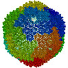 PDB-1gw8: |
