Movie
Movie Controller

[English] 日本語
 Yorodumi
- EMDB-1006: A cryo-electron microscopic study of ribosome-bound termination f... -
+ Open data

- Basic information
| Entry | Database: EMDB / ID: EMD-1006 | |||||||||
|---|---|---|---|---|---|---|---|---|---|---|
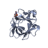
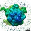
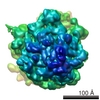
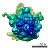
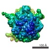
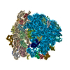
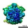
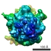
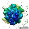
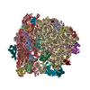
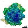
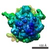
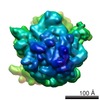
| Title | A cryo-electron microscopic study of ribosome-bound termination factor RF2. | |||||||||
 Map data Map data | Ribosome-bound termination factor RF2 | |||||||||
 Sample Sample |
| |||||||||
| Function / homology |  Function and homology information Function and homology informationtranslation release factor activity, codon specific / translational termination / small ribosomal subunit / large ribosomal subunit rRNA binding / cytosolic large ribosomal subunit / tRNA binding / rRNA binding / structural constituent of ribosome / translation / viral translational frameshifting / cytosol Similarity search - Function | |||||||||
| Biological species |  | |||||||||
| Method | single particle reconstruction / cryo EM / Resolution: 11.3 Å | |||||||||
 Authors Authors | Rawat U / Gao H / Zavialov A / Gursky R / Ehrenberg M / Frank J | |||||||||
 Citation Citation | Journal: Nature / Year: 2003 Title: A cryo-electron microscopic study of ribosome-bound termination factor RF2. Authors: Urmila B S Rawat / Andrey V Zavialov / Jayati Sengupta / Mikel Valle / Robert A Grassucci / Jamie Linde / Bente Vestergaard / Måns Ehrenberg / Joachim Frank /  Abstract: Protein synthesis takes place on the ribosome, where genetic information carried by messenger RNA is translated into a sequence of amino acids. This process is terminated when a stop codon moves into ...Protein synthesis takes place on the ribosome, where genetic information carried by messenger RNA is translated into a sequence of amino acids. This process is terminated when a stop codon moves into the ribosomal decoding centre (DC) and is recognized by a class-1 release factor (RF). RFs have a conserved GGQ amino-acid motif, which is crucial for peptide release and is believed to interact directly with the peptidyl-transferase centre (PTC) of the 50S ribosomal subunit. Another conserved motif of RFs (SPF in RF2) has been proposed to interact directly with stop codons in the DC of the 30S subunit. The distance between the DC and PTC is approximately 73 A. However, in the X-ray structure of RF2, SPF and GGQ are only 23 A apart, indicating that they cannot be at DC and PTC simultaneously. Here we show that RF2 is in an open conformation when bound to the ribosome, allowing GGQ to reach the PTC while still allowing SPF-stop-codon interaction. The results indicate new interpretations of accuracy in termination, and have implications for how the presence of a stop codon in the DC is signalled to PTC. | |||||||||
| History |
|
- Structure visualization
| Movie |
Movie viewer |
|---|---|
| Structure viewer | EM map: SurfViewMolmilJmol/JSmol |
| Supplemental images |
- Downloads & links
-EMDB archive
| Map data | emd_1006.map.gz | 7.9 MB | EMDB map data format | |
|---|---|---|---|---|
| Header (meta data) | emd-1006-v30.xmlemd-1006.xml | 11.4 KB 11.4 KB | Display Display | EMDB header |
| Images |  1006.gif 1006.gif | 29.8 KB | ||
| Archive directory |  http://ftp.pdbj.org/pub/emdb/structures/EMD-1006ftp://ftp.pdbj.org/pub/emdb/structures/EMD-1006 http://ftp.pdbj.org/pub/emdb/structures/EMD-1006ftp://ftp.pdbj.org/pub/emdb/structures/EMD-1006 | HTTPS FTP |
-Related structure data
| Related structure data | 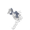 1mi6MC 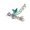 1mvrMC 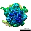 1007C 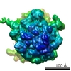 1008C 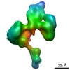 1009C  1010C M: atomic model generated by this map C: citing same article ( |
|---|---|
| Similar structure data |
-Links
| EMDB pages | EMDB (EBI/PDBe) / EMDataResource |
|---|---|
| Related items in Molecule of the Month |


-Map
| File | Download / File: emd_1006.map.gz / Format: CCP4 / Size: 8.2 MB / Type: IMAGE STORED AS FLOATING POINT NUMBER (4 BYTES) | ||||||||||||||||||||||||||||||||||||||||||||||||||||||||||||
|---|---|---|---|---|---|---|---|---|---|---|---|---|---|---|---|---|---|---|---|---|---|---|---|---|---|---|---|---|---|---|---|---|---|---|---|---|---|---|---|---|---|---|---|---|---|---|---|---|---|---|---|---|---|---|---|---|---|---|---|---|---|
| Annotation | Ribosome-bound termination factor RF2 | ||||||||||||||||||||||||||||||||||||||||||||||||||||||||||||
| Projections & slices | Image control
Images are generated by Spider. | ||||||||||||||||||||||||||||||||||||||||||||||||||||||||||||
| Voxel size | X=Y=Z: 2.82 Å | ||||||||||||||||||||||||||||||||||||||||||||||||||||||||||||
| Density |
| ||||||||||||||||||||||||||||||||||||||||||||||||||||||||||||
| Symmetry | Space group: 1 | ||||||||||||||||||||||||||||||||||||||||||||||||||||||||||||
| Details | EMDB XML:
CCP4 map header:
| ||||||||||||||||||||||||||||||||||||||||||||||||||||||||||||
 Z (Sec.)
 Y (Row.)
 X (Col.)


-Supplemental data
- Sample components
-Entire : E.coli 70s ribosome
| Entire | Name: E.coli 70s ribosome |
|---|---|
| Components |
|
-Supramolecule #1000: E.coli 70s ribosome
| Supramolecule | Name: E.coli 70s ribosome / type: sample / ID: 1000 / Number unique components: 4 |
|---|---|
| Molecular weight | Theoretical: 2.5 MDa |
-Supramolecule #1: E.coli 70s ribosome
| Supramolecule | Name: E.coli 70s ribosome / type: complex / ID: 1 / Recombinant expression: No / Ribosome-details: ribosome-prokaryote: LSU 50S |
|---|---|
| Source (natural) | Organism: |
| Molecular weight | Theoretical: 2.5 MDa |
-Macromolecule #1: peptidyl P-tRNA
| Macromolecule | Name: peptidyl P-tRNA / type: ligand / ID: 1 / Recombinant expression: No |
|---|---|
| Source (natural) | Organism: |
-Macromolecule #2: E-tRNA
| Macromolecule | Name: E-tRNA / type: ligand / ID: 2 / Number of copies: 1 / Recombinant expression: No |
|---|---|
| Source (natural) | Organism: |
-Macromolecule #3: MFTI-mRNA
| Macromolecule | Name: MFTI-mRNA / type: ligand / ID: 3 Details: Zavialov et al., A posttermination ribosomal complex is the guanine nucleotide exchange factor for peptide release factor RF3. Cell. 107,1-20 (2001). Number of copies: 1 / Recombinant expression: Yes |
|---|---|
| Source (natural) | Organism: |
-Experimental details
-Structure determination
| Method | cryo EM |
|---|---|
 Processing Processing | single particle reconstruction |
| Aggregation state | particle |
-Sample preparation
| Concentration | 0.09 mg/mL |
|---|---|
| Buffer | pH: 7.5 Details: Polymix buffer, containing at final concentration 5 mM potassium phosphate, 5 mM magnesium acetate, 5mM ammonium chloride, 95 mM potassium chloride, 0.5 mM calcium chloride, 8mM putrescine, 1mM dithioerythritol |
| Vitrification | Cryogen name: ETHANE / Chamber humidity: 58 % / Chamber temperature: 36 K / Instrument: HOMEMADE PLUNGER Details: Vitrification instrument: Two side blotting plunger. Rapid-freezing in liquid ethane Method: Blot and Plunge |
- Electron microscopy
| Microscope | FEI TECNAI F20 |
|---|---|
| Temperature | Average: 93 K |
| Date | Aug 9, 2001 |
| Image recording | Category: FILM / Film or detector model: KODAK SO-163 FILM / Digitization - Scanner: ZEISS SCAI / Digitization - Sampling interval: 14 µm / Number real images: 34 / Average electron dose: 20 e/Å2 / Bits/pixel: 12 |
| Tilt angle min | 0 |
| Tilt angle max | 0 |
| Electron beam | Acceleration voltage: 200 kV / Electron source:  FIELD EMISSION GUN FIELD EMISSION GUN |
| Electron optics | Calibrated magnification: 49696 / Illumination mode: FLOOD BEAM / Imaging mode: BRIGHT FIELD / Cs: 2.0 mm / Nominal defocus max: 5.975 µm / Nominal defocus min: 1.39 µm / Nominal magnification: 50000 |
| Sample stage | Specimen holder: Oxford,cryo-transfer 3500 / Specimen holder model: GATAN LIQUID NITROGEN |
| Experimental equipment |  Model: Tecnai F20 / Image courtesy: FEI Company |
-Image processing
| CTF correction | Details: Wiener filtration of defocus groups |
|---|---|
| Final reconstruction | Applied symmetry - Point group: C1 (asymmetric) / Algorithm: OTHER / Resolution.type: BY AUTHOR / Resolution: 11.3 Å / Resolution method: FSC 0.5 CUT-OFF / Software - Name: SPIDER/WEB Details: Frank, J. (2000) Three-Dimensional Cryoelectron Microscopy of Ribosomes, Methods of Enzymology (Ch.18) 317, 276-291. Number images used: 14965 |
-Atomic model buiding 1
| Initial model | (PDB ID: , ) |
|---|---|
| Details | manual fitting using O |
| Refinement | Protocol: RIGID BODY FIT |
| Output model | PDB-1mi6: PDB-1mvr: |